Pain guide

Ankle pain: where it hurts, what helps, and when to get it checked

Ankle pain is common: in a review of 31 studies, about 1 in 7 adults aged 45 and over had frequent ankle pain. Most of it is short-term and eases with looking after it yourself. Sprains are the commonest ankle injury in sport, and most recover with early, protected movement rather than rest.

Start with the warning signs and the X-ray rules below. Then find where it hurts: if it points to a clear problem, go to that guide. If you're not sure, follow the plan on this page from stage 1.

When to get help first

Exercise is for pain that has been checked or is clearly mild and settling.

Get urgent medical help if

  • Call 999 or go to A&E: after an injury, you heard a crack, the foot or ankle has changed shape or points at an odd angle, a bone is sticking out, there is a bad cut over it, you're in severe pain, or the toes look blue or white, feel numb or are cold to touch. These can mean a broken bone. Don't drive yourself.
  • Call 999: calf or leg pain or swelling with breathlessness or chest pain. It could be a clot that has reached the lungs.
  • Call 999 or go to A&E: a foot or leg that is suddenly cold, pale, numb, weak or very painful without an injury. A sudden loss of blood supply is a vascular emergency.
  • Same day (urgent GP appointment or NHS 111): after an injury, you can't put weight on it or walk more than a few steps, the pain is very bad or getting worse, there is a lot of swelling or bruising or it is getting worse, or it feels very stiff. If you think it's broken, don't treat it yourself.
  • Same day: a sudden snap or pop at the back of the heel, then being unable to walk on tiptoe or climb stairs. It could be an Achilles tendon rupture or a broken heel bone.
  • Same day: a sudden, very painful, hot, swollen, red joint, often the big toe, especially with a high temperature, feeling shivery or being unable to eat. It may be gout, but it could be a joint infection (septic arthritis), which is urgent.
  • Same day (GP or NHS 111): throbbing pain and swelling in one calf, with red or darker skin. It could be a blood clot (DVT). Watch for it after an ankle injury or time in a cast or boot.
  • Same day: you have diabetes and a new foot problem, such as an ulcer or wound, signs of infection, or a hot, swollen foot with a change in colour, even if it doesn't hurt. Contact your diabetes foot team or GP: NICE asks for these to reach a specialist foot service within 1 working day, and a suspected Charcot foot is kept off weight until it has been checked.
  • Same day: in a cast or boot, pain that is getting worse, toes that are numb or burning, a cast that feels too tight, toes that are swollen, blue or white, or a bad smell from under the cast.

See a doctor or physiotherapist if

  • Pain that isn't better after 2 weeks of looking after it yourself, is getting worse, keeps coming back, or stops you doing normal things.
  • An ankle that keeps giving way months after a sprain.
  • Tingling or loss of feeling in the foot, or a foot that feels weak.
  • Any foot pain if you have diabetes: foot problems can be more serious.
  • Flat feet that are painful, stiff, weak or numb, that you didn't have before, or that affect only one foot.
  • One spot on a foot bone that hurts with every step and later at night: a possible stress fracture.

Where does it hurt?

Location narrows the likely causes; it does not diagnose.

Outside of the ankle, after it rolled inwards

Swelling and bruising: a sprain of the outer ligaments, which is where most sprains happen. Check the X-ray rules below first.

Read: Sprained ankle →
Outside of the ankle that keeps rolling or giving way

Months or years after a sprain, with repeated sprains or giving way, and ongoing pain or swelling: chronic ankle instability.

Read: Chronic ankle instability →
Behind the bony bump on the outside, worse with activity

Sometimes with clicking or snapping: the peroneal tendons, which are easy to confuse with a ligament problem. See the notes below.

Inside of the ankle and arch, the arch slowly dropping

Hard to rise on tiptoe, one foot flatter than the other: tibialis posterior tendon dysfunction (adult flatfoot). A flat foot you didn't have before, or on one side only, needs a GP check.

Read: Flat feet →
Deep ache in the ankle joint, with stiffness and swelling

Often years after a bad sprain or a fracture: ankle arthritis. Most ankle arthritis follows an injury.

Read: Ankle arthritis and stiff ankles →
Back of the ankle, or the cord above the heel

Stiff in the morning: Achilles tendinopathy.

Read: Achilles tendinopathy →
Above the ankle, between the shin bones

After the foot was forced outwards: a high ankle sprain, which often comes with other ligament damage or a fracture. Get it assessed.

Read: Sprained ankle →
Sudden hot, red, swollen ankle without an injury

Gout or a joint infection. A fever or feeling unwell makes infection more likely: get help the same day.

Calf swelling and throbbing pain after an ankle injury or a cast

With red or darker skin, it could be a blood clot (DVT): get help the same day.

Read: Calf pain →
The foot rather than the ankle

Heel, arch, ball of the foot and toe pain have their own map.

Read: Foot pain →

Understand it

Do I need an X-ray? The Ottawa ankle rulesStrong evidence

After a twisted ankle, clinicians use the Ottawa ankle rules to decide who needs an X-ray. An ankle X-ray is needed if the back edge or tip of either ankle bone is tender to press, or you couldn't take four steps straight after the injury or when you are examined. A foot X-ray is needed if the navicular (the bony bump on the inner midfoot) or the base of the fifth metatarsal (the bump halfway along the outer edge of the foot) is tender, or the same four-step test fails.

The rules are very good at ruling fractures out. In the study that refined them, they picked up every fracture and could cut ankle X-rays by about a third; a review of 27 studies (15,581 people) found they missed almost no ankle or midfoot fractures, in children too, and could cut X-rays by 30 to 40%.

They are a guide for clinicians, not a self-test. If you can't walk four steps, if pressing on one of those bony points hurts, if the foot looks out of shape, or if you're unsure, get it checked the same day. Ligament damage is best judged 4 to 5 days after the injury, once the swelling has gone down.

Sources 20, 21, 19, 2

Ankle pain is common, and sprains often come backStrong evidence

About 15% of adults aged 45 and over report frequent ankle pain. Sprains are commoner in women and girls than in men and boys, in children than adults, and in indoor and court sports. Pain falls fast in the first 2 weeks after a sprain, but in a review of 31 studies 5% to 33% still had some pain a year later and 3% to 34% sprained the ankle again. That is why the sprain guide keeps going after the pain has gone.

Sources 22, 23, 24

Outer ankle tendon pain (the peroneal tendons)Mixed evidence

The peroneal tendons run behind the bony bump on the outside of the ankle. Tendon irritation, tears and tendons slipping out of their groove cause outer ankle pain that can be hard to tell from a ligament injury, and it can come with an unstable ankle or a high-arched foot. Treatment advice rests on case series and expert opinion: anti-inflammatories, rest, changing activity and orthoses first, then a boot or cast for 6 weeks in stubborn cases.

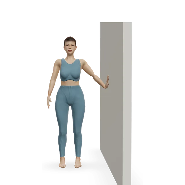
A sensible home plan borrows the tendon-loading approach (editorial): press the outside of the foot into a wall without moving (8 to 12 holds of about 6 seconds), then band eversion (3 sets of 10, as in a chronic ankle instability trial), single-leg calf raises, single-leg balance and side hops, keeping pain to about 5 out of 10 and settled by the next morning. Clicking, or a tendon you can feel slipping over the ankle bone, needs assessing.

Sources 38, 32, 39

Gout and a hot, swollen ankleStrong evidence

A sudden hot, red, swollen joint without an injury may be gout, which most often affects the big toe, or a joint infection (septic arthritis), which is urgent. See a GP for a first attack of gout, and get help the same day with a high temperature, shivering or being unable to put weight on it. There is no exercise plan for a hot, swollen joint: in a gout attack, the NHS advises rest, raising it and an ice pack for up to 20 minutes, and NICE says ice alongside medicine may help.

Sources 4, 5, 10

Diabetes and your ankles and feet

With diabetes, check your feet every day, especially after exercise. NICE asks for an ulcer, an infection, or an unexplained hot, swollen foot with a change in colour to reach a specialist foot service within 1 working day, and a suspected Charcot foot is kept off weight until it has been checked. Exercise is still recommended: a 2023 international guideline suggests a foot and ankle exercise programme for people at low to moderate risk, with suitable foot protection. The foot pain guide has more.

Sources 8, 13

Myths worth dropping

Many popular beliefs about ankles are weaker than they sound.

  • "If I can walk on it, it isn't broken." The X-ray rules also check for tenderness over the bones, because you can have a fracture and still walk. If a bony point is tender, or you're unsure, get it checked.
  • "Rest a sprain until it stops hurting." Early, protected weight-bearing and exercise beat immobilisation, and exercise in the first week improved function in a trial.
  • "Once it stops hurting, the sprain is fixed." Up to a third of people sprain the same ankle again, and in one cohort 40% had chronic instability a year later. Balance training cut repeat sprains in a large trial.
  • "Ice is the treatment." There isn't enough trial evidence to judge rest, ice, compression and elevation; ice may help comfort alongside exercise.
  • "Flat feet need fixing." Most cause no problems and need no treatment, and a flat foot is not a general injury risk.
  • "You need running shoes for your foot type." Choosing running shoes by foot type did not prevent injuries in a Cochrane review.

Sources 20, 2, 19, 26, 24, 27, 42, 7, 43, 44, 45
What helps, and how strong the evidence is

Sprains: early, protected movement beats restStrong evidence

A brace or tape with gradually more weight through the foot, plus exercise, beats immobilisation, and for a severe sprain up to 10 days in a below-knee cast or brace can speed early recovery. Starting exercise in the first week improved function in a trial of 101 people. The sprain guide has the full plan.

Sources 19, 25, 26

Balance training and a brace cut repeat sprainsStrong evidence

Exercise-based rehab after a sprain lowered the odds of another one over the next year (odds ratio 0.60 across 14 trials). In a trial of 522 athletes, an 8-week home balance programme cut repeat sprains from 33% to 22%. A brace worn for sport cuts them too, and lace-up braces cut ankle injuries in a trial of 1,460 school basketball players.

Sources 28, 27, 29, 30, 19

An ankle that keeps giving way: balance and strength training firstStrong evidence

The clinical guideline's top grade goes to balance and neuromuscular exercise for chronic ankle instability, and balance training gave the most consistent gains in a systematic review. In one trial, 6 weeks of balance training, or of band and heel-raise training, three times a week improved strength, balance and side-hopping.

Sources 19, 31, 32

Inner ankle tendon pain: strengthening helps, on thin evidenceEmerging evidence

In tibialis posterior tendon dysfunction, adding resistance exercise (a slow, lowering phase did best) to insoles and stretching improved pain and function in a small trial; a review of 3 trials (93 people) called the research thin. The flat feet guide has the plan.

Sources 35, 36

Ankle arthritis: ankle trials are thin, general arthritis advice appliesMixed evidence

Most ankle arthritis follows an injury: 78% in one series of 406 ankles. There are no completed ankle-arthritis exercise trials beyond a feasibility study, so NICE's general advice for osteoarthritis (tailored exercise, and weight loss if you need it) is the best guide.

Sources 40, 33, 34, 9

Calf stretching adds only a few degrees of ankle bendStrong evidence

In a systematic review, calf stretching increased ankle bend (dorsiflexion) by about 2 to 3 degrees. Whether that matters for pain or function is unclear, so the plans here use stretching alongside strength and balance, not instead.

Source 37

Insoles and shoe changes don't prevent repeat sprainsEmerging evidence

Foot orthoses and footwear changes haven't been shown to prevent repeat sprains, and a brace or tape on its own doesn't improve balance in an unstable ankle. For arthritis, NICE says not to routinely offer insoles, braces or supports unless the joint is unstable or loaded abnormally and exercise alone won't do.

Sources 19, 9

How much pain is OK while you exercise?

The foot and ankle pain rule

Some discomfort during exercise is fine. Aim for pain you would call mild to moderate, no more than about 5 out of 10, that settles within an hour, with no more swelling or stiffness the next morning. If it's worse the next day, do a little less (a smaller range, fewer reps, a lighter band), not nothing.

The exceptions: pain in one spot on a bone should be pain-free, because it could be a stress fracture; burning, tingling or numbness means stop that movement; and if your feet are numb, pain won't warn you, so check your skin after exercise.

If pain climbs day after day, or you notice a warning sign, stop and get it checked. The 5 out of 10 figure is an editorial starting point that matches this site's other pain guides, not a tested rule.

Sources 12, 13

The exercise plan: 3 stages

Start at the first stage. Each move opens a full guide with a 3D demo.

Stage 1

Settle: gentle movement, little and often

Days 1 to 14. Supportive shoes, short walks, and moves in a comfortable range. Doses come from NHS hospital ankle leaflets where named; the rest are editorial starting points.

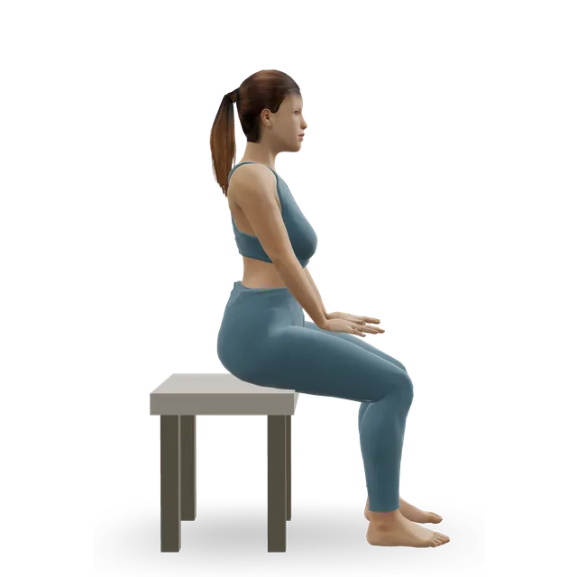
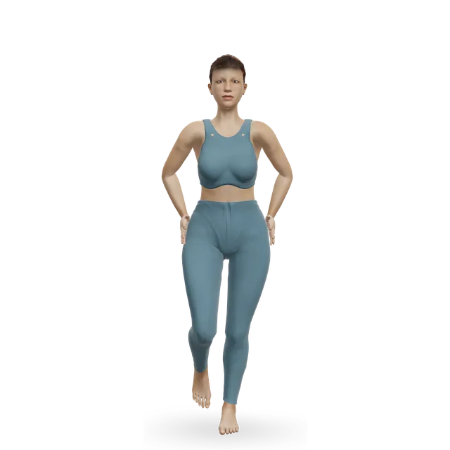
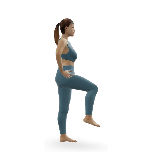
Ankle CARs demonstrated on the 3D figure3D demoAnkle CARsBeginner · No equipment

10 circles each way, 3 to 4 times a day · A comfortable range; the dose in a King's College Hospital leaflet

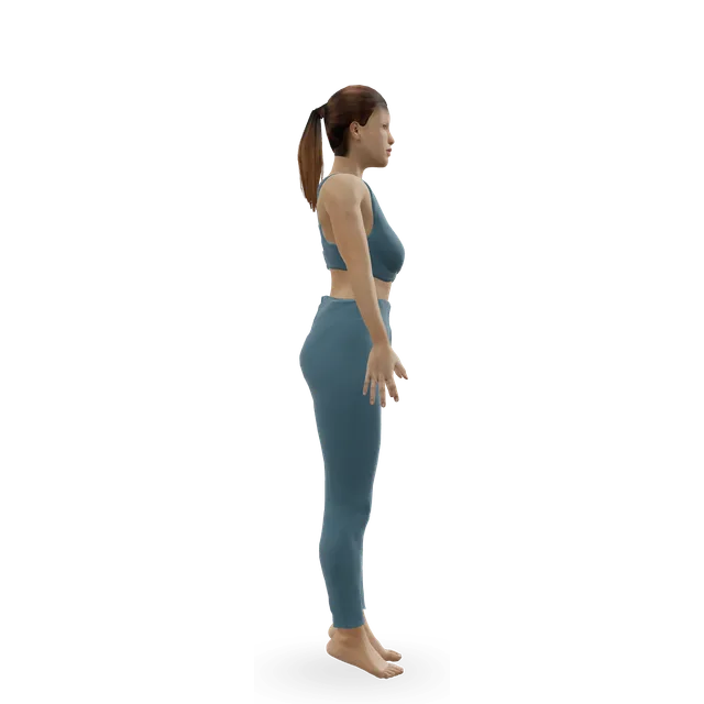
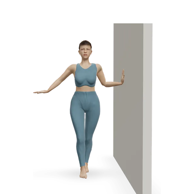
Single-leg stand demonstrated on the 3D figure3D demoSingle-leg standBeginner · A wall

Up to 30 s, a few times a day · Holding a support; the leaflet's first level

Easier: Tandem stance and heel-to-toe walk · Harder: Single Leg Reach Matrix

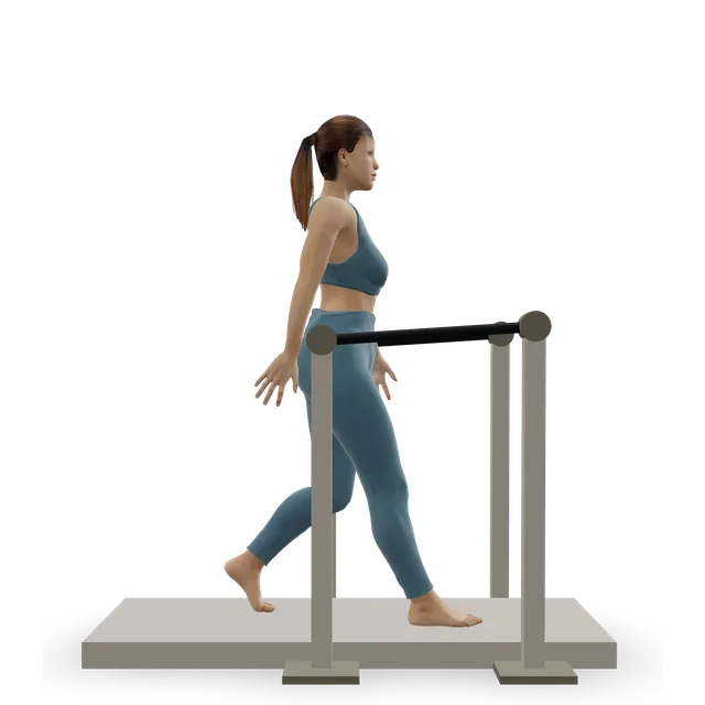
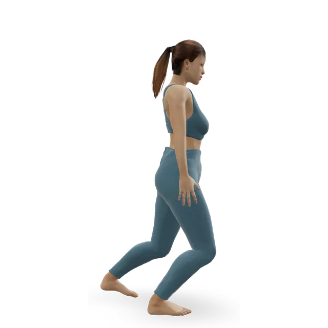
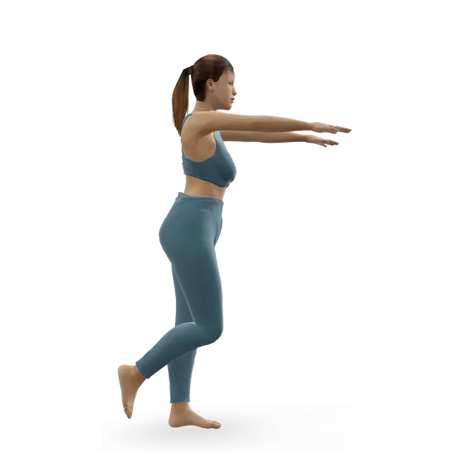
Treadmill walking demonstrated on the 3D figure3D demoTreadmill walkingBeginner · Cardio machine

A few minutes, often · Outdoors or around the house; avoid long spells on your feet at first

Harder: Treadmill jogging

Move on when: You can walk around normally with little pain, and any swelling is going down.

Follow along: a timer, a cue and the next move, one step at a time.
Stage 2

Build: strength in every direction, and balance

About weeks 2 to 6.

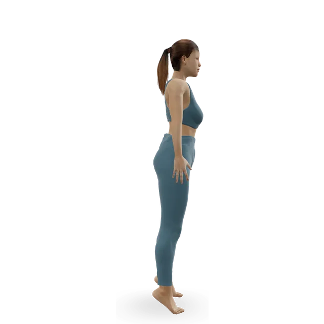
Slow calf raise demonstrated on the 3D figure3D demoSlow calf raiseBeginner · A wall

12 reps, 3 times a day · King's College Hospital leaflet

Easier: Seated bent-knee calf raise (soleus raise) · Harder: Standing dumbbell calf raise

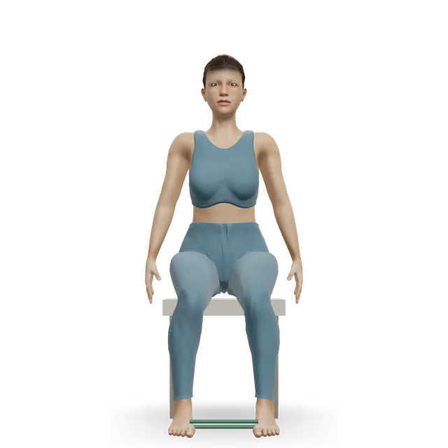
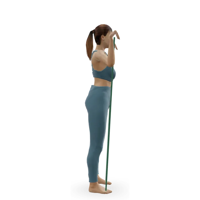
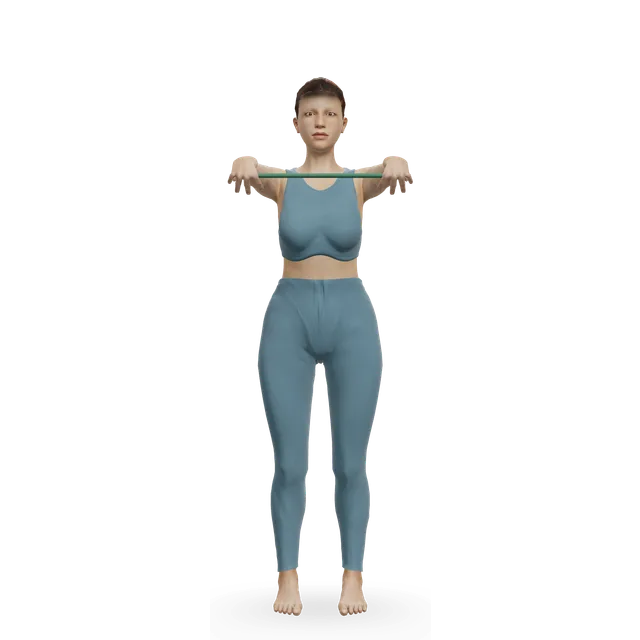
Band ankle eversion demonstrated on the 3D figure3D demoBand ankle eversionBeginner · A band + A bench or box

2 to 3 sets of 5 to 8 · United Lincolnshire Hospitals leaflet

Easier: Isometric ankle eversion against a wall · Harder: Pogo hops

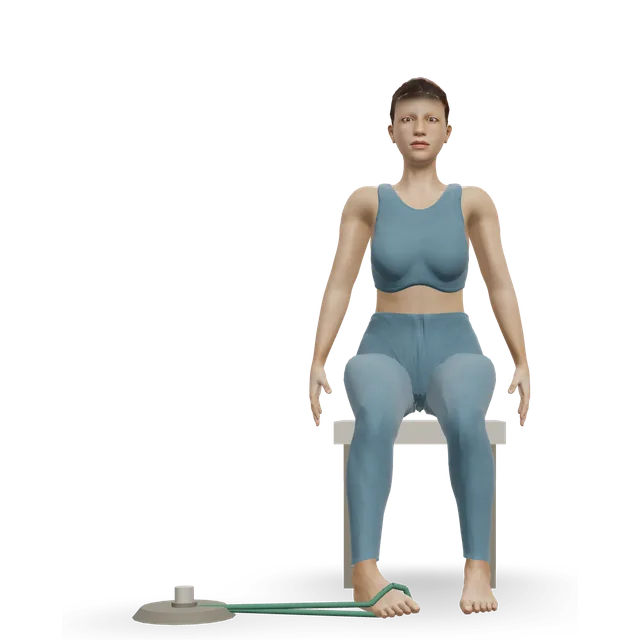
Band ankle inversion demonstrated on the 3D figure3D demoBand ankle inversionBeginner · A band + A bench or box

2 to 3 sets of 5 to 8 · United Lincolnshire Hospitals leaflet; a pain-free range

Harder: Single-leg calf raise from a step

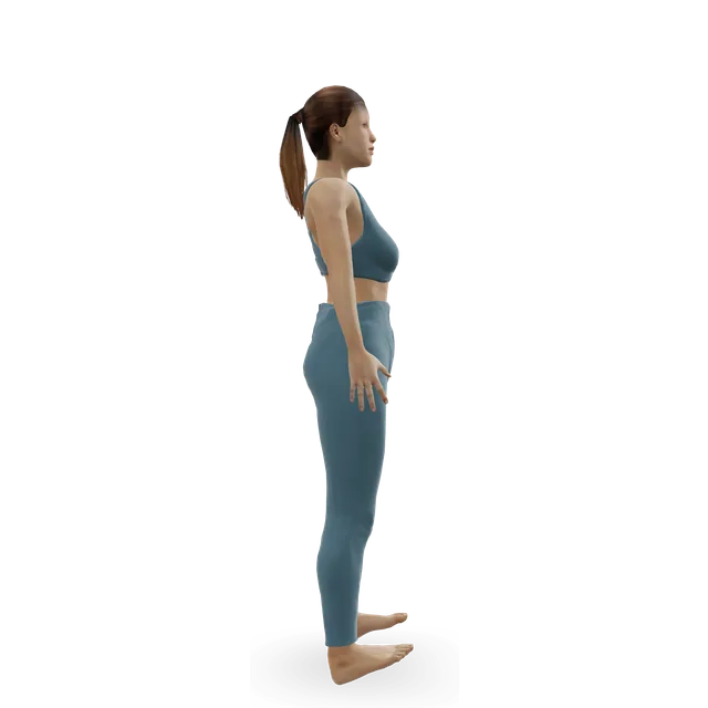
Tibialis raise demonstrated on the 3D figure3D demoTibialis raiseBeginner · No equipment

2 to 3 sets of 10 · The leaflet lifts the foot against a band; this swap is editorial

Harder: Single-leg tibialis raise

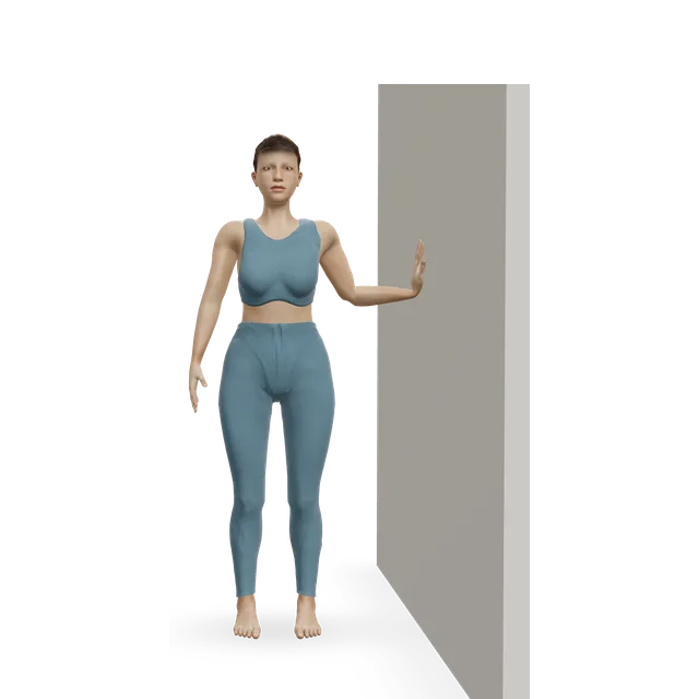
Knee-to-wall ankle rock demonstrated on the 3D figure3D demoKnee-to-wall ankle rockBeginner · A wall

10 slow rocks · A St George's Hospital leaflet exercise

Harder: Frog rock

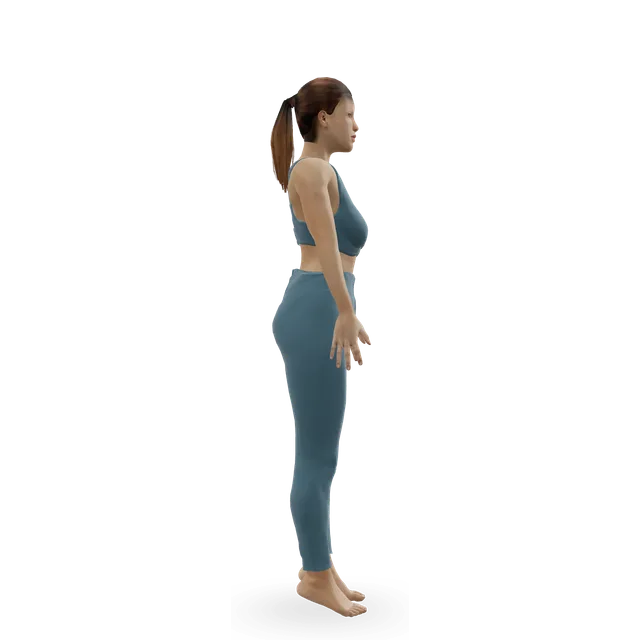
Single-leg stand demonstrated on the 3D figure3D demoSingle-leg standBeginner · A wall

Up to 30 s · Without support, then with your eyes closed: the leaflet's next levels

Easier: Tandem stance and heel-to-toe walk · Harder: Single Leg Reach Matrix

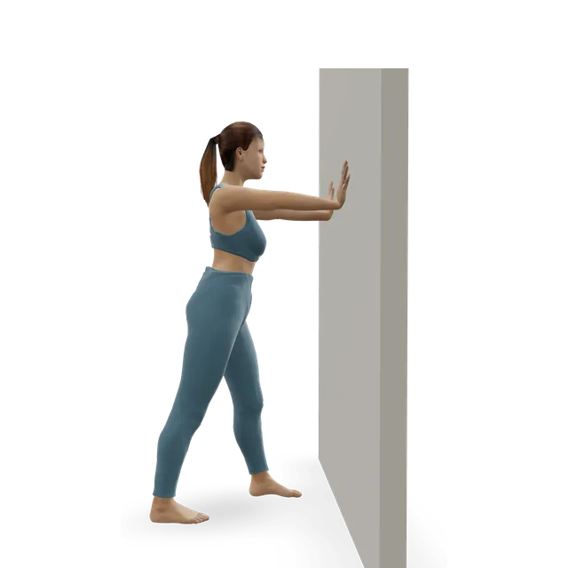
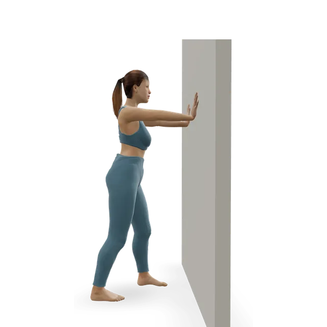
Wall calf stretch (gastrocnemius) demonstrated on the 3D figure3D demoWall calf stretch (gastrocnemius)Beginner · A wall

3 holds of 20 to 30 s · United Lincolnshire Hospitals leaflet

Easier: Seated calf stretch with a towel · Harder: Peroneals Stretch

Move on when: Twelve calf raises are easy, and you can stand on the sore leg for 30 seconds with your eyes closed.

Follow along: a timer, a cue and the next move, one step at a time.
Stage 3

Strengthen and return: one leg, then hops

From about week 6. Hop and jump every other day, not daily.

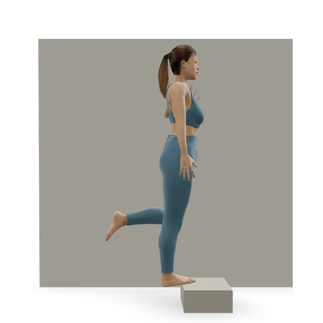
Single-leg calf raise from a step demonstrated on the 3D figure3D demoSingle-leg calf raise from a stepIntermediate · A step + A wall

12 reps, 3 times a day · King's College Hospital leaflet; or 2 sets of 10 building to 4 sets of 15 over 6 weeks, as in a chronic instability trial

Easier: Slow calf raise · Harder: Single-leg pogo hops

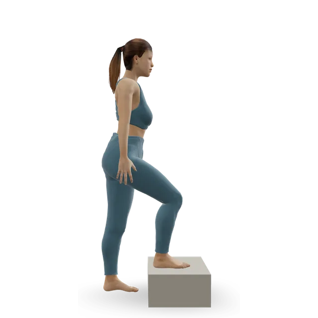
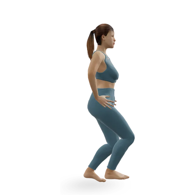
Step-up demonstrated on the 3D figure3D demoStep-upBeginner · A step

3 sets of 8 to 10 each side

Easier: Low step-up · Harder: Dumbbell step-up

Ankle bounces (mini pogo) demonstrated on the 3D figure3D demoAnkle bounces (mini pogo)Beginner · No equipment

3 sets of 8 to 12, every other day · The King's College Hospital leaflet's last stage

Harder: Pogo hops

Pogo hops demonstrated on the 3D figure3D demoPogo hopsIntermediate · No equipment

3 sets of 8 to 12, every other day · Once ankle bounces are easy

Easier: Ankle bounces (mini pogo) · Harder: Single-leg pogo hops

Move on when: Hopping and your usual activity are comfortable, with no extra swelling the next day.

Follow along: a timer, a cue and the next move, one step at a time.

Which stretch suits which pain

Hold 2 to 3 times for 20 to 30 seconds, a mild stretch, never sharp.

StretchReasonable forTake care or skipWhy
Wall calf stretch (gastrocnemius)Plantar heel pain, Sever's disease, a stiff ankle, ball-of-foot painInner ankle tendon pain: keep it gentle. After a sprain, start once you can take your full weightCalf stretching adds a few degrees of ankle bend.
Soleus wall stretchAs for the wall calf stretchAs for the wall calf stretchReaches the deeper calf muscle, with the knee bent.
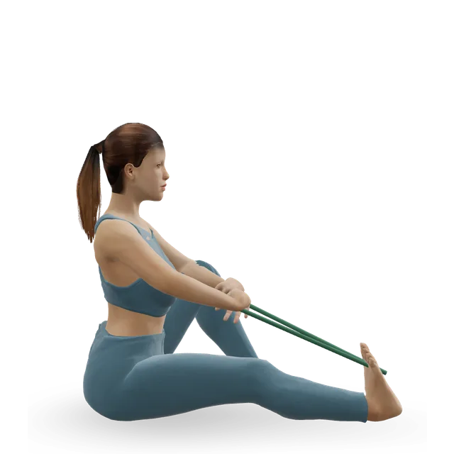
Seated calf stretch with a towelAnyone who can't stand to stretch; from about week 2 after a sprainNone within comfortNHS hospital leaflets start here after a sprain.
Knee-to-wall ankle rockA stiff ankle after a sprain or fracture; ankle arthritis within comfortA pinch at the front of the ankle: use a smaller rangeBending the ankle with your weight on it is how the sprain guideline measures ankle stiffness.
Ankle CARsEarly after a sprain; arthritis; morning stiffnessNone within comfortGentle movement in every direction.
Peroneals StretchNot needed by most peopleThe first weeks after an outer ankle sprainIt turns the sole inwards, the movement that sprains the outer ankle (an editorial caution).
Posterior Tibialis StretchNot needed by most peopleInner ankle tendon pain: don't force itThe tendon trials strengthened it rather than stretching it, and holding the ankle at the end of its range may squeeze the tendon.

Ankle pain: guides by cause

Routines to follow

More moves for this area

Questions people ask

Why does my ankle hurt with no injury?

Common causes without an injury include tendon problems (the Achilles at the back, the tibialis posterior on the inside, the peroneal tendons on the outside) and arthritis, which often follows an old injury. A sudden hot, red, swollen ankle with no injury needs a same-day check, because it could be gout or an infection.

Why does my ankle hurt when I walk?

Use the map above: the outside after a twist points to a sprain, the inside with a dropping arch to the tibialis posterior tendon, a deep ache with stiffness to arthritis, and the back of the ankle to the Achilles. If it is no better after 2 weeks of self-care, see a GP.

Inside or outside ankle pain: what's the difference?

Outer ankle pain is usually a sprained ligament, an ankle that keeps giving way, or the peroneal tendons. Inner ankle pain with a flattening arch is often the tibialis posterior tendon, and a new or one-sided flat foot needs a GP check.

Should I wear an ankle brace?

After a sprain, a brace or tape with early walking is the guideline's first choice, and a brace for sport afterwards cuts repeat sprains. A brace doesn't train your balance, so do balance work alongside it. For arthritis, NICE says braces aren't routinely needed unless the joint is unstable.

Is my ankle sprained or broken?

Clinicians use the Ottawa ankle rules: an X-ray if the back edge or tip of either ankle bone is tender, the bony points on the inner midfoot or outer edge of the foot are tender, or you can't take four steps. You can have a fracture and still walk. A crack, an odd shape, or toes that are blue, white, numb or cold need A&E.

Why are my ankles stiff in the morning?

Stiffness after rest is typical of osteoarthritis, which in the ankle usually follows an old injury, and stiffness is common after a sprain or fracture too. Gentle ankle circles and knee-to-wall rocks help many people loosen up. If it is getting worse or comes with swelling, see a GP.

How do I strengthen my ankles?

Calf raises, band work in both directions, lifting the front of the foot, and balance on one leg, progressing to hops. That is what this page's plan builds, from NHS hospital leaflets and a trial in which 6 weeks of band and heel-raise training or balance training improved strength, balance and hopping.

Other pain guides

Sources

  1. NHS · Ankle pain
  2. NHS · Sprains and strains
  3. NHS · Broken ankle
  4. NHS · Gout
  5. NHS · Septic arthritis
  6. NHS · Deep vein thrombosis (DVT)
  7. NHS · Flat feet
  8. NICE NG19 · Diabetic foot problems: prevention and management
  9. NICE NG226 · Osteoarthritis in over 16s: diagnosis and management (2022)
  10. NICE NG219 · Gout: diagnosis and management (2022)
  11. NHS Scotland Right Decision Service · Intermittent claudication: vascular surgery pathway
  12. Health New Zealand Te Whatu Ora · Stress fractures of the foot and shin bone
  13. Bus 2024 · Guidelines on the prevention of foot ulcers in people with diabetes (IWGDF 2023 update)
  14. St George's University Hospitals NHS · Ankle sprains (patient leaflet, 2025)
  15. King's College Hospital NHS · Ankle sprain: information for patients (2021)
  16. United Lincolnshire Hospitals NHS · Ankle sprain exercises (2025)
  17. Arthritis UK · Foot and ankle pain
  18. Arthritis UK · Osteoarthritis of the foot and ankle
  19. Martin 2021 · Lateral ankle ligament sprains: JOSPT clinical practice guideline, revision 2021
  20. Bachmann 2003 · Accuracy of the Ottawa ankle rules to exclude fractures of the ankle and midfoot (systematic review, BMJ)
  21. Stiell 1993 · Decision rules for X-rays in acute ankle injuries: refinement and prospective validation (JAMA)
  22. Thomas 2011 · Foot and ankle pain in middle and old age (systematic review, Pain)
  23. Doherty 2014 · The incidence and prevalence of ankle sprain injury (meta-analysis of 181 studies, Sports Medicine)
  24. van Rijn 2008 · What is the clinical course of acute ankle sprains? (systematic review, American Journal of Medicine)
  25. Lamb 2009 · Mechanical supports for acute, severe ankle sprain: the CAST trial (RCT, Lancet)
  26. Bleakley 2010 · Accelerated rehabilitation after ankle sprain (RCT, BMJ)
  27. Hupperets 2009 · Unsupervised home balance training and repeat ankle sprains: the 2BFit trial (RCT, BMJ)
  28. Wagemans 2022 · Exercise-based rehabilitation reduces reinjury after ankle sprain (meta-analysis update, PLoS One)
  29. Janssen 2014 · Bracing vs neuromuscular training to prevent repeat ankle sprains (RCT, BJSM)
  30. McGuine 2011 · Lace-up ankle braces and injury rates in high school basketball players (RCT, AJSM)
  31. Kosik 2017 · Treatments to improve function in chronic ankle instability (systematic review, BJSM)
  32. Hall 2018 · Balance and strength training protocols for chronic ankle instability, part I: clinical outcomes (RCT, Journal of Athletic Training)
  33. Tejero 2021 · Conservative treatment of ankle osteoarthritis (review, Journal of Clinical Medicine)
  34. Smith 2024 · Education plus exercise for ankle osteoarthritis (feasibility RCT, Musculoskeletal Science and Practice)
  35. Kulig 2009 · Orthoses and resistive exercise for posterior tibial tendon dysfunction (RCT, Physical Therapy)
  36. Ross 2018 · Exercise for posterior tibial tendon dysfunction: a systematic review of trials and guidelines (BMJ Open Sport & Exercise Medicine)
  37. Radford 2006 · Does stretching increase ankle dorsiflexion range of motion? (systematic review, BJSM)
  38. Davda 2017 · Peroneal tendon disorders (review, EFORT Open Reviews)
  39. Silbernagel 2007 · Continued sports activity with a pain-monitoring model in Achilles tendinopathy (RCT, AJSM)
  40. Valderrabano 2009 · Etiology of ankle osteoarthritis (406 ankles, Clinical Orthopaedics and Related Research)
  41. Hertel 2019 · An updated model of chronic ankle instability (Journal of Athletic Training)
  42. van den Bekerom 2012 · Rest, ice, compression and elevation for ankle sprains in adults (systematic review, Journal of Athletic Training)
  43. Neal 2014 · Foot posture as a risk factor for lower limb overuse injury (meta-analysis, Journal of Foot and Ankle Research)
  44. Nielsen 2014 · Foot pronation and injury risk in novice runners in neutral shoes (1-year cohort, BJSM)
  45. Relph 2022 · Running shoes for preventing running injuries in adults (Cochrane review)

Exercise education, not a diagnosis or treatment plan. It is written from the guidelines and studies listed above; review by a clinician is pending. If your pain follows an injury, is severe, is getting worse, or comes with any warning sign listed on this page, get it assessed. Health disclaimer.