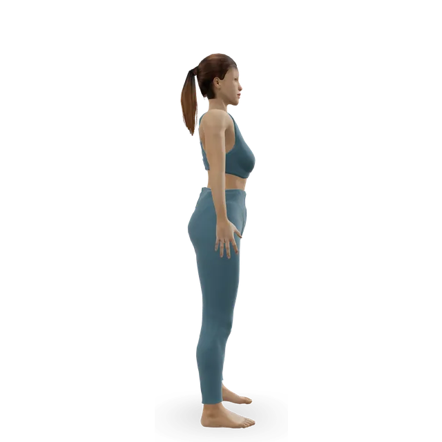
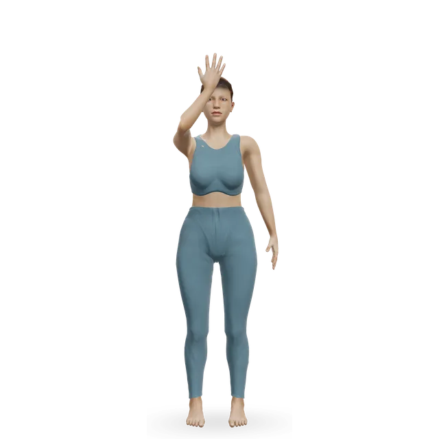
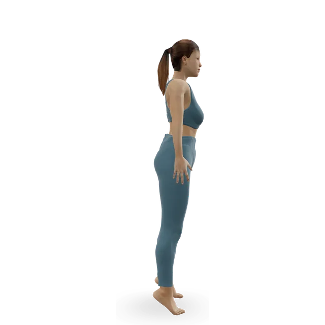
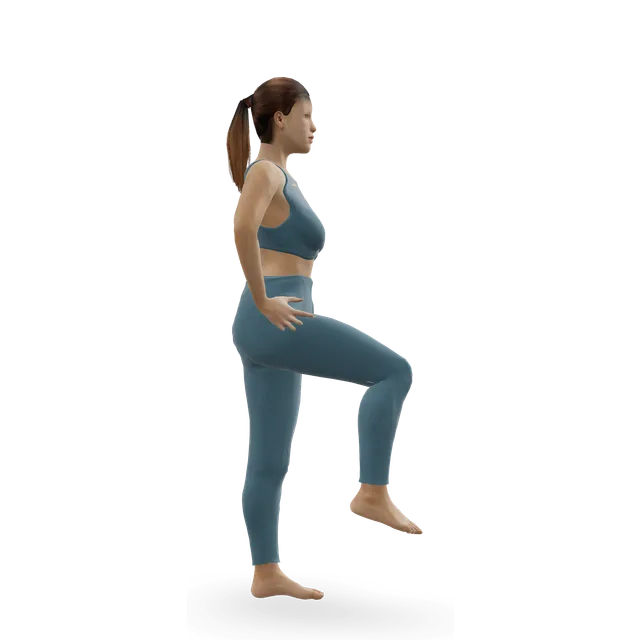
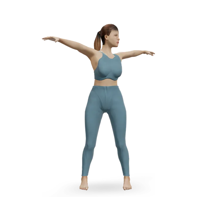
Also called suboccipitals, muscles at the base of the skull, rectus capitis posterior major and minor, obliquus capitis superior and inferior
The suboccipital muscles are four small pairs of muscles tucked under the base of the skull: the rectus capitis posterior major and minor and the obliquus capitis superior and inferior. They join the skull to the top two neck vertebrae, beneath the larger muscles at the back of the neck.
They make the small nodding and turning movements of the head and keep it finely balanced. They are often blamed for headaches at the base of the skull: studies link them with some headaches, but have not shown that they cause them.
Where the suboccipital muscles is and what it does
What it does
Tip the head back at the very top of the neck: the small upward nod, or lifting your eyes to a high shelf.Extension of the head at the atlanto-occipital joint
Turn the head at the top joint: the small turn of a 'no'.Rotation of the head to the same side at the atlantoaxial joint
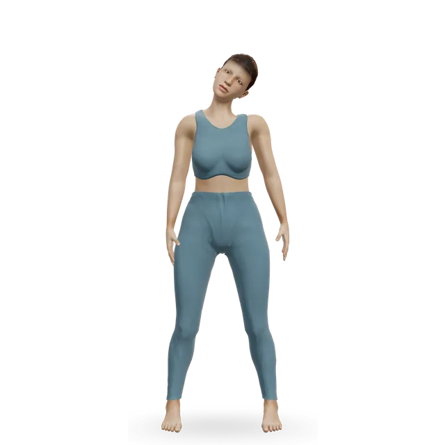
Tilt the head slightly to the side.Lateral flexion of the head (obliquus capitis superior)
Keep the head finely balanced on the neck.Postural control of head position
Nerve supply
The suboccipital nerve, the back branch (dorsal ramus) of the first neck nerve (C1). It only moves these muscles; feeling from the back of the scalp travels in a different nerve, the greater occipital nerve from C2, which passes right beside them.
Fine-tuning and sensing head positionEmerging evidence
They are described as the head's postural muscles that also let it nod and turn. Small muscles that work alongside much larger ones have a much higher density of muscle spindles, the sensors that detect stretch, which led researchers to suggest they mainly feed position information back to the brain. That idea comes from comparing published spindle counts, not from testing these muscles directly.
Working harder when the head pokes forwardEmerging evidence
In fine-wire electromyography (EMG), which records muscle activity, of 20 people, the rectus capitis posterior muscles worked at about 11 to 14% of their maximum with the head in a relaxed position and about 35 to 39% with the head pushed forward. In another EMG study, the rectus capitis posterior major also worked harder as the head was drawn back into a chin tuck. These show muscle activity, not harm or a training effect.
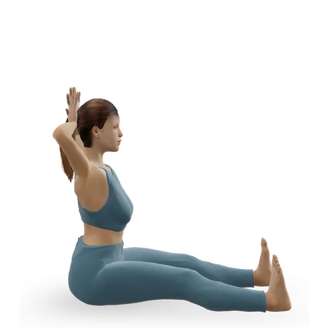
One EMG study, which we read as an abstract, found the rectus capitis posterior major working harder as the head is drawn back; that may be the muscle steadying the head rather than being trained. The two presses come next because pressing the back of the head into your hands loads these muscles' main action, tipping the head back; that order follows from their actions. No study has tested exercises to strengthen these muscles.
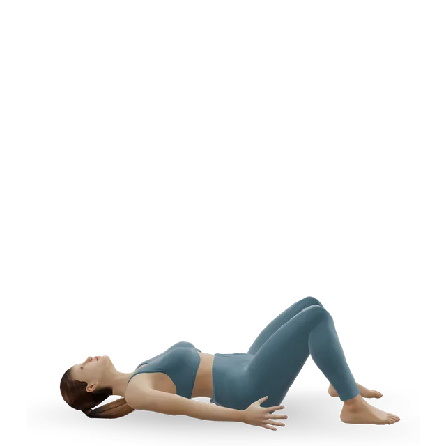
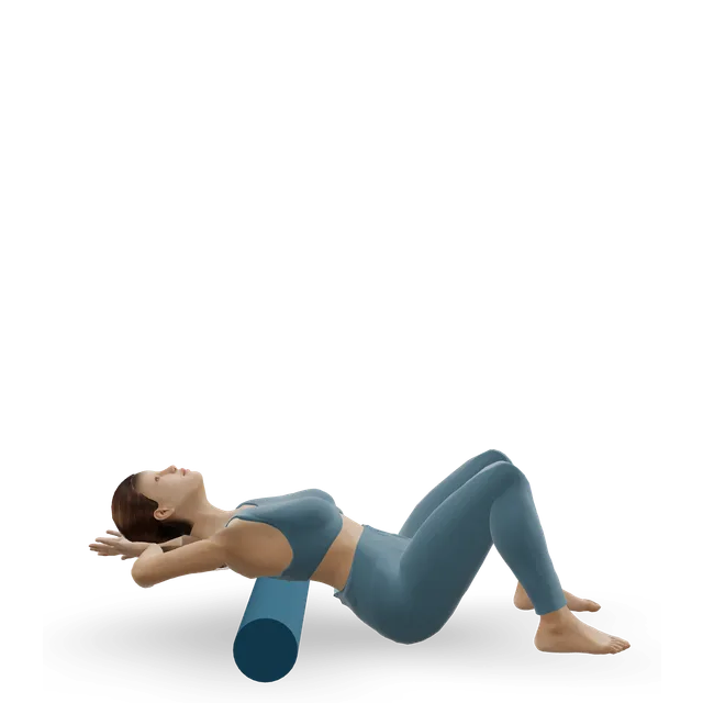
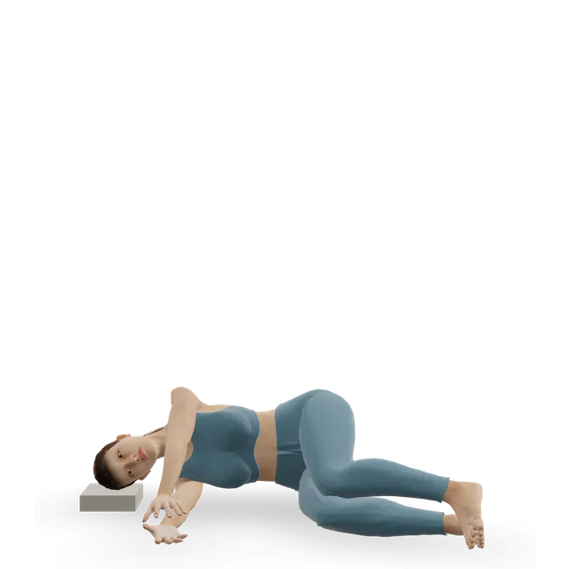
Nodding your chin gently toward your throat bends the head forward at the top joint, the opposite of what these muscles do. No study has measured a stretch for them. 'Suboccipital release', with a clinician's fingertips held under the base of the skull, is a hands-on technique; no trial has tested releasing these muscles yourself with balls or rollers, and you should never press on the front or sides of the throat.
Headache from the neck is pain referred from structures supplied by the top three neck nerves, most often the joint between the second and third neck vertebrae, and physiotherapy is the first treatment. In 20 people with this kind of headache, the suboccipital muscles and upper trapezius had higher tone and stiffness than in 20 people without, measured with a hand-held device.
The rectus capitis posterior minor is joined to the dura, the tough covering of the spinal cord, by a bridge of connective tissue found in every specimen in two cadaver studies. Whether tension on it causes headaches is a hypothesis. A headache that starts suddenly and is extremely painful, or comes with weakness, confusion or loss of vision, needs 999.
In a small blinded study, 13 of 20 people with chronic tension-type headache had active trigger points in the suboccipital muscles, while only latent ones were found in 6 of 20 people without headaches; those with active points had more intense and frequent headaches. Examining for trigger points is not reliable, though, and the headache classification says the mechanisms are not known.
In a review, hands-on soft-tissue treatment reduced headache pain and frequency compared with no treatment, on low-quality evidence. In a trial of 76 people, suboccipital pressure techniques improved some measures of quality of life.
On magnetic resonance imaging (MRI) scans, 79 women with persistent whiplash had more fat inside the neck extensor muscles than 34 women without, most of all in the rectus capitis posterior minor and major. It was not a feature of neck pain that began without an injury, and the amount did not track pain or disability. These are findings, not a treatment target.
In a pilot study of 7 people with chronic neck pain and 7 without, MRI showed marked shrinking and fatty infiltration of the rectus capitis posterior muscles, and standing balance was worse. It was a very small study.
Brief, stabbing, electric-shock pain over the back of the head, lasting seconds to minutes, comes from an irritated occipital nerve, usually the greater occipital nerve, which passes the obliquus capitis inferior. It is diagnosed with a nerve block, and a constant ache suggests something else. A Dutch study found it in 3.2 per 100,000 people.
The tissue bridge to the dura is proven to cause headachesMixed evidence
The bridge between the rectus capitis posterior minor and the dura is real, and after concussion a smaller rectus capitis posterior minor on MRI went with worse symptoms and headache. But no study shows that tension on the bridge causes headaches, or that treating it helps.
Suboccipital release has proven, lasting benefitsEmerging evidence
Small studies found short-term changes. Added to one session of a chin-nod exercise, it improved head position more than the exercise alone. In young adults, it improved hamstring flexibility tests compared with a placebo technique, and adding it to hamstring stretching for 4 weeks gave bigger gains in two of three tests.
These were short-term scores, mostly in healthy people. No study has shown a lasting effect or a benefit for any problem.
Looking at your phone overloads your suboccipitals, and that is what gives you neck painMixed evidence
A forward head does make the rectus capitis muscles work about three times harder. But forward head posture is only modestly linked with neck pain in adults, in studies that measured people once and so can't show cause, and in 396 adults followed for a year, bending the neck to text did not predict neck pain, while poor sleep and low activity did.
Four small pairs of muscles under the base of the skull: the rectus capitis posterior major and minor and the obliquus capitis superior and inferior. They join the skull to the top two neck vertebrae, under the bigger muscles at the back of the neck.
What do the suboccipital muscles do?
They tip the head back and turn it at the top two joints of the neck, the small nodding and turning movements, and keep the head finely balanced. Small muscles like these are packed with stretch sensors, which may make them important for sensing head position.
Can tight suboccipital muscles cause headaches?
They are linked with some headaches: they were stiffer in people with headaches from the neck, and trigger points in them were common in chronic tension-type headache. Those studies can't show that the muscles cause the headaches. A sudden, extremely painful headache, or one with weakness, confusion or loss of vision, needs 999.
How do you stretch the suboccipital muscles?
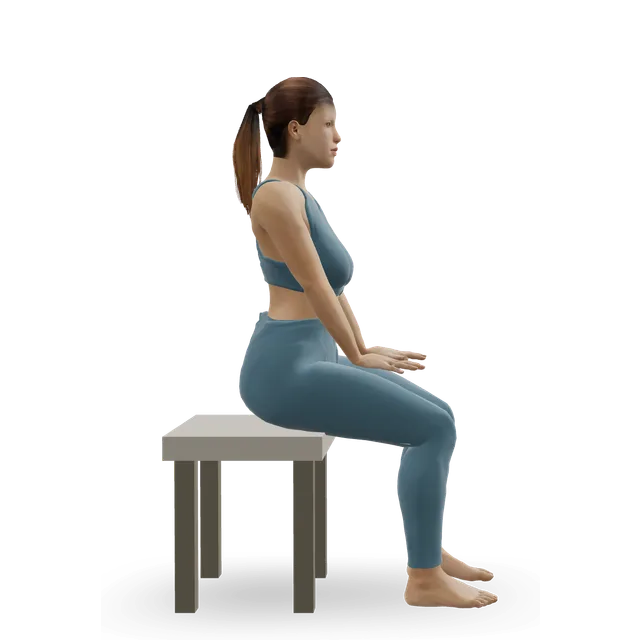
Nod your chin gently toward your throat, lying down or sitting tall, which bends the head forward at the top joint, the opposite of what these muscles do. Hold for a few seconds and keep it gentle; no study has tested a stretch specifically for them.
What is suboccipital release, and does it work?
It is a hands-on technique: a clinician holds their fingertips under the base of your skull for a few minutes. Small studies found short-term changes, such as a better head position when it was added to a chin-nod exercise, and better hamstring flexibility scores in young adults. Lasting benefits have not been shown.
Can I do suboccipital release myself with tennis balls?
No trial we found has tested releasing these muscles yourself with balls or tools. If you try it, keep the pressure light, stay on the back of the neck, never press on the front or sides of the throat, and stop for dizziness, tingling or a worsening headache.
Why do my suboccipitals ache after looking at my phone?
Poking your head forward made the rectus capitis muscles work about three times harder in one study. But bending the neck to text did not predict neck pain over a year in 396 adults, while poor sleep and low activity did, so regular movement matters more than a perfect posture.
What is the suboccipital triangle?
A small triangle at the base of the skull outlined by three of these muscles: the rectus capitis posterior major and the two obliquus capitis muscles. It holds the vertebral artery and the suboccipital nerve, which is why surgeons use it as a landmark.
What nerve supplies the suboccipital muscles?
The suboccipital nerve, the back branch of the first neck nerve (C1), which moves only these muscles. Feeling from the back of the scalp travels in a different nerve, the greater occipital nerve from the second neck nerve, which passes right beside them.
Anatomy and movement education, not a diagnosis or treatment plan. If you have pain that follows an injury, is severe or getting worse, or comes with numbness, weakness or swelling, get it assessed. Health disclaimer.