Also called VMO, vastus medialis oblique, teardrop muscle, inner quad
The vastus medialis is the quadriceps muscle on the inner front of your thigh. Its lowest, most angled fibres form the teardrop just above the inner side of the kneecap, often called the VMO (vastus medialis oblique).
It straightens the knee with the other three quads and helps steady the kneecap, mostly by pulling on the ligament on its inner side. It can't be trained on its own, and it doesn't need to be: strengthening the whole thigh and hip is what helps kneecap pain.
Shaded: the quads area. It marks the region, not the exact outline of the vastus medialis.
Starts at (origin)
The inner side and back of the thigh bone, from just below the hip almost to the knee. Its lowest fibres start partly from the tendon of the big inner-thigh muscle.Lower intertrochanteric line and medial lip of the linea aspera; VMO fibres also from the adductor magnus tendon (one StatPearls chapter says the femoral neck)
Attaches to (insertion)
The inner top edge of the kneecap, the quadriceps tendon, and the sheet of tissue on the inner side of the kneecap.Medial base and border of the patella, medial side of the quadriceps tendon and medial patellar retinaculum; deep fibres into the vastus intermedius aponeurosis
What it does
Straightens the knee with the other quads, giving about a quarter of their strength.Knee extension
Helps steady the kneecap. Its own pull on the kneecap is modest; its lowest fibres blend into the ligament that holds the kneecap's inner side, which provides more than half of the static hold against the kneecap sliding outward.Indirect patellar stabilisation, via the medial retinaculum and the medial patellofemoral ligament
Nerve supply
Femoral nerve, from the L2 to L4 nerve roots (one source adds L1), through the nerve to vastus medialis, which runs down the inner thigh in the adductor canal. One or two branches reach the muscle, and the same nerve also supplies the knee joint.
The VMO's fibres run at a steeper angle than those above them, and in a 3D study of 12 cadaver legs it had shorter fibres and a smaller cross-section, suggesting it does more to steady the kneecap. But none of 374 cadaver legs had a layer of muscle covering fully separating the two parts, though some had a fascial plane, and a review found too little good evidence to call them separate muscles.
In one small 1984 lab study that injected fluid into 10 healthy knees, which we read as an abstract, the vastus medialis was inhibited at 20 to 30 millilitres, before the rectus femoris and vastus lateralis at 50 to 60. A review describes this arthrogenic muscle inhibition after knee arthritis, surgery and injury, and says it can last a long time.
In a 12-week trial it grew about the same with the leg extension and the leg press, and its activity, recorded with electromyography (EMG), was similar in both: 32% against 35% of maximum. Ten weeks of squats grew the quads, apart from the rectus femoris, in another trial. On the inclined leg press it was the most active quad whatever the feet did, and single-leg squats drew more activity from it than lunges or step-ups. A 6-week quad strengthening programme changed its fibre angle on ultrasound in 21 men, in a study with no control group.
EMG shows how hard a muscle is working, not whether it will grow.
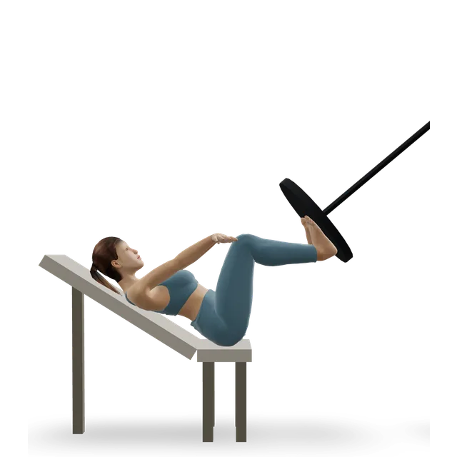
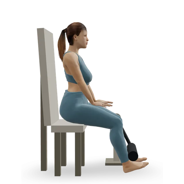
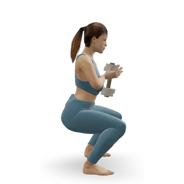
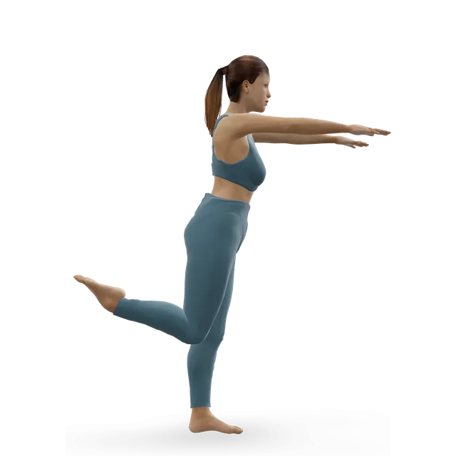
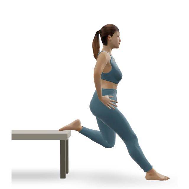
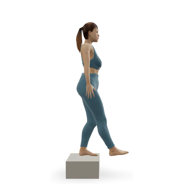
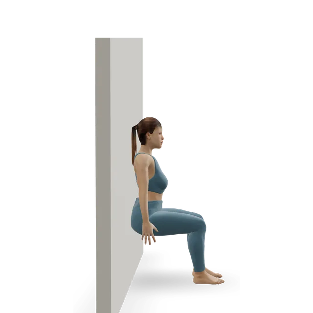
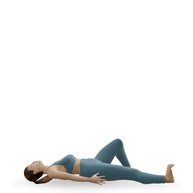
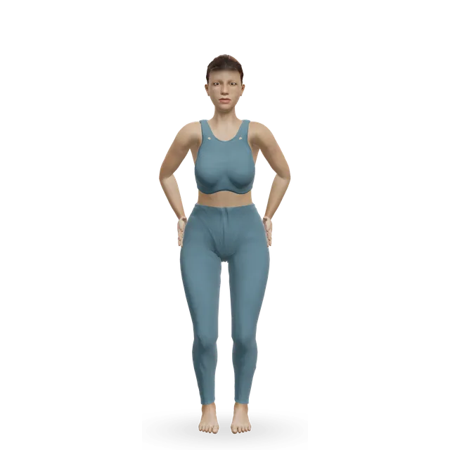
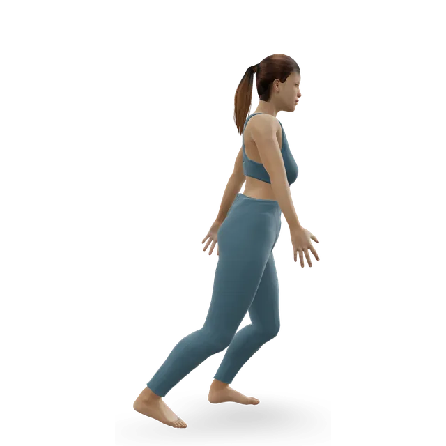
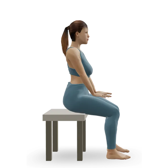
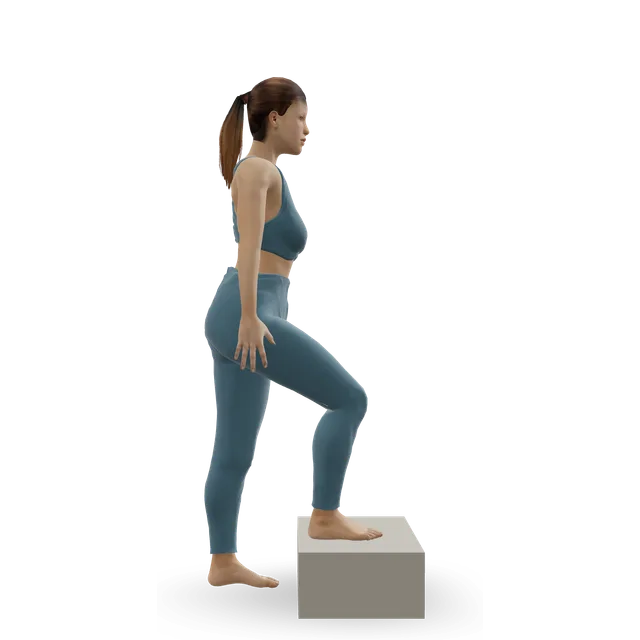
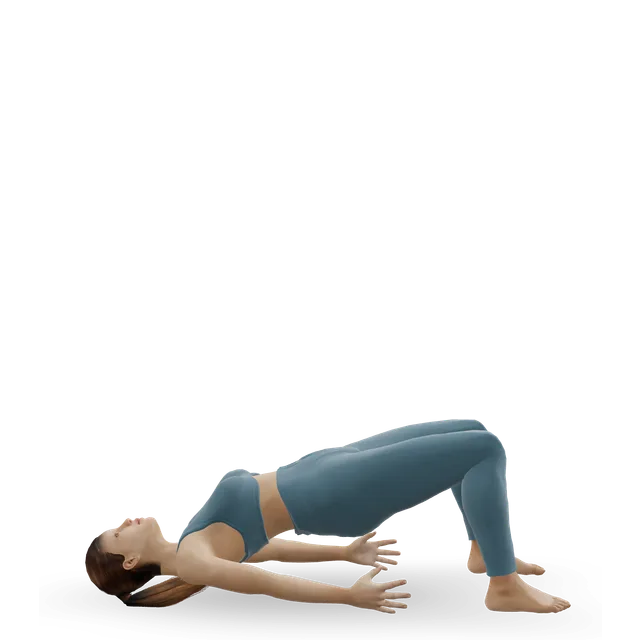
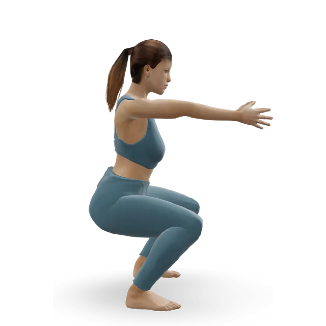
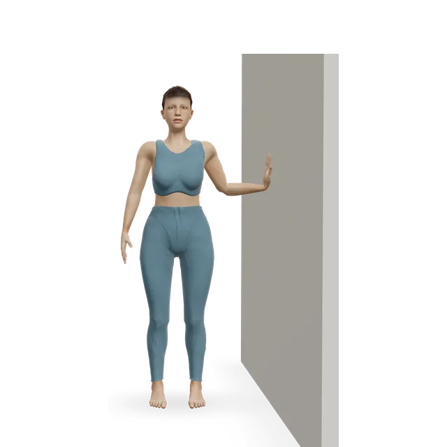
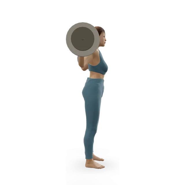
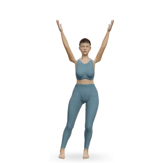
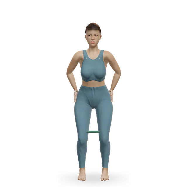
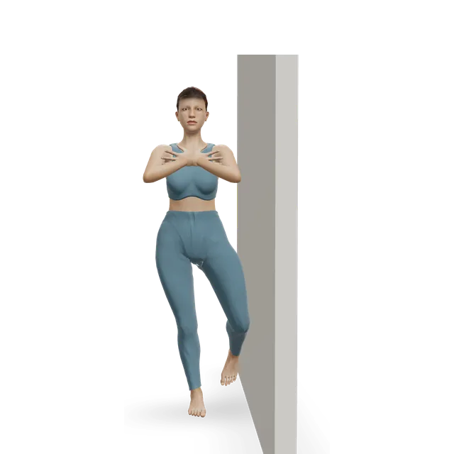
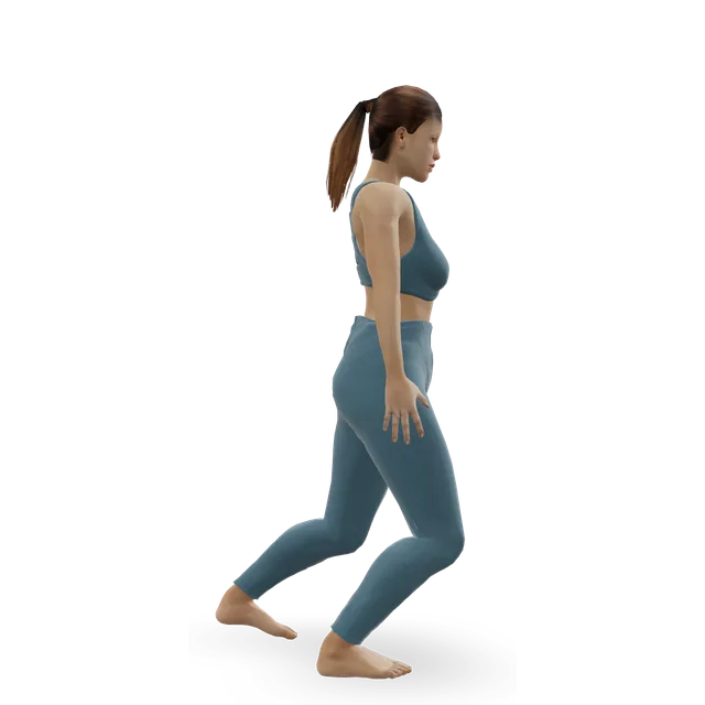
The order rests on measurements of the whole thigh, because no exercise has been shown to isolate this muscle: the leg press and leg extension grew it in a 12-week trial, squats grew the quads in another, and one-leg squats drew high activity in EMG studies, which show activity rather than growth. The skater squat is the closest move on our figure to the one-leg squat measured. The split squat, step-down, wall sit and quad set follow from its knee-straightening action; the quad set is the usual first step when swelling has switched the thigh off. Turning the feet, changing the knee angle or squeezing a ball did not shift work toward it.
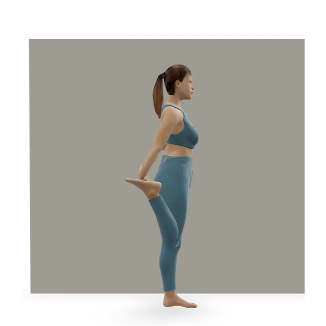
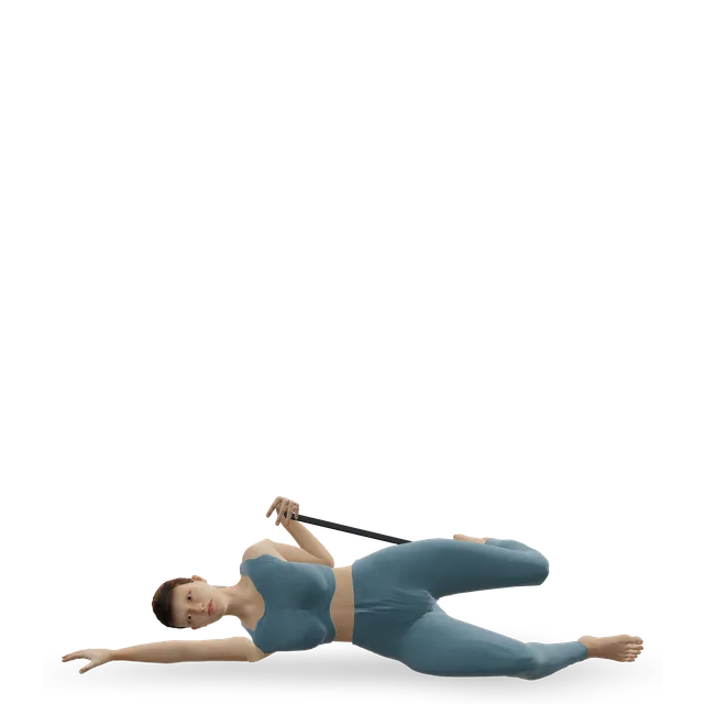
It crosses only the knee, so bending the knee fully stretches it whatever the hip does; these follow from its anatomy rather than from a study. For kneecap pain, stretching is an optional extra to strengthening.
Pain around the kneecap (runner's knee)Strong evidence
The 2019 American physiotherapy guideline gives its top grade to combining hip and knee exercises, and advises against EMG biofeedback, a live display of the inner quad's activity, for training it. In a trial of 69 people, training aimed at the VMO and general quad strengthening eased pain equally.
People with one painful knee had smaller thigh muscles on that side, all four quads alike, with no selective wasting of the VMO.
The main inner restraint is the medial patellofemoral ligament (MPFL), which provided more than half of the hold in a cadaver study, and the VMO's fibres blend into it. After a first dislocation, exercises aimed at the VMO were no better than general quad strengthening in a trial of 50 people; the general group scored slightly better at a year, though not by a meaningful amount, and about half dropped out.
Wasting after knee swelling, injury or surgeryEmerging evidence
Fluid in the knee switches the quads off, and in the small lab study the vastus medialis switched off first. A review says the inhibition can last after knee injury, surgery or arthritis, and clinicians look for its wasting when examining kneecap pain.
Weak knee-straightening muscles went with a higher risk of developing painful knee osteoarthritis across five cohorts of 5,707 people, and in an analysis of 48 trials, exercise aimed at the quadriceps eased pain more than general leg exercise. The knee arthritis guide treats the thigh-focused point as unsettled.
Strengthening the whole thigh and hip is what helps. Training aimed at the VMO was no better than ordinary quad strengthening for kneecap pain or after a first dislocation, and the 2019 American guideline advises against biofeedback on the inner quad.
Turning the toes out, squeezing a ball between the knees or working the last few degrees of straightening is often said to isolate it. In a review of 20 EMG studies, changing foot position or knee angle, or adding a squeeze, did not shift work toward it; a squeeze during squats did not raise its workload on muscle scans; and foot position on the leg press made no difference. In the leg extension, all the quads work hardest near full straightening.
A weak or late-firing VMO causes kneecap painMixed evidence
A meta-analysis found a trend toward the VMO switching on later than the outer quad in people with front-of-knee pain, with so much variation between studies that its meaning is unclear, and not everyone with the pain shows it. The wasting seen in kneecap pain involves all four quads, not the VMO alone.
The vastus medialis oblique: the lowest fibres of the vastus medialis, angled toward the kneecap, which form the teardrop above the inner knee. Whether it is a separate muscle is debated: its fibres run differently, but in 374 cadaver legs none had a layer fully separating the two parts.
What does the vastus medialis do?
It straightens the knee with the other three quads, providing about a quarter of their strength. It also helps steady the kneecap, mainly through the ligament on the kneecap's inner side, into which its lowest fibres blend.
Can you isolate the VMO?
No. Changing foot position or knee angle, or adding a squeeze, did not shift work toward it in 20 studies of muscle activity. Squeezing a ball during squats did not raise its workload on scans either.
What are the best vastus medialis exercises?
Heavy knee-straightening work in general: it grew about the same with the leg press and the leg extension over 12 weeks, and single-leg squats drew high activity. Pick moves you can load and progress.
Do VMO exercises help knee pain?
Thigh and hip strengthening helps kneecap pain, and it is what the 2019 American physiotherapy guideline recommends. Training aimed at the VMO was no better than ordinary quad strengthening in a trial, and the same guideline advises against VMO biofeedback.
Why has my VMO shrunk after a knee injury?
Fluid in the knee switches the quads off, and in one small lab study the vastus medialis switched off first, with 20 to 30 millilitres of fluid. This inhibition can last after injury or surgery, so rebuilding often starts with simple thigh tightening and moves on to loaded knee exercises.
Does a weak VMO make the kneecap dislocate?
The main restraint is the ligament on the kneecap's inner side, which the VMO blends into. After a first dislocation, exercises aimed at the VMO were no better than general thigh strengthening in a trial of 50 people.
How do you build the teardrop muscle?
With any progressive knee-straightening training: leg presses, squats and leg extensions all grew the vastus medialis in trials. There is no special teardrop exercise.
Which nerve supplies the vastus medialis?
A branch of the femoral nerve, from the second to fourth lumbar nerve roots (L2 to L4), that runs down the inner thigh in the adductor canal. The same nerve also supplies the knee joint.
Anatomy and movement education, not a diagnosis or treatment plan. If you have pain that follows an injury, is severe or getting worse, or comes with numbness, weakness or swelling, get it assessed. Health disclaimer.