Posterior tibial tendonitis: exercises for inner ankle pain and a dropping arch
The posterior tibial tendon runs behind the bony bump on the inside of the ankle and holds up the arch. When it is overloaded it hurts and swells along the inner ankle, rising onto tiptoe gets hard, and over time the arch can drop, often on one side. It's called posterior tibial tendonitis or posterior tibial tendon dysfunction (PTTD), and surgeons now group it under progressive collapsing foot deformity.
A new or one-sided flat foot needs a GP check. Most people improve without surgery, with insoles or a brace and exercise, but the evidence for the exercises is thin: two small trials of 36 and 39 people. This guide explains the single-leg heel raise test, what the trials found and a staged plan with 3D demos.
Exercise is for pain that has been checked or is clearly mild and settling.
Get urgent medical help if
Same day: you have diabetes and a new foot problem, such as an ulcer or wound, signs of infection, or a hot, swollen foot with a change in colour, even if it doesn't hurt. Contact your diabetes foot team or GP: NICE asks for these to reach a specialist foot service within 1 working day, and a suspected Charcot foot is kept off weight until it has been checked.
Contact NHS 111: after an injury, severe ankle pain, feeling faint, dizzy or sick from the pain, a snap, grinding or popping noise at the time of the injury, the ankle or foot at an odd angle, or not being able to walk. These might be signs of a broken ankle or an infection.
Same day: a sudden pop on the inside of the ankle followed by a change in the shape of the foot. The tendon may have torn (our suggestion).
Same day: a sudden, very painful, hot, swollen, red joint, often the big toe, especially with a high temperature, feeling shivery or being unable to eat. It may be gout, but it could be a joint infection (septic arthritis), which is urgent.
See a doctor or physiotherapist if
Flat feet that are painful, stiff, weak or numb, that you didn't have before, or that affect only one foot.
You can't do a single-leg heel raise on the painful side, or the foot is visibly flattening.
Pain on the outside of the ankle as well as the inside, which can mean the foot shape is changing.
Tingling or loss of feeling in the foot, or a foot that feels weak.
Any foot pain if you have diabetes: foot problems can be more serious.
Not improving after about 6 weeks of insoles and exercise (our suggestion).
Understand it
What it is
The tibialis posterior is a deep calf muscle. Its tendon runs behind the bony bump on the inside of the ankle and into the arch, and it is the main support of the inner arch. When the tendon is irritated, worn or torn, it can't hold the arch up as well: the arch can flatten, the heel tips outwards and the front of the foot drifts out.
Clinicians have long used four stages: 1, a sore tendon that still works; 2, a failing tendon with a flat foot that can still be corrected by hand; 3, a stiff flat foot with arthritis below the ankle; and 4, arthritis in the ankle too.
In 2020, a consensus of 9 surgeons voted unanimously to rename the whole range progressive collapsing foot deformity, because the problem is a complex change in foot shape that isn't always caused by the tendon. Only 3 of the 9 thought MRI (magnetic resonance imaging) findings should be part of the new stages.
Mostly women over 40. When women over 40 at an English GP practice were surveyed and examined, 3.3% had stage 1 or 2, and none had been diagnosed, despite long-standing symptoms. A clinical review suggests it may affect up to 10% of older women.
Risk factors include flat feet, high blood pressure, diabetes, a steroid injection near the tendon, inflammatory arthritis and being overweight. It is often missed: the same review says most people are first told they have a sprain or arthritis.
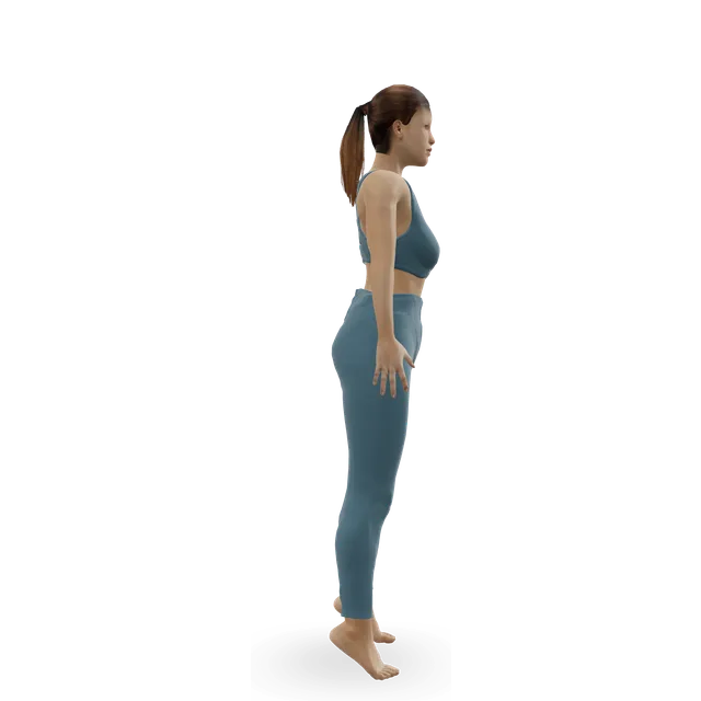
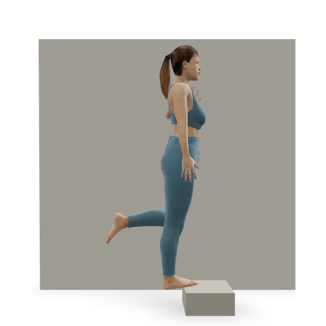
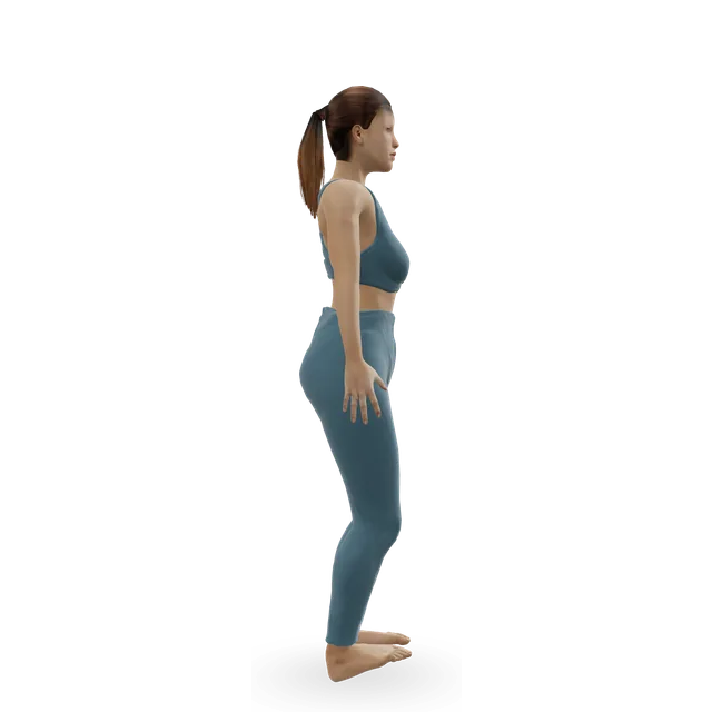
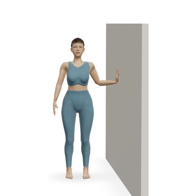
Standing on one leg and rising onto tiptoe needs a working tendon. A clinical review says someone with a normal foot can do about 8 to 10 single heel raises; with this problem it's hard or impossible, and the heel doesn't swing inwards. From behind, more toes show on the outside of the affected foot (the too many toes sign).
The test has limits. In 50 people, those with stage 2 didn't rise as high, but healthy older adults also rose less high than young adults, so expectations need adjusting for age. In a two-leg heel raise, people with stage 2 turned the heel in about as much as people without the problem, but their midfoot sagged instead of lifting (6.5 degrees of midfoot bend against 12 in controls), so a heel that turns in doesn't rule it out. Strength turning the foot in was 20% to 30% lower on the affected side.
The diagnosis is mainly clinical. X-rays taken standing show the shape of the foot and any arthritis, and an MRI scan can show inflammation or a tear. In one group of 64 people, a split tear on MRI went with treatment without surgery not working. No study of tendon changes on MRI in people without symptoms was found, so we can't say how often scans show changes that don't matter.
The foot shape can slowly change if nothing is done, but most people improve without surgery. An NHS-commissioned booklet says most cases resolve without surgery within 3 to 12 months. The American Academy of Orthopaedic Surgeons (AAOS) warns that pain can last more than 3 months even with early treatment, and that after many months of pain it is not unusual for it to last another 6 months once treatment starts. In 33 feet treated with a brace, about 70% were free of the brace without surgery 7 to 10 years later.
Insoles or a brace, with exercise, firstEmerging evidence
In a case series of 47 people with stage 1 or 2, a short ankle brace or a foot orthosis, with a high-repetition exercise programme at home and in clinic, led to good results in 83% after a median of 10 physiotherapy visits over about 4 months; 89% were satisfied and 11% needed surgery.
Across 4 small trials (186 people, most at high risk of bias), orthoses reduced pain in the early stages, especially with exercise, and orthoses with personalised arch support did better than flat insoles. In 64 people, 87.5% were treated without surgery over about 27 months, and wearing a brace went with success. The AAOS says most people in the early stages can be treated with orthoses and braces.
Strengthening the tendon: thin evidenceEmerging evidence
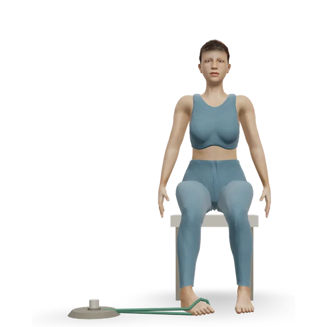
The evidence is thin. In a trial of 36 adults with stage 1 or 2, everyone wore orthoses and stretched for 12 weeks; adding resistance exercise helped, and the group whose exercise had a slow lowering phase improved most in pain and function. Their exercise swept the front of the foot inwards against resistance with the foot slightly pointed: 3 sets of 15 at a load they could only just manage, twice a day.
In a trial of 39 adults with stage 2, a home programme of heel raises and band work, from 2 sets of 10 to 3 sets of 30, twice a day, added little to an orthosis and stretching, and strength didn't change. A review of these and one other trial (93 people in all) called the research sparse, found people did about 68% to 79% of their sessions, and noted that 15 to 30 repetitions trains endurance rather than strength.
Every trial included calf stretches (3 to 10 holds of 30 s, 2 to 4 times a day), and the AAOS lists calf stretching as part of physiotherapy. But a review points out that holding the ankle fully bent up squeezes and stretches the tendon behind the ankle bone, which may work against easing the pain. So the plan here keeps stretching optional and gentle.
For a bad flare, a 2004 clinical review advises 4 to 8 weeks in a cast or removable boot, then orthoses and lace-up shoes. The AAOS mentions 6 to 8 weeks in a cast or boot, but warns that a cast weakens the leg muscles and keeps it for when other treatment hasn't worked. The trials above used orthoses and exercise without routine casting.
Steroid injections into the tendon are avoided: the 2004 review calls them contraindicated, the AAOS says they are not normally done because of the risk of the tendon rupturing, and an NHS-commissioned booklet says they are rarely advised.
Surgery is considered if pain persists after several months of good treatment, and an NHS-commissioned booklet says only after 6 months, and rarely. Operations range from cleaning the tendon sheath to moving another tendon to do its job, cutting and shifting the heel bone, and fusing joints. The AAOS says it takes 6 months or more before healing is complete and pain is much better.
Up to 5 out of 10, with the arch held: borrowed, and editorialEmerging evidence
Aim for discomfort no more than about 5 out of 10, settling within an hour and no worse the next morning. The 5 out of 10 figure is editorial, as on this site's other tendon pages, borrowing the pain-monitoring approach from an Achilles trial; it hasn't been tested in this tendon.
An NHS-commissioned booklet adds two practical rules: if an exercise is painful, do fewer or do them less often, and stop a heel raise or squat if you feel you're losing your foot position, then reset.
Start at the first stage. Each move opens a full guide with a 3D demo.
Stage 1
Support the arch and start loading the tendon (about weeks 0 to 2)
Wear the orthoses, brace or supportive lace-up shoes you've been given: the trials ran for 12 weeks with insoles worn throughout. Doses are marked with their source or are editorial.
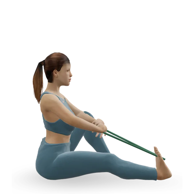
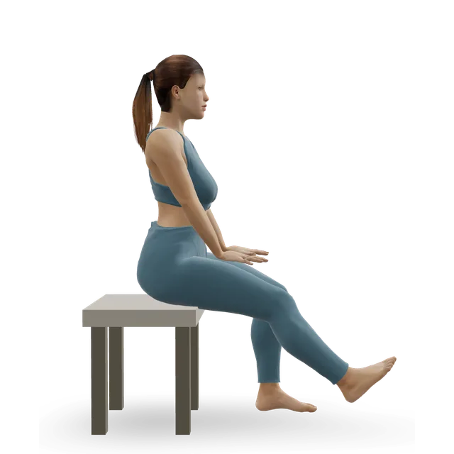
3 sets of 15 with a light band, once or twice a day · Sweep the front of the foot inwards with the foot slightly pointed: the format from the trial of 36 people; the light start is ours
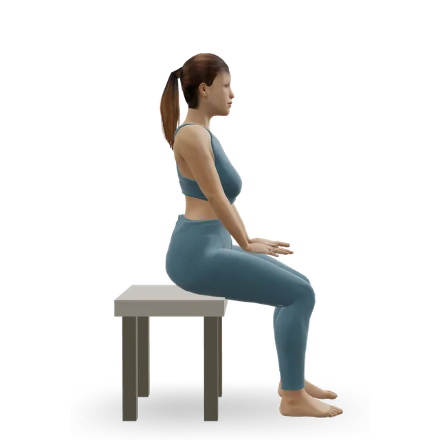
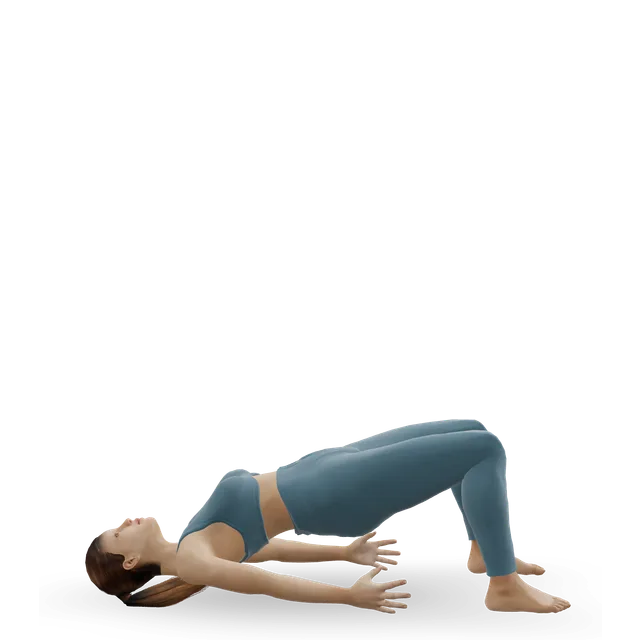
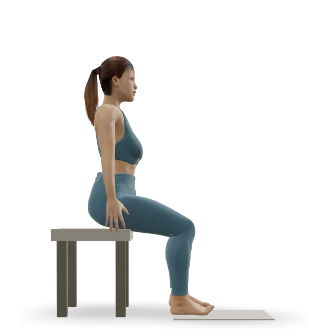
2 sets of 10 on both feet, twice a day · The starting dose from the trial of 39 people. A soft ball held between the heels helps keep them turning in (an NHS-commissioned booklet)
Build from 2 sets of 10 to 3 sets of 30, twice a day · The trial of 39's progression; holding the top for up to 10 s is an NHS-commissioned booklet's version
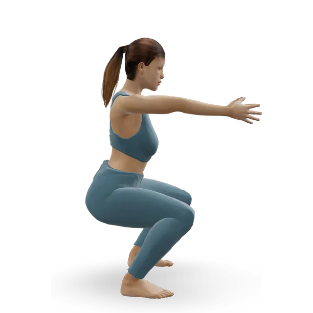
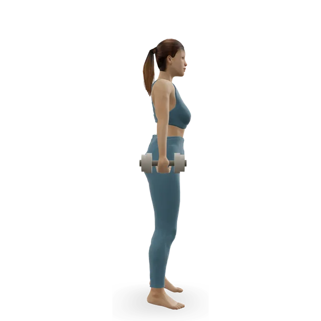
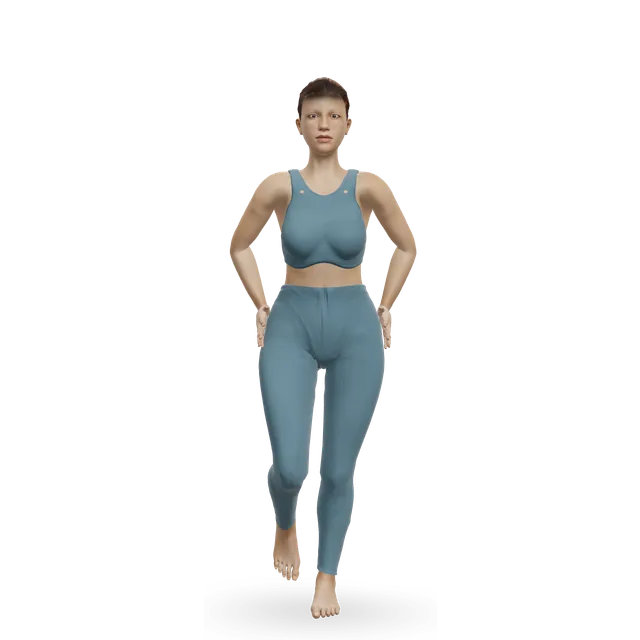
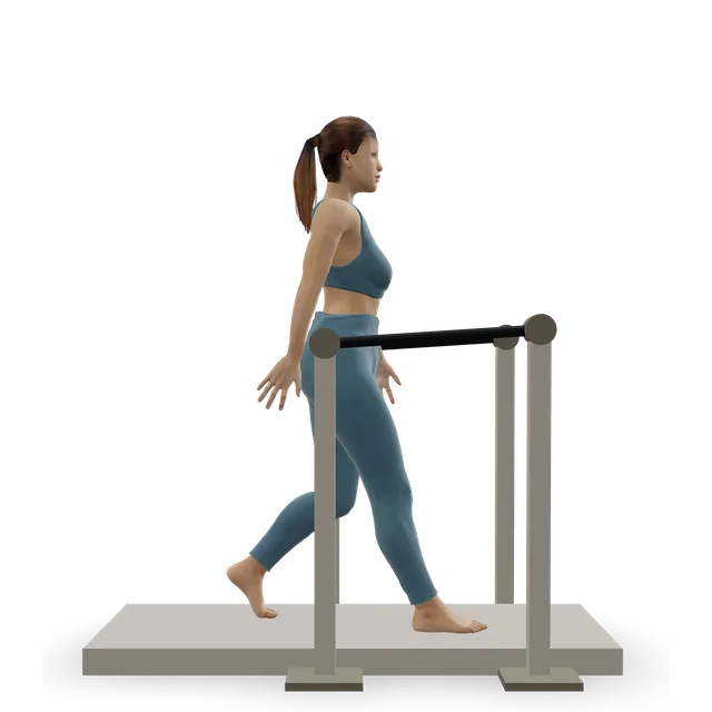
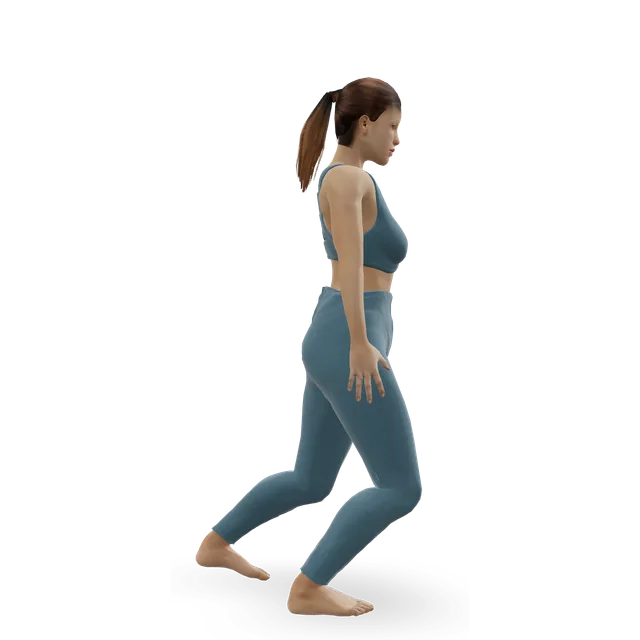
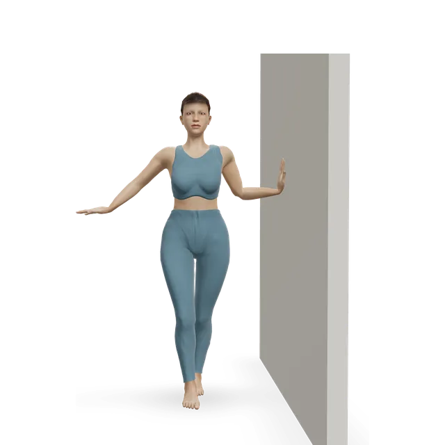
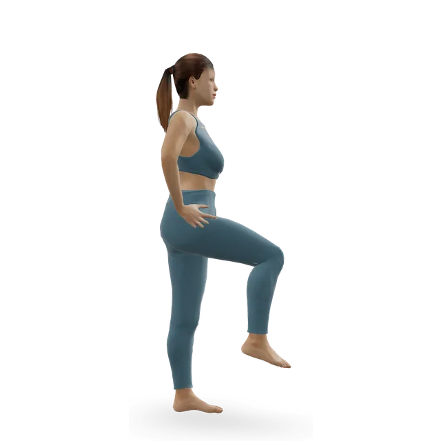
2 to 3 sets of 10, building to 30 · On flat ground first; the trial of 39's single-leg raises. Watch that the heel turns in and the arch stays lifted as you rise
Move on when: Single-leg raises are comfortable. In one case series of 47 people, the targets for finishing physiotherapy were 50 single-leg heel raises with little or no pain and walking about 30 m on tiptoe: a long-term goal, not a test. Keep strength work going twice a week (our suggestion), and if pain persists after about 6 months of good treatment, ask about a referral.
Follow along: a timer, a cue and the next move, one step at a time.
What to change for now
The arch lift (no 3D demo)
This NHS-commissioned exercise suits stages 1 and 2. The site doesn't demonstrate it yet, so here are the steps.
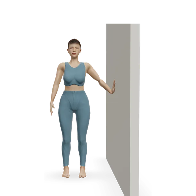
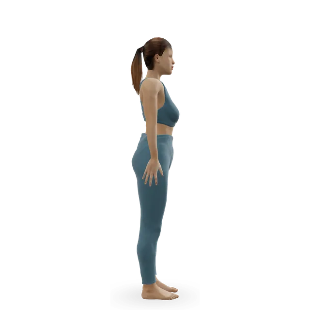
1. Stand with your feet hip-width apart.
2. Keep your toes relaxed and flat on the floor, and lift the arches of your feet. You may feel your thighs turn out a little.
3. Hold for 10 seconds, then relax.
4. Reset your feet and repeat 10 times, for 3 sets. Do fewer, or do them less often, if it's painful.
Supportive lace-up shoes, or walking boots that support the arch and ankle if you're on your feet for long; leave out flip-flops and other unsupportive footwear. A podiatrist can provide orthoses for your foot. A simple lace-up ankle brace may help mild to moderate cases, and heavier custom braces can help people with severe, stiff flat feet avoid surgery.
Cut back the standing, walking, running and jumping that hurt, and swap to cycling, an elliptical trainer or swimming. A cold pack for 20 minutes, 3 or 4 times a day, can ease swelling. Losing weight, if you need to, takes stress off the arch.
A painful, weakened tendon on the inside of the ankle that normally holds up the arch. Over time the arch can drop, which surgeons now call progressive collapsing foot deformity.
What are the symptoms?
Pain and swelling behind the inner ankle bone and along the instep, aching on long walks, difficulty rising onto tiptoe, and a foot that looks flatter than the other. Later, pain can spread to the outside of the ankle as the foot shape changes.
What is the single-leg heel raise test?
Standing on the painful leg and rising onto tiptoe. Most people manage about 8 to 10; with this problem it's hard or impossible, and the heel doesn't swing in. Older adults rise less high anyway, so a clinician should judge it.
What exercises help posterior tibial tendonitis?
Sweeping the front of the foot inwards against a band and letting it back slowly, alongside insoles and stretching, helped most in a trial of 36 people. Heel raises on both feet and then on one were part of another trial's home programme, which added little to insoles and stretching. The evidence is thin, but these are the best-tested exercises.
Do I need insoles or a brace?
Usually, yes, early on. Arch-supporting orthoses eased pain in small trials, and wearing a brace went with avoiding surgery in one group of 64 people.
How long does it take to get better?
The trials ran for 12 weeks, and one centre's programme took about 4 months of physiotherapy. Most people improve within 3 to 12 months, though pain can last more than 3 months even with early treatment.
Should I stretch it?
Gently, if at all. Hard stretches that bend the ankle fully up may squeeze the tendon.
Will I need surgery?
Most people don't: in one group, about 70% were free of their brace without surgery 7 to 10 years later. Surgery is considered after about 6 months of good treatment without surgery.
Is it the same as flat feet?
No. Most flat feet are normal and need no treatment. This is a painful tendon that makes a foot flatten, often on one side, and it needs assessing.
Can I keep walking and exercising?
Yes, in supportive shoes and your insoles, within the pain rule. Swap running and jumping for cycling or swimming during a flare.
Exercise education, not a diagnosis or treatment plan. It is written from the guidelines and studies listed above; review by a clinician is pending. If your pain follows an injury, is severe, is getting worse, or comes with any warning sign listed on this page, get it assessed. Health disclaimer.