The peroneal tendons run behind the bony bump on the outside of the ankle and steady it against rolling over. When they are overloaded they ache behind or below the ankle bone, worse with walking, running and jumping and easing with rest. Most people call it peroneal tendonitis; doctors prefer tendinopathy, because the tendon isn't necessarily inflamed.
It often follows ankle sprains, goes with an ankle that keeps giving way, and is easily mistaken for a sprain. No trial has tested exercises for it, so the plan here is borrowed from ankle instability and Achilles tendon trials and from NHS programmes, and the page says when clicking, snapping or a tear needs a clinician.
Exercise is for pain that has been checked or is clearly mild and settling.
Get urgent medical help if
Call 999 or go to A&E: after an injury, you heard a crack, the foot or ankle has changed shape or points at an odd angle, a bone is sticking out, there is a bad cut over it, you're in severe pain, or the toes look blue or white, feel numb or are cold to touch. These can mean a broken bone. Don't drive yourself.
Contact NHS 111: after an injury, severe ankle pain, feeling faint, dizzy or sick from the pain, a snap, grinding or popping noise at the time of the injury, the ankle or foot at an odd angle, or not being able to walk. These might be signs of a broken ankle or an infection.
Same day: after an injury, a pop or snap behind the outer ankle bone with swelling, or a tendon you can see or feel slipping over the bone. The tendons may have come out of their groove (dislocated), which is often mistaken for a sprain; experts advise a specialist review.
Same day: a sudden, very painful, hot, swollen, red joint, often the big toe, especially with a high temperature, feeling shivery or being unable to eat. It may be gout, but it could be a joint infection (septic arthritis), which is urgent.
See a doctor or physiotherapist if
Tenderness on the bony bump halfway along the outer edge of the foot after rolling the ankle: clinicians use the Ottawa ankle rules to decide whether it needs an X-ray for a fracture.
Clicking or snapping behind the outer ankle bone that keeps happening or hurts.
An ankle that keeps giving way months after a sprain, or that goes over more often than before.
Tingling or loss of feeling in the foot, or a foot that feels weak.
Pain that hasn't started to improve after about 12 weeks of the plan, or ankle pain that stops you doing normal activities, is getting worse, or hasn't improved after 2 weeks of looking after it at home.
Understand it
The tendons and what they do
Two muscles run down the outside of the lower leg, the peroneus longus and peroneus brevis (also called the fibularis muscles). Their tendons share a sheath behind the outer ankle bone, held in a groove by a band of tissue. They turn the sole of the foot outwards, and they are the first muscles to fire when the ankle is suddenly forced to roll inwards.
Specialists sort their problems into three kinds: tendinopathy, tears, and tendons that slip out of place (subluxation or dislocation). A small bone inside the longus tendon (the os peroneum) can also become painful, under the outer edge of the midfoot.
Pain around the outside of the ankle, above, behind or below the ankle bone, that is worse with activity such as walking, running and jumping and eases with rest. There may be swelling and tenderness along the tendons, sometimes up the side of the leg to just below the knee. NHS Lanarkshire adds feeling unsteady on uneven ground and poorer balance on that leg.
A clinician feels the tendons while you push the foot up and out against resistance, checks for pain when you turn the foot out against resistance or when it is stretched inwards, and checks the ankle ligaments and the shape of the foot.
It often goes with an unstable ankle: in 61 people having surgery for chronic ankle instability, 77% had inflamed peroneal tendon sheaths and 25% a tear in the brevis tendon. A review says chronic peroneal problems are often preceded by an unstable ankle or by a foot shape that loads the tendons, such as a high-arched foot with the heel tipped in; in one series of people operated on for peroneal tears, 82% had that foot shape. Crowding in the tendons' tunnel, such as from a low-lying muscle or an extra small muscle, may play a part, but these variants are common in people with no symptoms: in 65 volunteers, 17% had the extra muscle and 61% a flat or rounded groove.
NHS leaflets add age, being overweight, diabetes, inflammatory arthritis, long spells of standing, walking or running or a sudden increase in them, unsupportive footwear, training without enough recovery, long-term steroid tablets or certain antibiotics (fluoroquinolones), previous foot or ankle injury or surgery, and smoking.
In one study, only 60% of 40 peroneal tendon problems were diagnosed correctly at the first visit, and acute dislocations of the tendons are missed in up to 40% of cases, usually taken for a sprain. On an X-ray after an injury, a small flake of bone pulled off the outer ankle bone points to a dislocation.
The tendons can be forced out of their groove when the muscles contract hard as the ankle is pushed upwards, tearing the band that holds them. There is swelling and tenderness behind the outer ankle bone, and the slip can often be brought on by turning the foot out against resistance. Painful clicking when you circle the ankle can be the two tendons swapping places inside their sheath. Both need assessing, because treatment without surgery often fails to stop a dislocation happening again.
What a scan does and doesn't showEmerging evidence
Ultrasound and MRI (magnetic resonance imaging) are both suitable when a scan is needed. But tendon changes are common in people without outer ankle pain, so a scan finding needs to match the examination:
In 294 foot and ankle MRI scans of people with no outer ankle injury, 35% showed some peroneal tendon change. The authors advise reassurance when the scan doesn't match the examination.
In 487 ankle MRI scans of people without outer ankle symptoms, 47% showed tendon changes.
Tears are found in 11% to 38% of ankles in studies of donated bodies, and an international consensus advises treating only tears that cause symptoms.
NHS services say most people improve within about 12 weeks of following their advice, though some need 6 to 9 months, or up to a year. Flare-ups are common and usually settle within 6 weeks.
Treatment without surgery comes first, on expert opinionEmerging evidence
An international consensus of 16 specialists from 9 countries says treatment without surgery should be considered for everyone with a peroneal tendon problem. After an injury, it treats the injury that came with it, then adds weight-bearing, movement and tendon-loading exercises once it is painless, and it advises avoiding keeping the ankle still. Shockwave therapy can be considered if symptoms last more than 3 months; there isn't enough evidence for platelet-rich plasma injections.
A specialist review adds rest, activity changes, anti-inflammatory painkillers and insoles that tilt the outer forefoot for mild cases, with 6 weeks in a cast or walking boot for stubborn ones. No trials of treatment without surgery were found: this is expert opinion and small case series.
The doses in the plan below come from these three NHS services. They haven't been tested in trials.
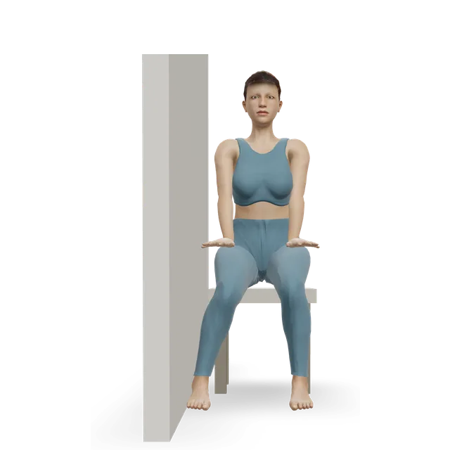
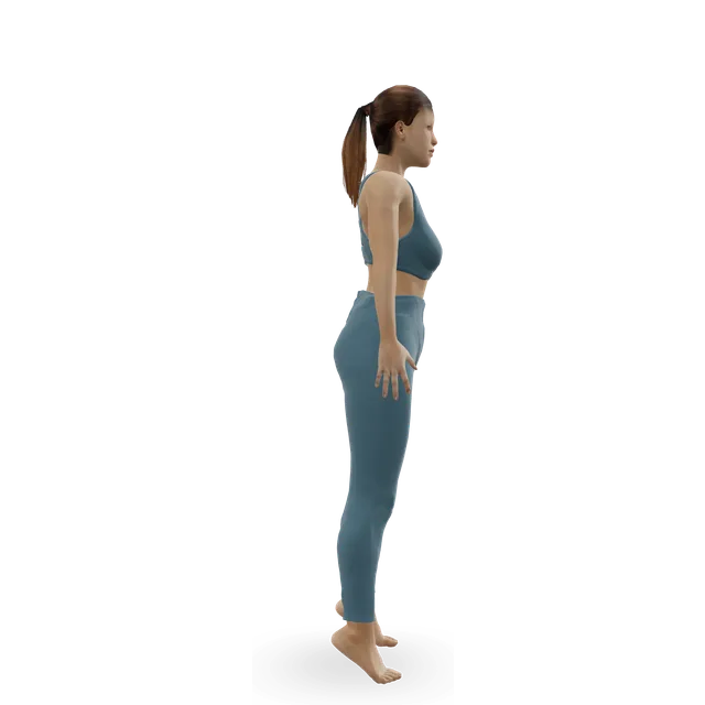
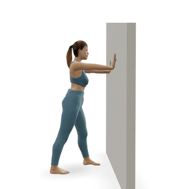
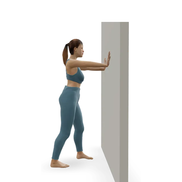
NHS Borders: supportive running-style trainers or boots with a slight heel. Two-leg heel raises up to 10, 3 times a day, stopping if the ankle feels tired or shaky, then single-leg raises; pushing the foot out against a belt and holding for a few seconds, up to 10, 3 times a day; calf stretches with the knee straight and bent, 30 s, twice. Low-impact activity rather than running for now.
NHS Lanarkshire: early-stage exercises first, progressing over time, staying in the green zone of a 0 to 10 pain scale (sometimes amber) during exercise and for 2 days after. See a physiotherapist if there's no improvement after 6 to 12 weeks.
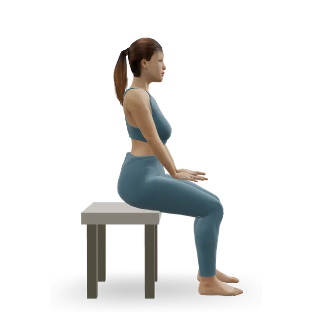
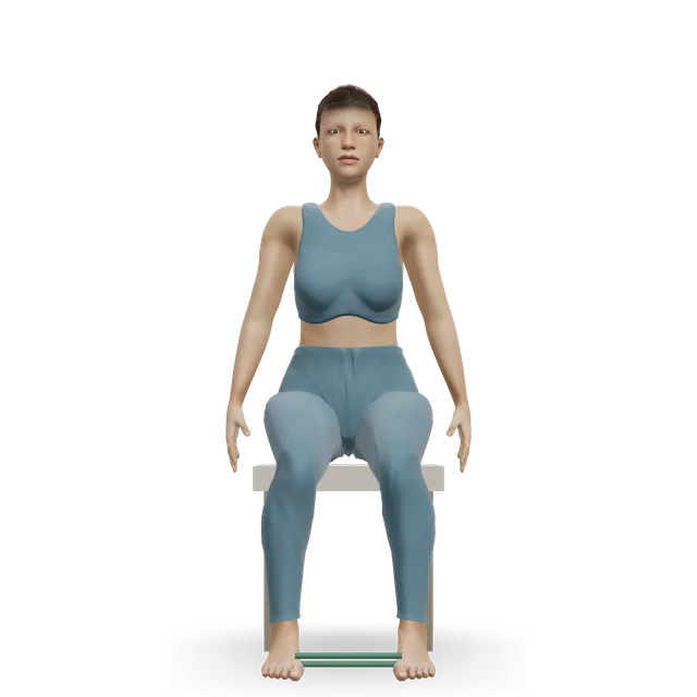
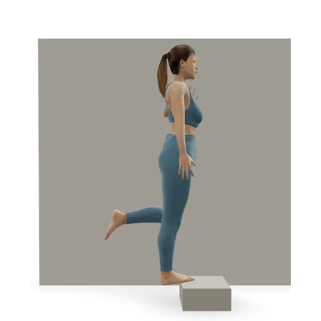
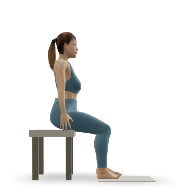
Livewell Southwest (NHS podiatry in Plymouth): calf raises with the toes turned in and the heels apart, seated, then standing, then on one leg, then on a step, then with weight, and band exercises pushing the foot out, each taking 4 s up and 4 s down, to tiredness. 1 to 3 sets every other day, or 2 to 3 times a week.
Borrowed from an ankle instability trialEmerging evidence
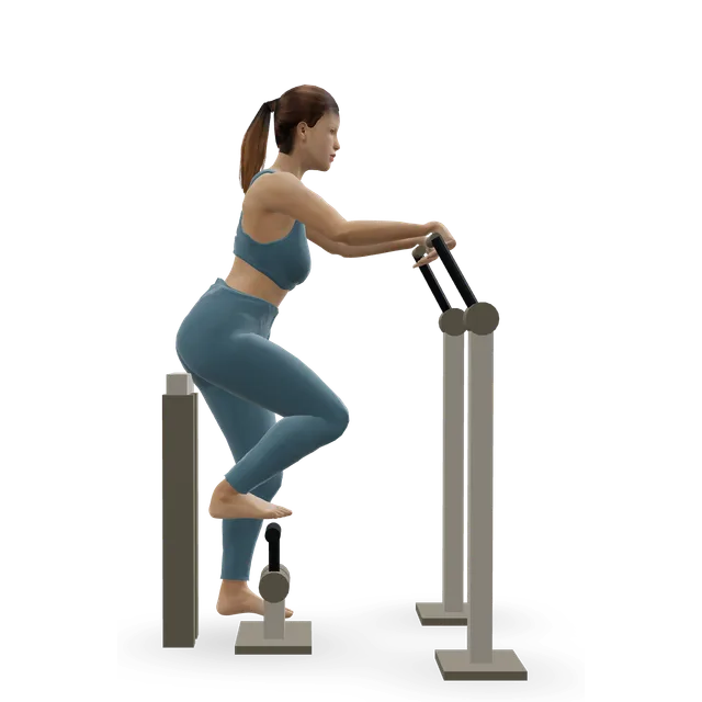
In a trial of 39 people with ankles that kept giving way (a related problem), 6 weeks of three 20-minute sessions a week of band work (turning the foot out, in and up, 3 to 5 s a repetition, from 3 sets of 10 to 4 sets of 10 with heavier bands) and single-leg heel raises on a step (from 2 sets of 10 to 4 sets of 15) improved strength, balance and side hopping. Only that group got stronger at turning the foot out under a lengthening load. This was a different condition, so its use here is borrowed.
Borrowed from Achilles and other tendon researchEmerging evidence
A review of tendon exercise trials notes that managing the load on a tendon is the most important part of rehab, and that exercise has established benefit for the Achilles, kneecap, shoulder and elbow tendons. In an Achilles trial of 38 people, carrying on with running and jumping under a pain-monitoring rule did no harm compared with 6 weeks of rest from them. Neither was a peroneal study.
Tears are treated only if they cause symptoms, starting without surgery; operations repair the tendon and deal with what caused it. After a first dislocation, people who aren't athletes may try 6 weeks in a cast or a boot with a heel wedge, but the consensus warns that this fails about half the time; athletes are usually offered early surgery, which deepens the groove. A review reports success rates of 26% to 57% without surgery.
Steroid injections: avoided by one specialist centreMixed evidence
A specialist review says steroid injections carry a risk of the tendon rupturing and aren't used at its centre, and the international consensus couldn't agree on them.
Up to 5 out of 10, settled by the next morning: borrowed, and editorialEmerging evidence
Some discomfort during exercise is fine: aim for no more than about 5 out of 10, settling within an hour and no worse the next morning. The 5 out of 10 figure is editorial, as on this site's other tendon pages. It borrows the pain-monitoring approach from an Achilles trial, in which carrying on with running and jumping under the rule did no harm, and it hasn't been tested in these tendons.
NHS services put it in their own words: stay mostly in the green zone of a pain scale during exercise and for the next 2 days, sometimes amber, never red; some discomfort is fine, but stop if it becomes more than tolerable; and stop a set if the ankle feels tired or shaky.
If it's worse the next morning, or flares up, lighten the load for a few sessions rather than stopping.
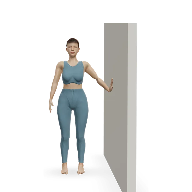
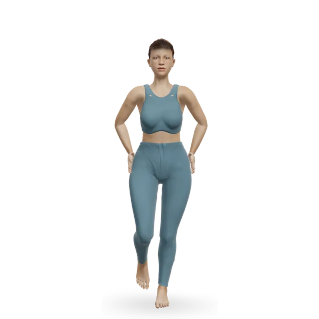
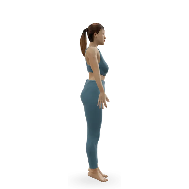
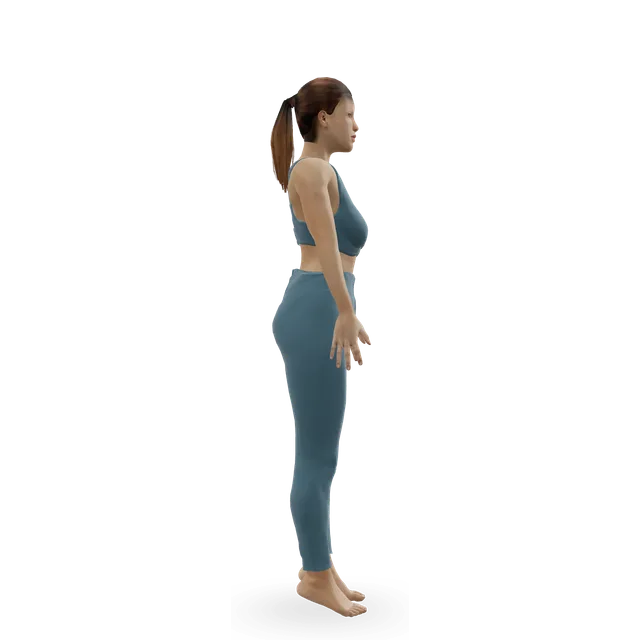
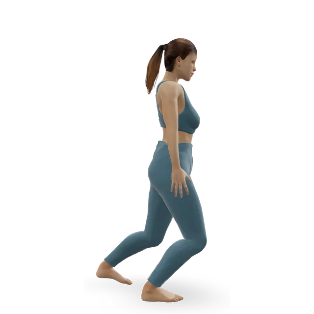
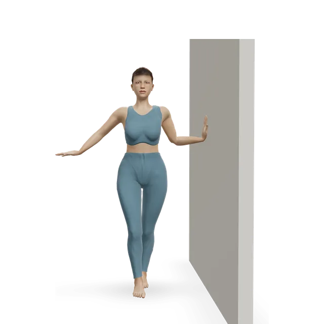
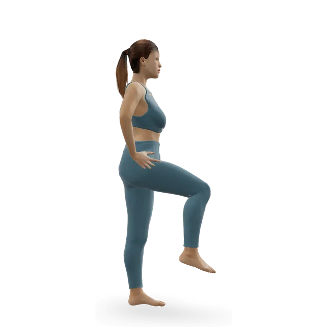
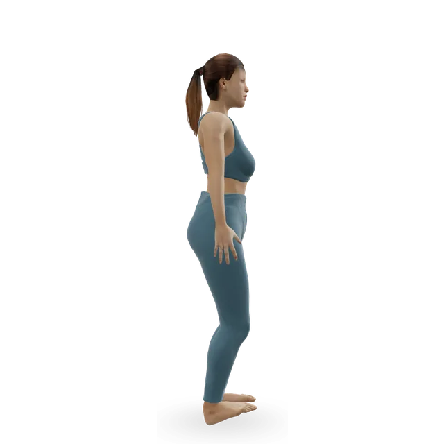
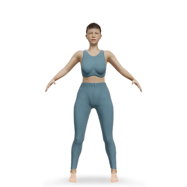
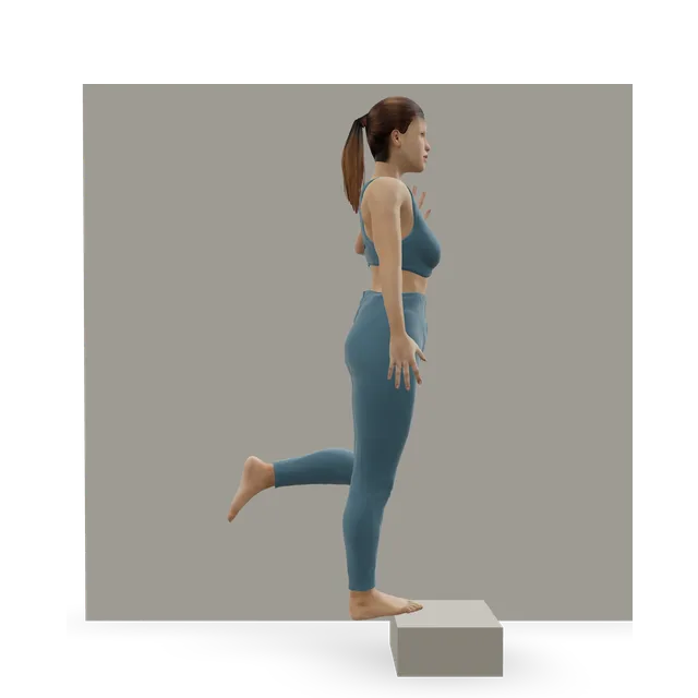
Start at the first stage. Each move opens a full guide with a 3D demo.
Stage 1
Settle and start loading (about weeks 0 to 2)
Keep walking, swap running and jumping for cycling or swimming, and wear supportive trainers with a slight heel. This plan is borrowed from ankle instability and Achilles tendon trials and from NHS programmes; doses are marked with their source or are editorial.
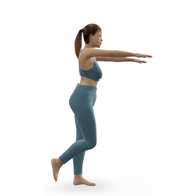
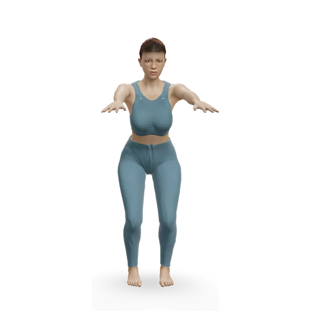
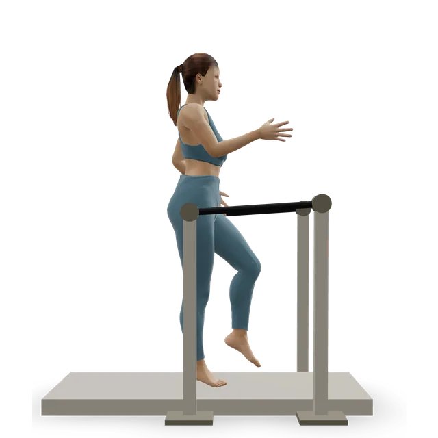
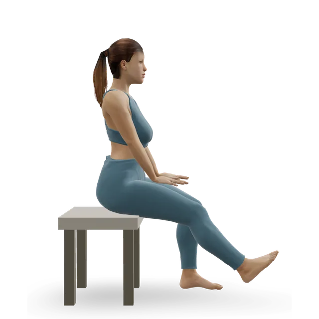
3 sets of 10, 3 to 5 s a repetition, rising to 4 sets of 10, with a heavier band every 2 weeks, 3 times a week · The ankle instability trial's progression; or 1 to 3 sets to tiredness, 4 s each way, every other day (an NHS podiatry leaflet)
Up to 10, building to 2 sets of 10, then 4 sets of 15 by about week 6 · On flat ground first. NHS Borders' dose, then the ankle instability trial's progression
Move on when: Hopping side to side and your usual activity stay within the pain rule the next morning. If the ankle keeps going over, follow the chronic ankle instability plan as well.
Follow along: a timer, a cue and the next move, one step at a time.
What to change for now
Shoes and surfaces
Comfortable running-style trainers or walking boots with a slight heel give the tendons the most support; hard, flat or slip-on shoes, sandals and very flexible soles give less. Keep to flat ground at first, and leave cambered roads and rough trails until later (our suggestion).
NHS services suggest changing activities rather than stopping all movement: break long walks or spells of standing into shorter ones with breaks, and swap running for swimming or cycling for now. For a flare-up, ice wrapped in a damp towel for 10 to 20 minutes, 2 to 3 times a day, for the first 5 to 7 days can ease pain and swelling.
Behind, below or just above the bony bump on the outside of the ankle, sometimes along the outer edge of the foot or up the side of the leg. It is worse with activity and eases with rest.
What exercises help peroneal tendonitis?
Holds pushing the foot outwards, band exercises, heel raises, balance work and then hops, built up over weeks. They're borrowed from ankle instability and Achilles tendon trials and from NHS programmes, because no trial has tested exercises for this tendon.
How long does peroneal tendonitis take to heal?
NHS services say most people improve within about 12 weeks. Some need 6 to 9 months, or up to a year, and flare-ups along the way are normal.
Should I stop running?
Swap running for cycling or swimming at first. Return with walk-jog intervals once hopping is comfortable within the pain rule.
Should I wear an ankle brace or boot?
Supportive trainers come first. Experts advise against routinely keeping the ankle still; a boot is for stubborn cases or a dislocated tendon, on a clinician's advice.
Is it a sprain or a tendon problem?
A sprain follows a twist and hurts over the ligaments in front of and below the ankle bone. Tendon pain sits behind the bone and often builds gradually. The two often happen together, and peroneal problems are easily missed at first.
My ankle clicks or snaps on the outside. Is that serious?
If you can feel a tendon slipping over the ankle bone, especially if it hurts or followed an injury, get it assessed. A first dislocation treated without surgery comes back in about half of people.
Do I need an MRI scan?
Not usually to start with. Tendon changes show on MRI in many people without outer ankle pain, so a scan is used when the examination suggests a tear or the plan isn't working.
Are steroid injections a good idea?
Specialists are cautious. One centre avoids them because of the risk of the tendon rupturing, and an international consensus couldn't agree on their use.
Does a high arch matter?
A high-arched foot with the heel tipped in puts more load on these tendons, and it is common in people who need surgery for a tear. Insoles may help some people, so ask a podiatrist.
Exercise education, not a diagnosis or treatment plan. It is written from the guidelines and studies listed above; review by a clinician is pending. If your pain follows an injury, is severe, is getting worse, or comes with any warning sign listed on this page, get it assessed. Health disclaimer.