Pain guide · Hip pain

Avascular necrosis of the hip: causes, stages, treatment and safe exercise

Avascular necrosis (osteonecrosis) of the hip happens when part of the ball of the hip loses its blood supply and the bone starts to die. It most often affects adults aged 30 to 50. Steroid medicines and heavy drinking are behind most cases, and sickle cell disease, lupus and cancer treatment raise the risk too.

It needs a specialist: an X-ray can look normal early on, a magnetic resonance imaging (MRI) scan shows it, and treatment to save the hip works best before the bone collapses. This guide explains the risk factors and stages, what crutches, exercise, core decompression and a hip replacement can and can't do, the urgent signs, and how to stay active safely while it's assessed, with a 3D demo of every exercise.

When to get help first

Exercise is for pain that has been checked or is clearly mild and settling.

Get urgent medical help if

  • Severe hip pain after a fall or injury, or you cannot stand, walk or put weight on the leg. In an older adult this can be a broken hip; the leg may look shorter or turned outward.
  • Severe hip pain that started suddenly without a fall, a hip that is swollen and hot or where the skin has changed colour, or hip pain with a high temperature or feeling generally unwell: ask for an urgent GP appointment or get help from NHS 111. With known avascular necrosis, pain that suddenly gets much worse can mean the bone has given way.
  • With sickle cell disease, call 999 if you think you're having a crisis and have severe pain, breathing difficulties or are being sick, if you have a high temperature, or if you have an erection that is painful or doesn't go away (priapism). Don't drive yourself to A&E.
  • A child or teenager with sudden hip, thigh or knee pain, a limp, or who will not put weight on a leg. Hip problems in children can be felt only in the knee.
  • Buttock or leg pain with numbness around the genitals or bottom, new trouble controlling your bladder or bowels, or weakness or numbness in both legs.
  • Pain and swelling in your hip or leg with breathlessness or chest pain: call 999 or go to A&E, and do not drive yourself. It can be a blood clot that has reached the lungs.
  • After core decompression or a hip replacement: throbbing or cramping pain in your hip or leg, a high temperature or feeling hot, cold or shivery, oozing or pus from the wound, or redness, swelling or pain that is getting worse. Ask for an urgent GP appointment, get help from NHS 111 or contact your surgical team. These can be signs of a blood clot or an infection.

See a doctor or physiotherapist if

  • Hip, groin, buttock or thigh pain for more than 6 weeks with a normal X-ray, or new hip pain with a risk factor (avascular necrosis in the other hip, drinking a lot of alcohol, high-dose or repeated steroids, medicines that dampen the immune system, chemotherapy, sickle cell disease or another blood clotting disorder, HIV (human immunodeficiency virus), or a recent pregnancy): ask your GP about an MRI scan and a referral to a hip specialist.
  • New hip or knee pain in a teenager or young adult during or after treatment for leukaemia: tell the cancer team.
  • With known avascular necrosis: pain that is getting worse quickly, new pain at rest or at night, a new limp, or a hip that catches. Contact your specialist team before carrying on with exercise.
  • Pain in the other hip: it's often in both, and the second hip is most at risk in the first 2 years.
  • Hip pain that stops normal activities or disturbs sleep, keeps getting worse, or has not improved after 2 weeks of looking after it at home.
  • Stiffness lasting more than 30 minutes after waking.

Understand it

What it isStrong evidence

Avascular necrosis, also called osteonecrosis or AVN, happens when the blood supply to part of the ball of the hip (the femoral head) is cut off. The bone cells in that area die, the bone under the cartilage weakens, and over time the joint surface can crack and collapse, leading to arthritis.

It's uncommon. In UK GP records, the hip accounted for about three quarters of all osteonecrosis, and a UK review says it's the third most common reason for a hip replacement under the age of 50. In the US it accounts for about 1 in 10 hip replacements.

Sources 1, 2, 3, 4, 5

Who gets itEmerging evidence

It's more common in men and in adults aged 30 to 50, though a UK review estimates the average age when it's found at about 58, and in women the risk rises with age. Steroid medicines and heavy drinking are linked with more than 80% of cases.

  • Steroid medicines: across 57 studies (23,561 people), 6.7% of those given a total of more than 2 g of steroid (counted as prednisone) developed it. The rate rose by 3.6% for every extra 10 mg a day, and was higher above 20 mg a day. In a Japanese study, people who took steroid tablets but didn't drink had about 30 times the odds of people who did neither.
  • Alcohol: the more people drank, the higher the risk. The odds were about 3, 9 and 15 times those of non-drinkers in people drinking under about 40 UK units a week, about 40 to 99, and about 100 or more (our conversion from grams). Smoking raised the odds about 5 times.
  • Sickle cell disease: the highest risk of getting worse. In 121 hips without symptoms, 91% became painful and 77% collapsed over 14 years.
  • Lupus: 9% of people with lupus have avascular necrosis with symptoms, and 29% have it without symptoms.
  • Cancer treatment in young people: among children with leukaemia, 22% of those aged 10 to 18 developed it within 5 years, against 2% of those aged 1 to 9.
  • Others: a hip fracture or dislocation, radiotherapy, organ transplants, HIV, Gaucher disease, decompression sickness from diving, blood clotting disorders and inflamed blood vessels (vasculitis), Crohn's disease, a recent pregnancy, and medicines that dampen the immune system. In many people no cause is found.

Sources 5, 1, 3, 6, 7, 8, 9, 10, 11, 2, 12

How it feelsStrong evidence

A deep ache in the groin, buttock or side of the hip, sometimes down the thigh to the knee. It's worse walking, sitting, bending forwards or squatting, and later at rest. The hip stiffens, so putting on socks and shoes gets harder, mornings can be stiff, and you may limp. Early on there may be no symptoms at all, or pain only when you put weight on the leg.

Sources 2, 1

Why a normal X-ray doesn't rule it outStrong evidence

Early on, it may not show on an X-ray even when it hurts. An MRI scan picks up nearly all cases, and a UK review reports that nearly 1 in 5 new cases in one series could be seen only on MRI. That review advises an MRI and referral to a hip specialist for hip pain lasting more than 6 weeks with a normal X-ray, or hip pain with risk factors. A German guideline also recommends an MRI when the X-ray is normal.

NICE (the National Institute for Health and Care Excellence) advises against routine scans for hip arthritis, except when there are unusual features or signs of a different diagnosis. If you have risk factors, it's worth mentioning them (our suggestion). AVN is often in both hips, and an MRI may show it in the other hip before it hurts.

Sources 2, 3, 5, 13, 14, 1

The stagesStrong evidence

Specialists use the ARCO staging system (from the Association Research Circulation Osseous, revised in 2019), which a 2026 review calls the most useful in practice. Treatment depends on the stage, the size and position of the dead area, and your symptoms.

  • Stage I: the X-ray is normal, but an MRI or bone scan shows it.
  • Stage II: changes show on the X-ray, but there's no crack and the ball hasn't flattened.
  • Stage III: a crack under the joint surface. In IIIA the ball has sunk by up to 2 mm; in IIIB, by more.
  • Stage IV: arthritis, with the joint space narrowing and the socket affected.

Sources 15, 16, 3, 2

How it usually goesEmerging evidence

Untreated, the bone usually breaks down within about 2 years, and a UK review says early referral is essential because saving the joint becomes impossible after that. Across 16 studies of 664 hips without symptoms, 59% went on to cause pain or collapse, but small areas on the inner side of the ball collapsed in fewer than 10%. Sickle cell disease progressed most, and lupus least. Getting worse can take months to more than a year.

Sources 13, 5, 17, 1

Look-alikesEmerging evidence

Transient osteoporosis of the hip causes sudden, severe hip pain, mainly late in pregnancy and in middle-aged men; an MRI shows widespread swelling in the bone, and it usually settles in 6 to 12 months. A bone bruise, a stress fracture and arthritis can look similar too. In children, a loss of blood supply to the hip is called Perthes' disease, which the NHS irritable hip page covers.

Sources 13, 18, 19

What helps, and how strong the evidence is

Early specialist assessment comes firstEmerging evidence

A UK review says early diagnosis and referral are essential, because the bone normally breaks down within 2 years, after which the joint can't be saved, and early treatment raises the chance of keeping the hip to 70% to 80% over 5 to 7 years. It also advises telling the teams who prescribe steroids, chemotherapy or immune treatment, so they can review them.

Source 5

Crutches ease pain, but on their own don't stop itEmerging evidence

A main aim of care without surgery is to take load off the hip, usually with crutches or a walking aid, the Royal National Orthopaedic Hospital (RNOH) says. But reviews say restricting weight on its own doesn't stop the disease getting worse, and in older studies only 22.7% of 819 hips managed without surgery had good results.

One 2026 trial in China (56 people, 96 hips, early and middle stages) found that 3 months of daily crutch walking eased pain by 3.1 points out of 10 at 90 days, and improved hip function and walking, compared with no walking aid. Whether it protects the bone in the long run isn't known.

Sources 2, 1, 20, 3, 21, 22

Exercise: keeps you moving, can't restore the blood supplyEmerging evidence

The RNOH advises exercises that strengthen the muscles around the hip, the leg and the trunk, keep the hip moving and build fitness, choosing those that put less load through the hip, such as water exercise or a stationary bike. Exercise can't restore the blood supply or reverse collapse.

In a small trial in sickle cell disease (46 people randomised, 38 analysed), physiotherapy alone did as well as core decompression plus physiotherapy at about 3 years: 86% and 82% of hips hadn't needed further surgery, and hip scores rose by 15.7 and 18.1 points. The differences weren't significant, and the trial was too small to be sure.

Sources 2, 23

Medicines: no proven way to stop collapseEmerging evidence

Painkillers ease pain but don't change the disease. Bone-strengthening drugs (bisphosphonates) didn't prevent collapse or hip replacement in two randomised trials, of alendronate (65 hips) and zoledronate (110 hips), and a review of randomised trials found the same. A German guideline says iloprost and alendronate can be tried; other drug and physical treatments, such as shockwave, magnetic field therapy and high-pressure (hyperbaric) oxygen, are still experimental.

Sources 2, 24, 25, 26, 13, 3

Core decompression and other hip-saving surgeryEmerging evidence

Core decompression drills one or more channels into the ball of the hip to relieve pressure and let new blood vessels grow, sometimes with bone graft or your own bone-marrow cells. It's for early disease, before the ball collapses, and remains the main operation at that stage; a German guideline suggests it when the dead area is under 30% of the ball.

In older studies, 63.5% of 1,206 hips had good results after core decompression, against 22.7% with care that didn't involve surgery. A meta-analysis of 32 studies (2,441 hips) found 65% success after about 4.5 years. In 10 randomised trials (530 hips), adding bone-marrow cells gave better pain relief, function and hip survival than core decompression alone, though the studies varied a lot. Afterwards you'll use a walker or crutches, and the American Academy of Orthopaedic Surgeons (AAOS) says people typically walk without them at about 3 months.

Sources 1, 16, 3, 13, 21, 27

Hip replacementStrong evidence

Once the ball has collapsed and arthritis has set in, a hip replacement relieves pain and restores function in most people, and results are now similar to those for arthritis. Across 67 reports (3,277 hips), the rate of repeat surgery fell from 17% before 1990 to 3% after. It was higher with sickle cell disease, Gaucher disease and kidney failure or a kidney transplant, and being young is the main risk for a later repeat operation. The hip replacement guide covers recovery.

Sources 1, 3, 12, 13
How much pain is OK while you exercise?

Your team's limits first, then low pain and no limpEmerging evidence

If you've been told to use crutches or limit the weight on the leg, that comes first, ahead of any pain rule.

Our editorial rule, borrowed from this site's other hip guides and from pain-monitoring studies, and made stricter because the bone may be weak: keep hip pain at about 3 out of 10 or less during exercise, settling within an hour, no worse that night or the next morning, and with no new limp. If an exercise brings on pain or a limp, make it smaller or swap it for a sitting or lying version. It hasn't been tested in avascular necrosis.

Pain that is suddenly worse, new pain at rest or at night, a hip that catches, or not being able to take weight isn't covered by the rule: stop and contact your team. No running, jumping or impact sport unless your specialist agrees.

Sources 2, 1, 28, 29

The exercise plan: 3 stages

Start at the first stage. Each move opens a full guide with a 3D demo.

Stage 1

While it's assessed: protect the hip and keep moving (as your team advises)

Use crutches or a walking aid if you've been advised to. Keep the hip moving and the leg and trunk muscles working, with low-load fitness such as a stationary bike or water exercise, as the RNOH advises. Doses are editorial.

Recumbent exercise bike demonstrated on the 3D figure3D demoRecumbent exercise bikeBeginner · Cardio machine

10 to 20 min at an easy to moderate effort, building towards 30 · Within the weight limits your team sets. An upright bike works too

Harder: Stationary cycling

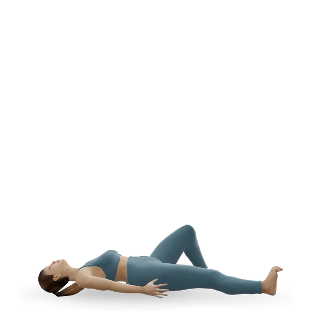
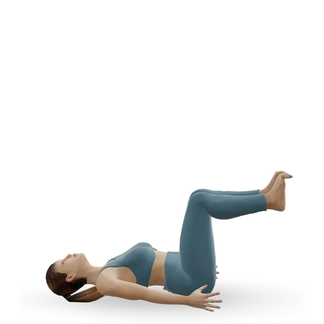
Supine heel slide demonstrated on the 3D figure3D demoSupine heel slideBeginner · No equipment

10 slow repetitions each side, once or twice a day · Within comfort; don't force the bend

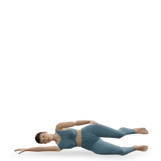
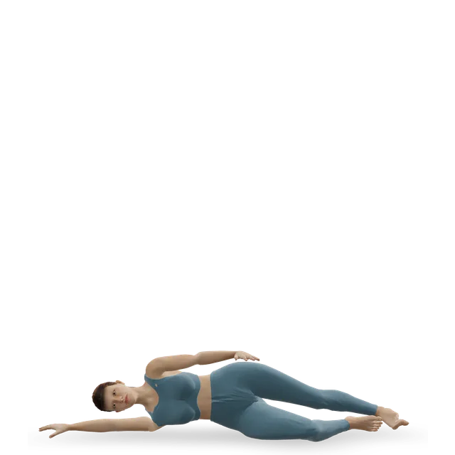
Harder: Hip windshield wipers

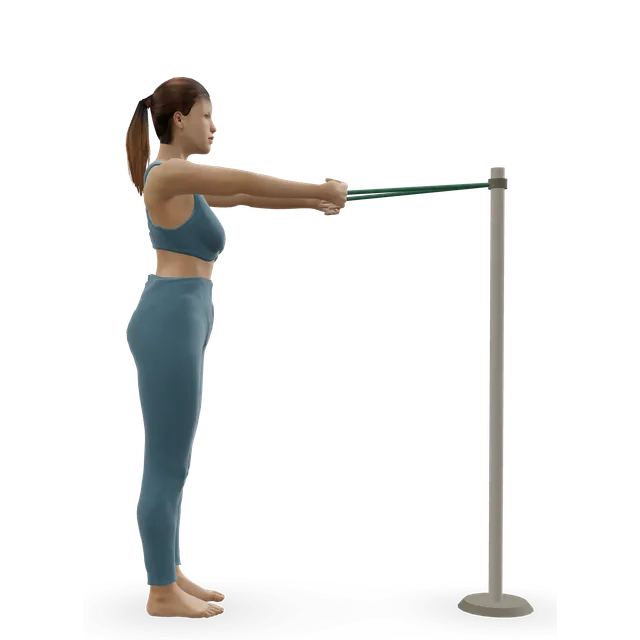
Band row demonstrated on the 3D figure3D demoBand rowBeginner · A band + An anchor point

3 sets of 10 to 15 · Upper-body strength while the hip is protected

Harder: Supported single-arm dumbbell row

Move on when: Your team has confirmed the stage and the plan, and told you how much weight the leg can take.

Follow along: a timer, a cue and the next move, one step at a time.
Stage 2

Build strength without impact (when your team allows full weight)

Strength for the hip, leg and trunk and steady fitness, without impact, and keeping your weight in a healthy range, as the RNOH advises. Two or three strength sessions a week. Doses are editorial.

Move on when: Keep going, stay in touch with your team, and tell them about any change in your pain.

Follow along: a timer, a cue and the next move, one step at a time.
Stage 3

After core decompression (as your team allows)

After core decompression you'll use a walker or crutches to protect the bone while it heals, and the AAOS says people typically walk without them at about 3 months. Your surgeon's protocol sets how much weight you can take and when, and protocols differ, so check these gentle starting points with your team. After a hip replacement, follow our hip replacement guide instead. Doses are editorial.

Quad set (static quads) demonstrated on the 3D figure3D demoQuad set (static quads)Beginner · No equipment

10 holds of 5 s, 3 times a day · From the first days, as your team allows

Harder: Straight-leg raise

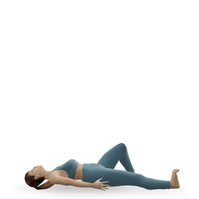
Supine heel slide demonstrated on the 3D figure3D demoSupine heel slideBeginner · No equipment

10 slow repetitions each side, once or twice a day · Within the range your team allows

Harder: Hip windshield wipers

Recumbent exercise bike demonstrated on the 3D figure3D demoRecumbent exercise bikeBeginner · Cardio machine

5 to 10 min, easy, building up · When your team allows weight through the leg

Harder: Stationary cycling

Move on when: Your team has cleared you to put full weight on the leg: then stage 2. For the long term, keep up two strength sessions and some low-impact fitness each week, with no running, jumping or impact sport unless your specialist agrees.

Follow along: a timer, a cue and the next move, one step at a time.

What to change for now

Day to day with a painful hip

The RNOH suggests losing weight if you need to, which could reduce your pain; pacing activities through the day; leading with the stronger leg going upstairs and the sore leg coming down; sitting with your hips slightly higher than your knees, and getting up regularly; and using aids for socks and shoes. Stopping smoking and cutting down on alcohol remove two risk factors.

Sources 2, 8

Water exercise

Walking in water and swimming put less load through the hip, and the RNOH suggests them. Start with 10 to 20 minutes and build up (our suggestion). The 3D figure can't show exercise in water, so it isn't in the plan.

Source 2

Steroids and your other teams

If you take steroids for another condition, don't change or stop them yourself: stopping suddenly can be dangerous (our caution). Tell the team who prescribe them about the diagnosis, so they can review the dose; the risk rises with the dose. The same goes for chemotherapy and treatments that dampen the immune system.

Sources 5, 6

Positions to swap, and why

Habit or positionWhy it mattersInstead
Skipping, jumping and runningThey load a bone that may be weak. Our osteonecrosis caution asks for a specialist's agreement first, and the RNOH recommends low-load exercise.A stationary bike or water exercise.Try: Recumbent exercise bike
Long walks without the crutches or aid you were advised to useTaking load off the hip is the aim of care without surgery, and daily crutch walking eased pain in a trial.Shorter walks with the aid, sitting to rest, and the bike for fitness.Try: Stationary cycling
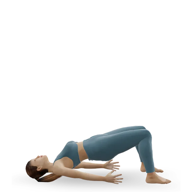
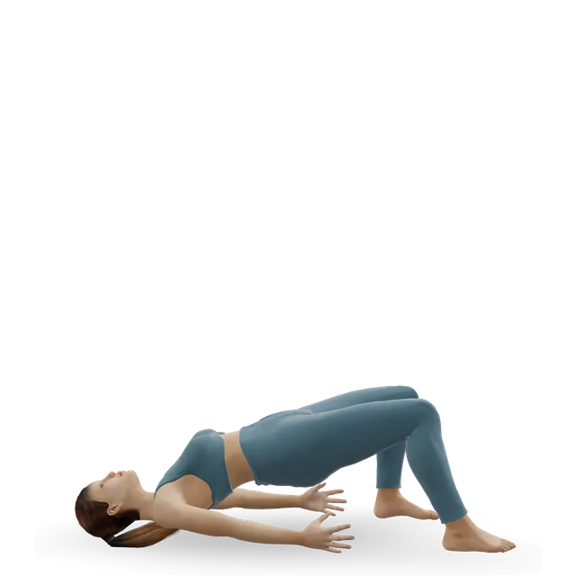
Heavy squats such as goblet squats, and deadlifts and lungesThey put a lot of load through the hip (our caution).Chair sit-to-stands and bridges.Try: Sit to stand from a chair
Forcing a deep squat hold or end-of-range hip stretchesSitting, bending and squatting are often painful, and nothing is gained by forcing the range.Gentle movement within comfort.Try: Supine heel slide
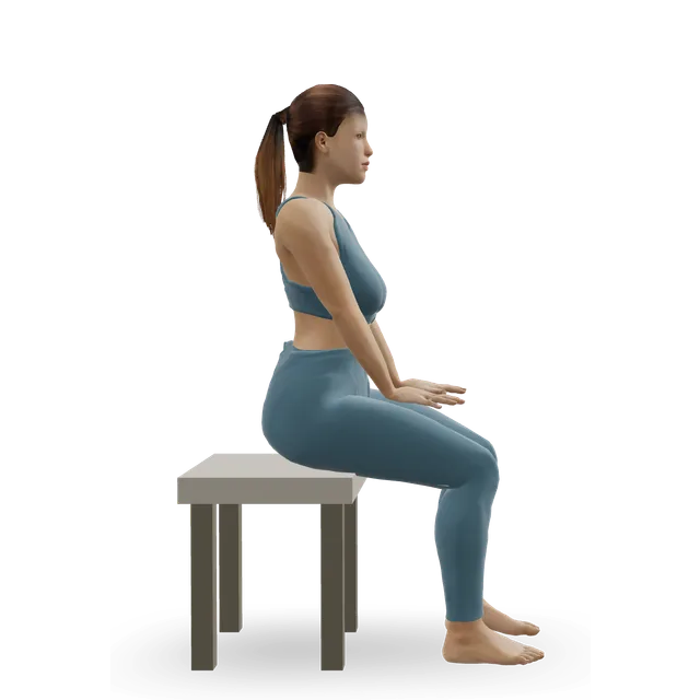
Low chairs and long spells of sittingThe RNOH advises sitting with your hips slightly higher than your knees and getting up regularly.A higher chair or a wedge cushion.
Changing or stopping steroid medicines yourselfSteroids are a major risk factor, but stopping suddenly can be dangerous (our caution).Ask the prescribing team to review them, and tell them about the diagnosis.
Waiting it out when hip pain lasts more than 6 weeks and the X-ray was normalEarly avascular necrosis may show only on an MRI scan.Ask your GP about an MRI and a hip specialist.
Relying on tablets or supplements to stop collapseBone-strengthening drugs didn't in two randomised trials.Follow your specialist's plan.

Routines to follow

More moves for this area

Questions people ask

What is avascular necrosis of the hip?

Part of the ball of the hip loses its blood supply and the bone dies. Without treatment, the surface can crack and collapse, leading to arthritis. It's also called osteonecrosis or AVN.

What causes it?

Most cases are linked with steroid medicines or heavy drinking. Others follow sickle cell disease, lupus, chemotherapy, HIV, organ transplants, radiotherapy, a hip fracture or dislocation, or a recent pregnancy, and sometimes no cause is found.

Do steroid medicines cause it?

They're one of the commonest causes, and the risk rises with the dose and the total amount: across 57 studies, 6.7% of people given a large total amount developed it, and daily doses above 20 mg carried more risk. Don't change your steroids yourself, but tell your doctor about any new hip pain.

What does AVN hip pain feel like?

A deep ache in the groin, buttock or side of the hip, sometimes down the thigh to the knee, worse with walking and weight-bearing and, later, at rest. Many people are stiff, especially putting on socks and shoes, and some limp.

Will it show on an X-ray?

Not early on. An X-ray can be normal for months while an MRI scan shows it. A UK review advises an MRI and a hip specialist for hip pain lasting more than 6 weeks with a normal X-ray, especially if you have risk factors.

What are the stages?

Stage I shows only on an MRI; stage II shows on an X-ray without a crack; stage III has a crack under the surface, with the ball sinking by up to 2 mm (IIIA) or more (IIIB); stage IV is arthritis. Treatment depends on the stage and the size of the dead area.

Can it heal without surgery?

Small areas on the inner side of the ball sometimes do well, but most larger ones get worse, and treatments without surgery haven't been shown to stop that. That's why early specialist assessment matters.

What exercises can I do?

Low-load exercise that keeps the hip moving and the leg and trunk muscles strong, such as a stationary bike, water exercise, bridges and gentle range-of-movement work, within your team's weight-bearing limits. Avoid running and jumping unless your specialist agrees.

Should I use crutches?

If your team advises it. Taking load off is a main aim of care without surgery, and in a recent trial daily crutch walking eased pain over 3 months, though on its own it doesn't stop the disease.

What is core decompression?

An operation that drills channels into the ball of the hip to relieve pressure and let new blood vessels grow, sometimes with bone graft or your own bone-marrow cells. It's for early stages, before the ball collapses, and you'll use a walker or crutches for a few months afterwards.

Will I need a hip replacement?

If the ball has collapsed and arthritis has set in, a hip replacement is the most reliable treatment, and results are now similar to those for arthritis. Earlier stages can sometimes be saved.

Can it affect the other hip?

Yes, it's often in both hips, and the second hip is most at risk in the first 2 years, so your specialist may scan both. An MRI scan can show it in a hip that doesn't hurt yet.

Related exercise guides

Other pain guides

Sources

  1. AAOS OrthoInfo · Osteonecrosis of the hip
  2. Royal National Orthopaedic Hospital NHS Trust · A patient's guide to avascular necrosis (2025)
  3. Mont 2020 · Non-traumatic osteonecrosis of the femoral head: a 5-year update (review, J Bone Joint Surg Am)
  4. Cooper 2010 · Osteonecrosis in two UK GP record databases (case-control study, Osteoporos Int)
  5. Lamb 2019 · Avascular necrosis of the hip: easily missed (clinical review, BMJ)
  6. Mont 2015 · Steroid dose and the risk of hip osteonecrosis (meta-analysis of 57 studies, J Arthroplasty)
  7. Fukushima 2013 · Alcohol, steroid tablets and hip osteonecrosis in Japan (case-control study, Bone Joint J)
  8. Hirota 1993 · Alcohol, smoking and the risk of hip osteonecrosis (case-control study, Am J Epidemiol)
  9. Hernigou 2006 · Silent hip osteonecrosis in adults with sickle cell disease (cohort of 121 hips, J Bone Joint Surg Am)
  10. Nevskaya 2017 · Avascular necrosis in lupus (meta-analysis, Clin Exp Rheumatol)
  11. Arnold 2026 · Osteonecrosis in children with acute lymphoblastic leukaemia (population study, EJHaem)
  12. Johannson 2011 · Hip replacement outcomes in osteonecrosis (systematic review of 67 reports, Int Orthop)
  13. Roth 2016 · German S3 guideline on non-traumatic femoral head necrosis in adults (guideline, Arch Orthop Trauma Surg)
  14. NICE NG226 · Osteoarthritis in over 16s: diagnosis and management (2022)
  15. Yoon 2020 · The 2019 revised ARCO staging of femoral head osteonecrosis (consensus, J Arthroplasty)
  16. Sharp 2026 · Osteonecrosis of the femoral head: how management has changed (review, Curr Rev Musculoskelet Med)
  17. Mont 2010 · What happens to untreated hip osteonecrosis without symptoms (systematic review of 16 studies, J Bone Joint Surg Am)
  18. Leung 2025 · Hip and pelvis conditions in pregnancy (review, Curr Rev Musculoskelet Med)
  19. NHS · Hip pain in children (irritable hip)
  20. Klumpp 2015 · Conservative treatment of hip osteonecrosis in adults (review, Clin Cases Miner Bone Metab)
  21. Mont 1996 · Core decompression vs non-operative care for hip osteonecrosis (review, Clin Orthop Relat Res)
  22. Zhang 2026 · Crutch walking in early and middle-stage hip osteonecrosis (RCT of 56, J Orthop Translat)
  23. Neumayr 2006 · Physiotherapy alone vs core decompression in sickle cell disease (RCT, J Bone Joint Surg Am)
  24. Chen 2012 · Alendronate to prevent collapse in hip osteonecrosis (placebo-controlled RCT, Arthritis Rheum)
  25. Lee 2015 · Zoledronate to prevent collapse in hip osteonecrosis (RCT, J Bone Joint Surg Am)
  26. Villa 2016 · Treating hip osteonecrosis before collapse (systematic review of RCTs, HSS J)
  27. Xiang 2026 · Bone-marrow cells added to core decompression (meta-analysis of 10 RCTs, Arthroscopy)
  28. Ageberg 2010 · NEMEX-TJR neuromuscular training for severe hip or knee osteoarthritis, and its pain rule (BMC Musculoskelet Disord)
  29. Silbernagel 2007 · Continued sports activity with a pain-monitoring model in Achilles tendinopathy (RCT, AJSM)
  30. NHS · Hip pain in adults
  31. NHS · Sickle cell disease
  32. NHS · DVT (deep vein thrombosis)
  33. NHS · Sciatica

Exercise education, not a diagnosis or treatment plan. It is written from the guidelines and studies listed above; review by a clinician is pending. If your pain follows an injury, is severe, is getting worse, or comes with any warning sign listed on this page, get it assessed. Health disclaimer.