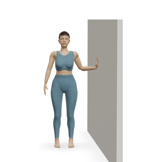
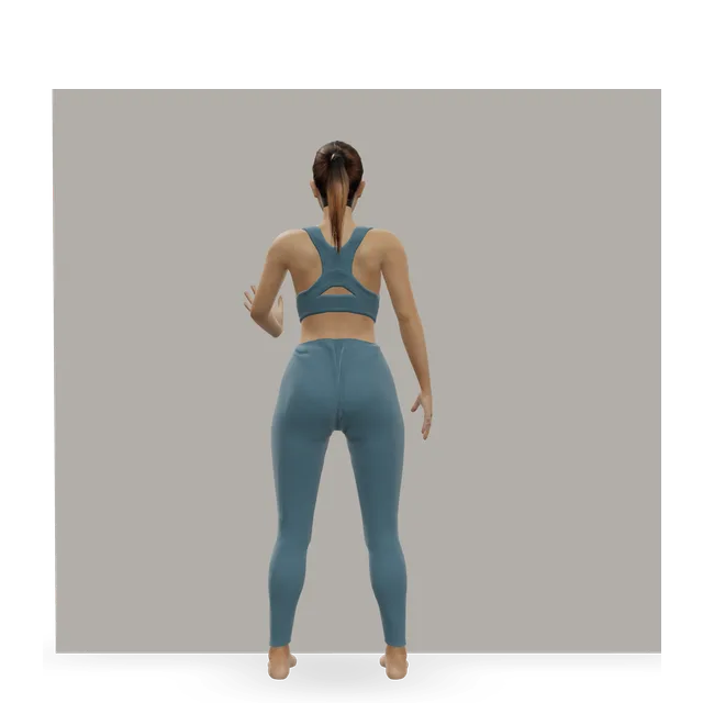
Pain guide · Ankle pain

High ankle sprain: how to spot it, why it takes longer, and when it needs a specialist

A high ankle sprain injures the ligaments that hold your two shin bones together just above the ankle, usually when the foot is planted and forced to turn outwards in a tackle or a twist. It hurts above the ankle, at the front between the bones, especially when you push off or turn the foot out, and it takes about twice as long as a common sprain or more, often 6 weeks or longer. Get it checked the same day: it often comes with other ligament damage or a fracture, sometimes high up the outer leg bone near the knee.

Most high ankle sprains are stable and heal with a boot or brace and rehab; unstable ones, where the bones spread apart, need a specialist and usually surgery. This guide explains the tests and scans, what your team is looking for, and a staged plan with 3D demos from protecting the ankle to hopping, running and cutting, moving on by milestones rather than dates.

When to get help first

Exercise is for pain that has been checked or is clearly mild and settling.

Get urgent medical help if

  • Call 999 or go to A&E: after an injury, you heard a crack, the foot or ankle has changed shape or points at an odd angle, a bone is sticking out, there is a bad cut over it, you're in severe pain, or the toes look blue or white, feel numb or are cold to touch. These can mean a broken bone. Don't drive yourself.
  • Same day: after the foot was forced outwards, pain above the ankle between the shin bones, or pain anywhere up the outer side of the leg to just below the knee, especially if you can't hop or walk normally. A high ankle sprain can be unstable, and the outer leg bone can break high up (a Maisonneuve fracture), so the whole lower leg needs examining and an X-ray.
  • Same day (urgent GP appointment or NHS 111): after an injury, you can't put weight on it or walk more than a few steps, the pain is very bad or getting worse, there is a lot of swelling or bruising or it is getting worse, or it feels very stiff. If you think it's broken, don't treat it yourself.
  • Same day (NHS 111), while you're recovering: the pain gets worse; a very high temperature, or feeling hot, cold or shivery, which could be an infection; the leg, foot or toes feel numb or burning; the cast is too tight, or the toes look swollen, blue or white; the cast or boot rubs or feels too loose; or there's a bad smell or discharge from under the cast.
  • After surgery, same day (urgent GP appointment or NHS 111): a very high temperature or feeling hot, cold or shivery, or a wound that is red, hot, swollen or oozing. These can be signs of an infection.
  • Call 999: calf or leg pain or swelling with breathlessness or chest pain. It could be a clot that has reached the lungs.
  • Same day (GP or NHS 111): throbbing pain and swelling in one calf, with red or darker skin. It could be a blood clot (DVT). Watch for it after an ankle injury or time in a cast or boot.

See a doctor or physiotherapist if

  • Pain that isn't better after 2 weeks of looking after it yourself, is getting worse, keeps coming back, or stops you doing normal things.
  • Still painful, swollen or unstable 6 to 8 weeks after the injury, or pain at the front of the ankle when you squat or push off: ask for a review. A missed instability, scarring, a pinch at the front of the ankle or a cartilage injury can cause lasting symptoms, and an MRI (magnetic resonance imaging) scan may be needed.
  • An ankle that keeps giving way months after a sprain.
  • Tingling or loss of feeling in the foot, or a foot that feels weak.

Understand it

What it is

Just above the ankle joint, the shin bone (tibia) and the outer leg bone (fibula) are held together by a group of ligaments called the syndesmosis: ligaments at the front and back, one between the bones, and a membrane running up the leg. They keep the socket of the ankle joint tight around the ankle bone. A high ankle sprain stretches or tears them, on its own or together with an outer ankle sprain, an injury to the strong ligament on the inner side (the deltoid), or a fracture. The National Institute for Health and Care Excellence (NICE) calls these the most severe ligament injuries of the ankle.

Sources 1, 2, 3

How it happens, and who gets it

Usually the foot is planted and turned outwards while the leg turns in: a player rolled on from the side, or a kneeling player whose heel is struck. Less often, the ankle is bent up hard. NHS Lothian lists football, rugby, skiing and basketball, falls and accidents.

In US college sport, high ankle sprains were commonest in men's American football, then wrestling and ice hockey; 60% came from contact, and 47% kept players out for a week or more. In elite European football, isolated high ankle sprains were 7% of 1,320 ankle ligament injuries, and players missed 39 days on average.

Sources 1, 2, 4, 5, 6

How it's recognised

Pain and swelling above the ankle at the front, between the bones, sometimes spreading up the leg; bruising and stiffness; and pain pushing off or pivoting. Most people can't walk normally without crutches at first.

No single test is reliable. In 87 people with ankle sprains checked against MRI, tenderness over the ligament picked up 92% of high ankle sprains and being unable to hop 89%, but both were also common in other sprains. The squeeze test (squeezing the shin bones together mid-calf) and pain out of proportion to the injury were rarer in other sprains: 88% and 79% of people without a high ankle sprain tested negative. So clinicians combine several tests, and a review advised MRI before a final diagnosis.

Sources 4, 1, 7, 8

Ruling out a break, all the way up the leg

Clinicians check for a fracture first. The Ottawa ankle rules decide who needs an ankle X-ray, and our ankle sprain guide explains them. With a suspected high ankle sprain, they also feel the whole outer leg bone: a Maisonneuve fracture breaks it high up, near the knee, with the syndesmosis torn, from the same twisting injury. It hurts mostly at the ankle and very little at the break.

In 5 emergency patients, a Maisonneuve fracture was missed despite ankle X-rays, and all of them needed surgery; the authors' message is to feel the whole fibula in every ankle injury. These injuries range from stable to very unstable, often come with other ankle fractures, and usually need the syndesmosis fixed.

Sources 9, 10, 11

Stable or unstable

A European consensus of specialist societies (ESSKA-AFAS) sorts isolated high ankle sprains into stable and unstable. Stable ones are treated without surgery, and unstable ones, where the two bones spread apart, with surgery. Specialists check with tests that move the outer leg bone and with X-rays that measure the gap between the bones; NHS Lothian says a standing X-ray may be taken.

Some injuries look stable at rest and spread apart only under load. In 64 elite athletes, a positive squeeze test made instability 9.5 times as likely, and an injury to the inner (deltoid) ligament 11 times; those that looked unstable had keyhole surgery to check. Clear instability without a fracture is uncommon, so most high ankle sprains are treated without surgery, but a review notes that unstable ones are often overlooked.

Sources 12, 4, 13, 1, 14

Scans: what they show

MRI shows the syndesmosis ligaments well, and other damage such as outer ligament sprains, bone bruises and cartilage injuries. Some findings are common and don't predict recovery: in professional ice hockey players, bone bruises were seen in 71% of 21 scans, and no MRI pattern predicted the time lost. In the National Football League (NFL), the pattern did matter: injuries with a fracture took a median of 250 days, those with a complete deep deltoid tear or a widened joint 175, and the rest 27. A scan finding guides your team; it isn't a timetable.

Sources 1, 15, 3

How it usually goes

It takes longer than a common sprain: a median of 62 days to full recovery, against 15 for an outer ankle sprain, in 63 young athletes; 55 days against 28 for severe outer sprains in cadets; and 45 days on average in National Hockey League players. In one NFL team, players with high ankle sprains missed more games and practices than those with outer sprains. Every step and push-off loads the injured ligaments, and other structures are often hurt too: only 13% of 83 NFL injuries were isolated.

Most people get back: 96% of 901 elite athletes returned to sport, after about 58 days on average. But among 96 cadets' ankle sprains, a high ankle sprain was the strongest predictor of lingering symptoms 6 months later. In 9 of 10 cadets, bone formed in the healing ligament, without instability, stiffness or arthritis at about 20 months.

Sources 16, 17, 18, 15, 19, 3, 20, 21
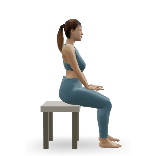
What helps, and how strong the evidence is

Stable injuries: protection, then staged rehabEmerging evidence

How much protection varies. The ESSKA-AFAS consensus recommends 3 weeks without weight in a below-knee cast, then balance exercises, but it rests on only 4 studies of non-surgical care. A rehab review prefers protection tailored to the injury: a boot, brace or tape that stops the foot turning outwards, and crutches with weight as comfort allows. NHS Lothian's leaflet says a short spell in a moon boot or brace may be used at the very start, with physiotherapy as the main treatment. In 64 elite athletes, stable injuries were treated with a boot and rehab, and returned at 45 days on average (23 to 63).

The rehab review describes three phases, moved through by milestones: protect the joint and keep the rest of the leg strong; restore range, strength, walking and balance; then hop, run, cut and practise sport. No randomised trial of rehab exists; the programme rests on clinical experience and case series. Your clinician decides how long you need protection.

Sources 22, 1, 4, 13

Unstable injuries: surgery, by a specialistEmerging evidence

Unstable injuries need the bones held in place while the ligaments heal, with a screw or a suture-button device (a strong cord between two small metal buttons). A 2021 review says unstable injuries are managed with surgery, and NICE says the evidence on suture fixation is adequate for routine use; screws are often removed later. After surgery, the consensus suggests partial weight through the foot from 6 weeks, though surgeons vary.

Return times after surgery: 64 days on average (27 to 104) in 64 elite athletes, against 45 for stable injuries treated without one; 55 days against 42 in a review of 312 athletes; and about 50 days in a review of elite athletes, most of whom had suture buttons. The groups had different injuries, so this doesn't mean surgery itself is slower. Injuries with a fracture take much longer.

Sources 22, 2, 14, 13, 23, 24, 3

Most athletes get back to sportStrong evidence

Return rates are high and consistent: 96% of 901 elite athletes returned, after about 58 days on average; 99% of 440 in another review, at 29 days without surgery and 50 with; and 93.8% to their previous level in a review of 312. The times vary a lot between groups, because the injuries and the sports differ, so treat them as a range rather than a target.

Sources 20, 24, 23

Injections: one small trialMixed evidence

In one small randomised trial of 16 elite athletes, an ultrasound-guided injection of plasma rich in growth factors (a kind of platelet-rich plasma, PRP) shortened the return (40.8 against 59.6 days), with less pain on return; a rugby pilot without a randomised comparison pointed the same way. That's too little to rely on, so injections aren't part of this plan.

Sources 25, 26

How much pain is OK while you exercise?

The foot and ankle rule, without forcing two positions earlyEmerging evidence

Mild to moderate pain, no more than about 5 out of 10, that settles within an hour, with no more swelling or stiffness the next morning, is fine; if it's worse the next day, do a little less, not nothing. That's our foot and ankle guides' rule, an editorial number. An NHS ankle sprain leaflet says to do the exercises without causing too much pain, and to move on once the current level is easy without discomfort.

Two positions are different. Early on, don't force the foot to turn outwards (pivoting or cutting on a planted foot) or to bend up hard (deep squats or lunges, with the knee pushed far over the toes): that's how the injury happens, and the rehab review avoids both until the second phase, then moves through the full range gently.

Move on by milestones, not dates: walking without crutches and with little discomfort; then jogging and hopping without pain or giving way; then sport drills at game speed with little or no pain. After surgery, your team's limits on weight and movement come first.

Sources 27, 1, 22

The exercise plan: 3 stages

Start at the first stage. Each move opens a full guide with a 3D demo.

Stage 1

Protect (weeks 0 to about 2, or longer if your team says)

Protect the ligaments in a boot or brace, on crutches with weight as comfort allows, or as your clinician advises; some teams use up to 3 weeks in a cast without weight. Avoid turning the foot outwards and forcing it to bend up, and keep the rest of the leg and your body strong. After surgery, this stage starts when your team allows. Doses are editorial unless named.

Move on when: You can walk without crutches and with little discomfort, and your team agrees.

Follow along: a timer, a cue and the next move, one step at a time.
Stage 2

Walk well, and restore range and strength (from when you're off crutches, often weeks 2 to 6)

A normal walk, full range without forcing, strength in every direction, and balance. Stationary cycling, 15 to 30 minutes, fits here too. Doses are an NHS leaflet's where named, otherwise editorial.

Ankle circles demonstrated on the 3D figure3D demoAnkle circlesBeginner · No equipment

10 slow circles each way, 3 to 4 times a day, within comfort · Don't force the turn outwards. An NHS leaflet's exercise

Harder: Ankle CARs (controlled articular rotations)

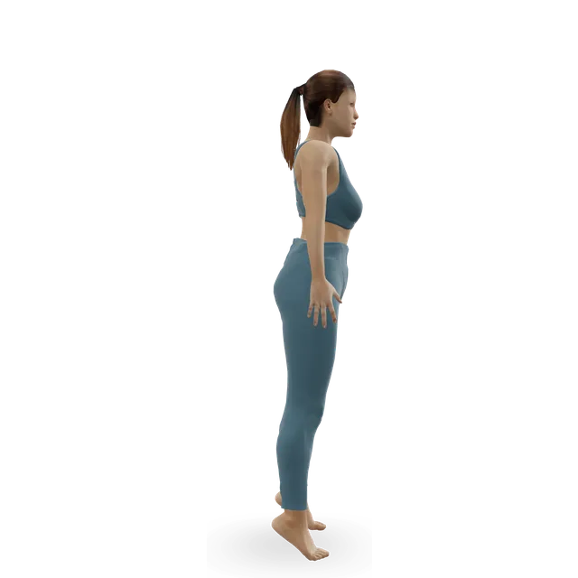
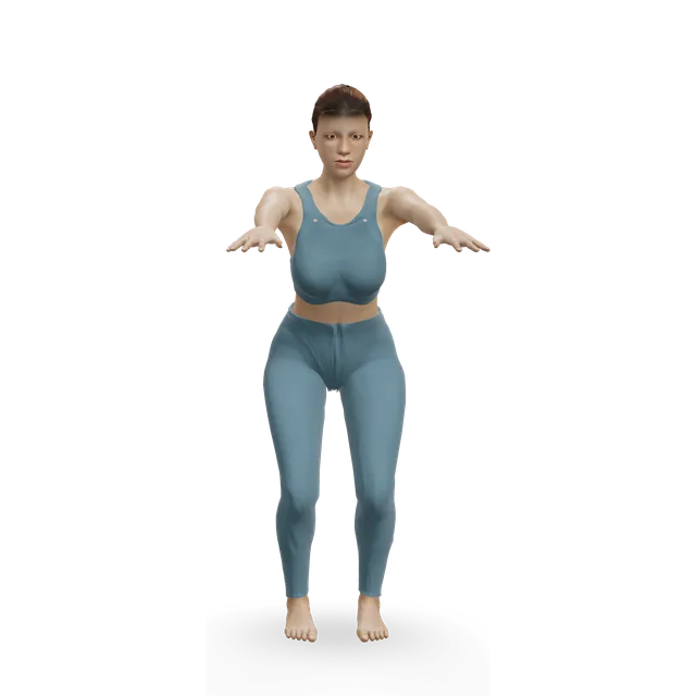
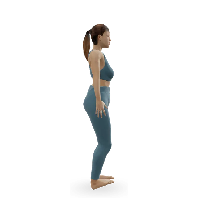
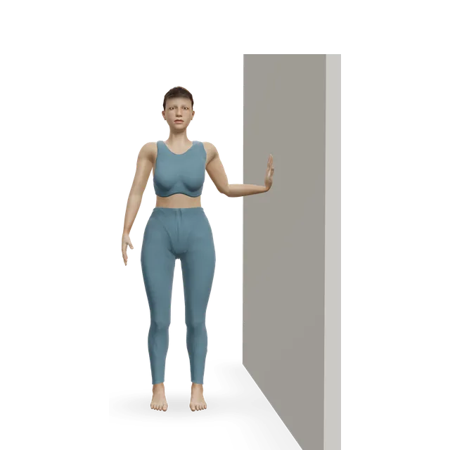
Slow calf raise demonstrated on the 3D figure3D demoSlow calf raiseBeginner · A wall

12 repetitions, 3 times a day, holding on · An NHS leaflet's dose

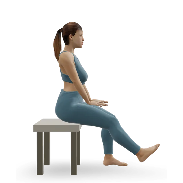
Easier: Seated bent-knee calf raise (soleus raise) · Harder: Standing dumbbell calf raise

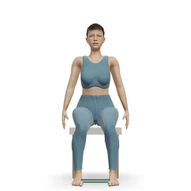
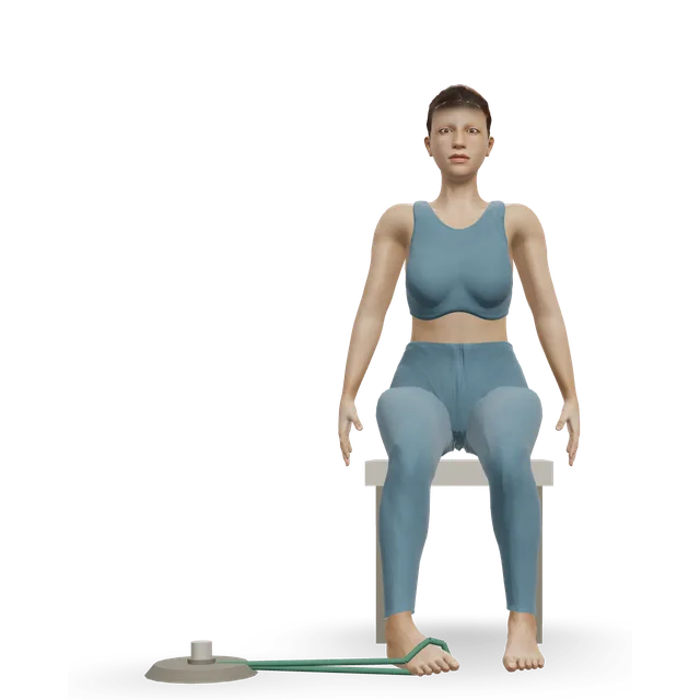
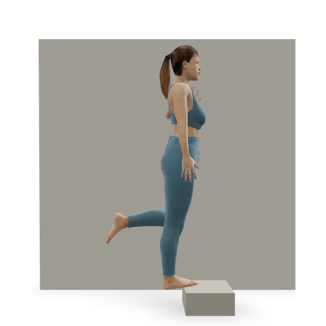
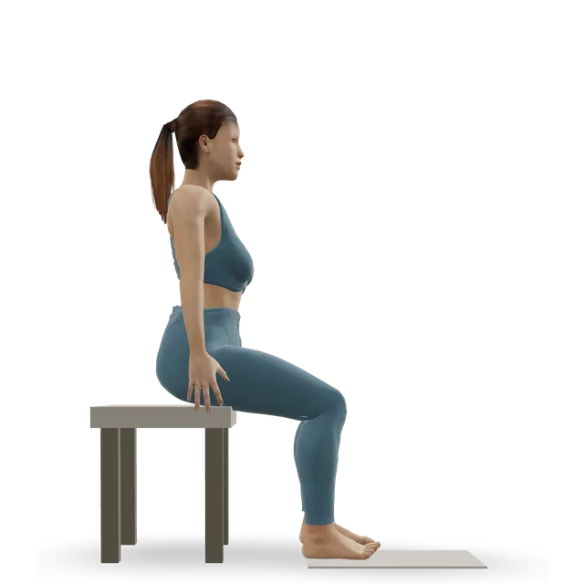
Band ankle inversion demonstrated on the 3D figure3D demoBand ankle inversionBeginner · A band + A bench or box

2 to 3 sets of 5 to 8, in a pain-free range · An NHS leaflet's dose

Harder: Single-leg calf raise from a step

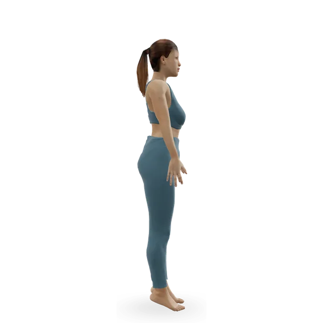
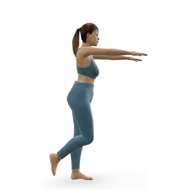
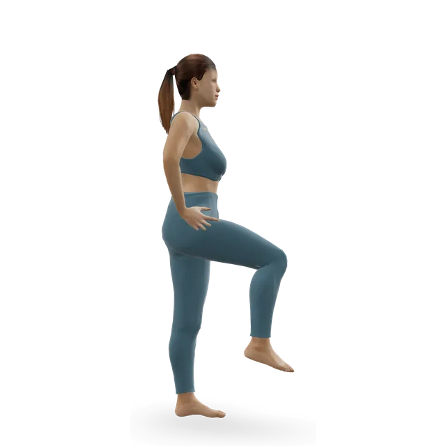
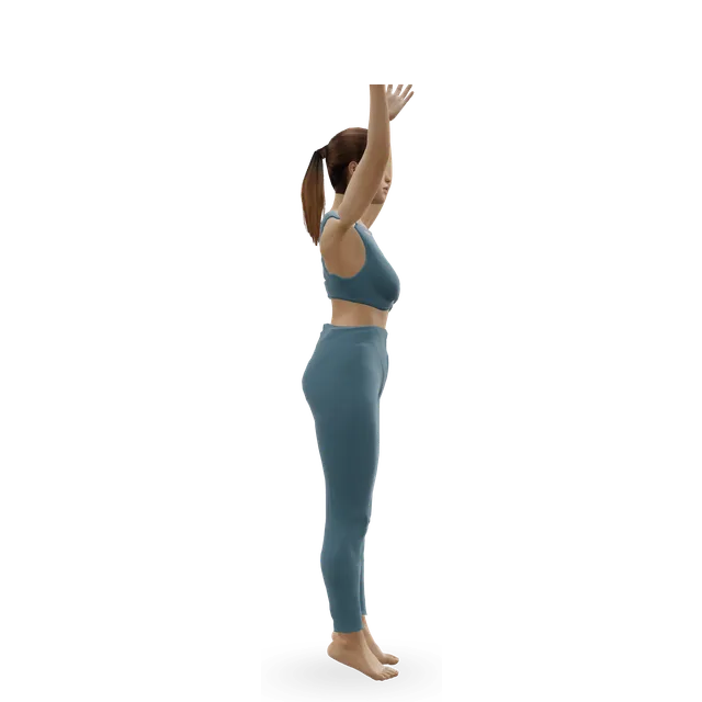
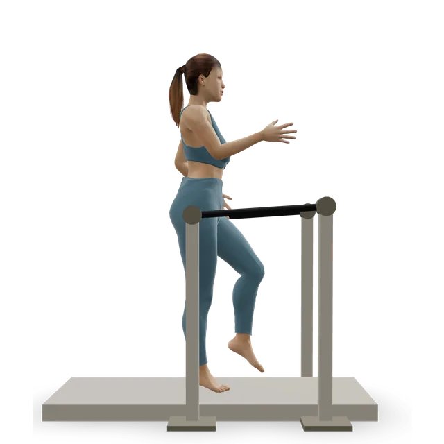
Single-leg stand demonstrated on the 3D figure3D demoSingle-leg standBeginner · A wall

Up to 30 s: holding on, then not, then with your eyes closed · An NHS leaflet's levels

Easier: Tandem stance and heel-to-toe walk · Harder: Single-leg reach matrix

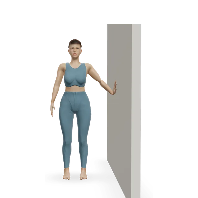
Knee-to-wall ankle rock demonstrated on the 3D figure3D demoKnee-to-wall ankle rockBeginner · A wall

Late in the stage: 10 slow rocks, without forcing the bend · The move page's dose

Move on when: You walk normally, can do 12 easy calf raises and stand on the injured leg for 30 s with your eyes closed, and then jog and hop without pain or giving way.

Follow along: a timer, a cue and the next move, one step at a time.
Stage 3

Hop, run and cut (often from weeks 4 to 8)

Hopping, running and agility, one direction at a time, then cutting, with tape or a lace-up brace, as in the rehab review's last phase. Doses are an NHS leaflet's where named, otherwise editorial.

Single-leg calf raise from a step demonstrated on the 3D figure3D demoSingle-leg calf raise from a stepIntermediate · A step + A wall

12 repetitions, 3 times a day · An NHS leaflet's dose

Easier: Slow calf raise · Harder: Single-leg pogo hops

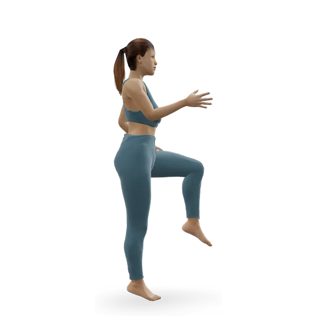
Pogo hops demonstrated on the 3D figure3D demoPogo hopsIntermediate · No equipment

3 sets of 8 to 12, every other day · Ankle bounces first. An NHS leaflet's first jumping level

Easier: Ankle bounces (mini pogo) · Harder: Single-leg pogo hops

Single-leg pogo hops demonstrated on the 3D figure3D demoSingle-leg pogo hopsAdvanced · No equipment

3 sets of 30 to 60 s, every other day · An NHS leaflet's second level

Easier: Pogo hops

Lateral cone hops demonstrated on the 3D figure3D demoLateral cone hopsIntermediate · Other / check instructions

3 sets of 30 to 60 s, every other day, last · Side to side once forwards and back are easy, as the NHS leaflet advises

Easier: Lateral bound (skater jump) · Harder: Single-leg cone hops

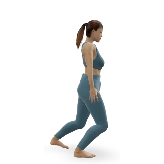
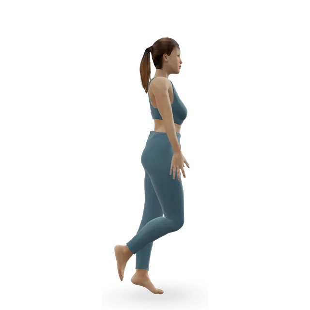
Shuttle runs demonstrated on the 3D figure3D demoShuttle runsAdvanced · No equipment

4 to 6 runs, wide turns first, then sharper over the sessions · The move page's caution: start with wide, unhurried turns

Easier: Treadmill jogging

Move on when: Sport drills at game speed are done well with little or no pain or giving way, and your clinician agrees.

Follow along: a timer, a cue and the next move, one step at a time.

What to change for now

How much protection: your team decides

Your clinician decides whether the injury is stable and how much protection you need: anything from a short spell in a boot or brace to 3 weeks in a cast without weight. After surgery, the stages above start when your team allows weight through the foot, often about 6 weeks after the operation in the consensus. Tape or a lace-up brace is usually used in the last phase and for sport.

Sources 22, 1, 4

Positions to swap, and why

Habit or positionWhy it mattersInstead
Pivoting or cutting on a planted foot earlyForcing the foot to turn outwards is how the injury happens, and rehab leaves it until late.Straight-line walking, then running.Try: Treadmill walking
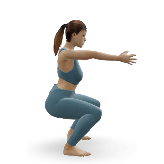
Deep squats and lunges earlyBending the ankle up hard wedges the shin bones apart, and the rehab review avoids forced bending early.Gentle ankle circles within comfort.Try: Ankle circles
Hopping or running at 2 weeks, as after a common sprainIt usually takes about twice as long or more, and hopping comes after pain-free walking and calf raises.Calf raises and balance first.Try: Slow calf raise
Coming off the boot or crutches before your team saysHow long you need protection depends on how stable the injury is, which your team judges.Follow the weight-bearing advice you were given.
Going back to sport without tape or a braceTape or a lace-up brace is usually used in the last phase and for sport.Tape or a lace-up brace for sport.
Ignoring pain high up the outer legA Maisonneuve fracture near the knee can be missed when only the ankle is examined.Ask for the whole leg to be examined.

Routines to follow

More moves for this area

Questions people ask

What is a high ankle sprain?

An injury to the ligaments that hold the two shin bones together just above the ankle (the syndesmosis), usually when the foot is planted and forced to turn outwards. It's different from the common sprain on the outer side of the ankle.

How do I know if it's a high ankle sprain?

Pain and swelling above the ankle at the front, between the bones, pain turning the foot outwards or pushing off, and being unable to hop. Squeezing the shin bones together mid-calf can hurt. No single test is reliable, so a clinician combines several, and an MRI scan confirms it.

Is a high ankle sprain worse than a normal sprain?

It usually takes longer: a median of 62 days against 15 for a common sprain in one study. It also often comes with other injuries, such as an inner ligament tear or a fracture.

How long does a high ankle sprain take to heal?

Often 6 to 8 weeks, with wide variation. Elite athletes with stable injuries returned at about 6 weeks, and those needing surgery at about 7 to 9 weeks, while injuries with a fracture take months.

Does a high ankle sprain need surgery?

Only if it's unstable, meaning the bones spread apart. Stable injuries are treated with a boot or brace and rehab; unstable ones are held with a screw or a suture-button device while the ligaments heal.

What is a Maisonneuve fracture?

A break high up the outer leg bone, near the knee, with the ligaments above the ankle torn, from the same twisting injury. It can be missed if only the ankle is examined, so tell the clinician about any pain up the outer leg.

Should I wear a boot?

Usually for a short time at first, or a brace; how long depends on how stable the injury is, and your clinician decides. A European consensus suggests up to 3 weeks in a cast without weight for stable injuries, while others use a boot with weight as comfort allows.

Can I walk on a high ankle sprain?

Many people need crutches at first, taking weight as comfort allows. Walking without crutches and with little discomfort is the sign to move on in rehab.

When can I run?

When you can walk normally, do calf raises easily and balance on the leg. Jogging and hopping should then be pain-free before cutting and sport.

Will it cause long-term problems?

Most athletes get back to sport (96% in a large review), but a high ankle sprain was the strongest predictor of lingering symptoms 6 months after an ankle sprain in one study. Get it reviewed if it's still painful, swollen or unstable at 6 to 8 weeks.

Do injections help?

One small trial of a platelet-rich plasma type injection in 16 elite athletes found a faster return, but that's too little to rely on. Injections aren't part of this plan.

Other pain guides

Sources

  1. Williams 2010 · Rehabilitation of high ankle sprains (review, Sports Health)
  2. NICE · Suture fixation of acute disruption of the distal tibiofibular syndesmosis (HealthTech guidance HTG374, 2015)
  3. Kuhn 2025 · High ankle injuries in the National Football League: MRI patterns and time out (cross-sectional study, Sports Health)
  4. NHS Lothian · Syndesmosis: what do I need to know? (patient information, 2026)
  5. Mauntel 2017 · High ankle sprains in US college sport (surveillance study, AJSM)
  6. Lubberts 2019 · Isolated high ankle sprains in 3,677 professional footballers (prospective cohort, Br J Sports Med)
  7. Sman 2015 · How accurate clinical tests are for a high ankle sprain, in 87 people (diagnostic study, Br J Sports Med)
  8. Sman 2013 · Clinical tests for a high ankle sprain (systematic review, Br J Sports Med)
  9. Taweel 2013 · Missed Maisonneuve fractures in 5 patients (case series, J Emerg Med)
  10. Bartoníček 2022 · Maisonneuve fractures of the ankle (critical review, JBJS Rev)
  11. Stufkens 2011 · Treating Maisonneuve fractures (evidence review, J Foot Ankle Surg)
  12. van Dijk 2016 · Classifying and diagnosing isolated high ankle sprains (ESSKA-AFAS consensus, Knee Surg Sports Traumatol Arthrosc)
  13. Calder 2016 · Stable or unstable grade II high ankle sprains in 64 athletes (prospective study, Arthroscopy)
  14. Corte-Real 2021 · Ankle and syndesmosis instability (review, EFORT Open Rev)
  15. Mollon 2019 · High ankle sprains in professional ice hockey players: MRI and return to play (case series, OJSM)
  16. Sman 2014 · Recovery after a high ankle sprain (prospective cohort, Med Sci Sports Exerc)
  17. Hopkinson 1990 · High ankle sprains in military cadets (case series, Foot Ankle)
  18. Wright 2004 · High ankle sprains in National Hockey League players (case series, AJSM)
  19. Boytim 1991 · High ankle sprains in a professional American football team (case series, AJSM)
  20. Li 2025 · Return to sport in elite athletes after high ankle sprains (systematic review and meta-analysis, J Orthop Surg Res)
  21. Gerber 1998 · Lasting problems after ankle sprains in cadets (prospective study, Foot Ankle Int)
  22. van Dijk 2016 · Non-surgical and surgical care of isolated high ankle sprains (ESSKA-AFAS consensus, Knee Surg Sports Traumatol Arthrosc)
  23. Vancolen 2019 · Return to sport after a high ankle sprain (systematic review, Sports Health)
  24. Bolia 2023 · Elite athletes' return to sport after high ankle sprains (systematic review and meta-analysis, Clin J Sport Med)
  25. Laver 2015 · A growth factor injection for high ankle sprains in 16 elite athletes (randomised trial, Knee Surg Sports Traumatol Arthrosc)
  26. Samra 2015 · A platelet-rich plasma injection for high ankle sprains in rugby players (pilot study, BMJ Open Sport Exerc Med)
  27. King's College Hospital NHS · Ankle sprain (patient leaflet)
  28. United Lincolnshire Hospitals NHS · Ankle sprain exercises (patient information)
  29. NHS · Broken ankle
  30. NHS · Ankle pain
  31. NHS · Sprains and strains
  32. AAOS OrthoInfo · Sprained ankle

Exercise education, not a diagnosis or treatment plan. It is written from the guidelines and studies listed above; review by a clinician is pending. If your pain follows an injury, is severe, is getting worse, or comes with any warning sign listed on this page, get it assessed. Health disclaimer.