Pain guide · Knee pain

Plica syndrome: a painful fold in the knee, and what helps

A plica is a fold in the knee's lining left over from before birth, and most knees have one. Plica syndrome is when the fold becomes thickened and irritated, usually on the inner edge of the kneecap, causing pain at one spot and often a click or snap as you bend, worse on stairs and after sitting.

Because plicae are so common, a scan showing one isn't a diagnosis: other causes are ruled out first. Most people start with about 3 months of thigh and hip strengthening and stretching, and many improve; if that doesn't work, removing the fold by keyhole surgery did better in two small trials. This guide covers how it's recognised, what the trials found and a staged plan with 3D demos.

When to get help first

Exercise is for pain that has been checked or is clearly mild and settling.

Get urgent medical help if

  • A hot, red or swollen knee with sudden severe pain, a very high temperature, or feeling hot, cold or shivery. This can be a joint infection.
  • You cannot move the knee or put any weight on it, or it is very painful, badly swollen or has changed shape.
  • The knee is locked and you cannot fully straighten it. A plica can catch, but a knee that won't straighten needs checking for a torn meniscus or a loose fragment.
  • Leg pain or swelling with breathlessness or chest pain: call 999 or go to A&E, and do not drive yourself. It can be a blood clot that has reached the lungs.
  • A child or teenager with sudden pain in the hip, thigh or knee, or who is limping or cannot put weight on one leg: ask for an urgent GP appointment or call NHS 111 the same day. Hip problems in children can be felt only in the thigh or knee.

See a doctor or physiotherapist if

  • New painful clicking, catching or giving way: get it checked (NHS 111 if it's sudden or severe). A meniscus tear or a loose fragment can cause the same symptoms. Painless clicking is normal.
  • Knee pain that has not improved within a few weeks of looking after it at home. Many areas offer NHS physiotherapy without a GP referral.
  • No clear improvement after about 3 months of a good exercise programme: ask about a referral to discuss an injection or keyhole surgery.

Understand it

What a plica isStrong evidence

Plicae are folds of the knee's lining (the synovium). They are left over from thin walls that divide the knee early in its development, before birth, and are only partly absorbed. There are four: above the kneecap, along its inner edge (the medial plica), below it, and rarely on the outer side. The medial plica is the one that usually causes trouble.

Sources 1, 2, 3

Most knees have oneEmerging evidence

In 3,889 knees seen at keyhole surgery in Japan, 80% had a medial plica. On MRI (magnetic resonance imaging) of 342 painful knees, 48% had one. A normal plica is thin, flexible and almost see-through: on dynamic ultrasound of 60 knees of volunteers without symptoms, 23 showed a plica, and none of them looked abnormal.

Sources 4, 5, 2, 6

When it becomes painfulEmerging evidence

A blow, a sudden increase in activity, or inflammation inside the knee can irritate the plica. It thickens, scars and loses its stretch, then rubs over the inner bump of the thigh bone or tugs on the lining, causing pain and sometimes wear of the cartilage next to it. An injury started it in 57% of 969 operated patients in a review, but only 16% of the 48 people in a Danish trial recalled one.

Sources 1, 7, 2

Who gets it, and how it feelsEmerging evidence

Mostly teenagers and young adults: the average age was 25 in the review of 969 patients and 27 in the Danish trial, and a British trial was in children and teenagers.

The main symptom is pain at one spot on the inner front of the knee. In the Danish trial, 67% also had clicking, 67% pain when sitting, 25% snapping, 25% a feeling of instability and only 8% swelling. Stairs, squatting, bending and getting up after sitting for a long time bring it on, and some people feel the knee catch.

Sources 1, 7, 3, 8, 2, 9, 10

How it's recognised, and what scans showEmerging evidence

It's a diagnosis of exclusion. Other causes of pain at the front of the knee, such as runner's knee, a meniscus tear, cartilage damage or tendon problems, are ruled out first, often with an MRI. The plica is then diagnosed from tenderness over a band on the inner edge of the kneecap, sometimes with a snap you can feel.

In a review of 7 studies (492 knees) checked against keyhole surgery, an examination test picked up 90% of painful plicae and was correctly negative in 89% of knees without one, and ultrasound did nearly as well (90% and 83%). MRI picked up 77% but wrongly flagged 42% of knees without plica syndrome. In another study, an examination method was confirmed at keyhole surgery in 44 of 48 people, and a 2026 study of 156 knees found MRI showed the plica well but classified it poorly. A scan can show a plica, but not whether it's the cause of your pain.

Sources 7, 2, 11, 12, 13

How common it is as a causeEmerging evidence

It's an uncommon cause of knee pain. In one Brazilian clinic, 63 of about 3,200 people with knee problems (2%) were thought to have it, and in one US series, 76 of nearly 2,000 keyhole examinations (4%) found a thickened plica as the only problem. Estimates range from 3 to 30%, depending on the group studied.

Sources 3, 14, 2, 7

How it usually goesMixed evidence

Many people improve with about 3 months of conservative care: 55 of 63 in one series. Results were more mixed in another, with 40% good, 20% average and 40% poor results in 136 people, and at a German surgical centre, 76 of 117 people (65%) went on to surgery after 3 months. People who stopped their exercises tended to slip back.

Sources 3, 15, 16, 7

How it differs from other front-of-knee pain

Runner's knee hurts around or behind the kneecap more widely; the Danish trial left out people with that kind of pain for this reason. Jumper's knee hurts on the tendon below the kneecap, a meniscus tear hurts on the joint line and can lock, and pes anserine pain sits lower, on the inner shin. Each has its own guide.

Source 7
What helps, and how strong the evidence is

Conservative care first, for about 3 monthsEmerging evidence

The usual first treatment is about 3 months of changing your activities, anti-inflammatory painkillers if they suit you, strengthening of the thigh and hips, and stretching of the hamstrings, calves and quadriceps, ideally with a physiotherapist. A US professional body says physical therapy is the most common treatment. This rests on case series and expert advice: no trial has compared exercise with no treatment.

In one series of 63 people, 90 days of changing activity, stretching and knee-straightening exercises, especially the last part of straightening, helped 55, and 8 had surgery. In another of 136, stretching gave good results in 40%, average in 20% and poor in 40%, which the authors judged enough to try it before surgery. The Danish trial's programme, which the plan below follows, used quadriceps strengthening, straight-leg raises, the leg press, an exercise bike and an elliptical trainer, with stretches.

Sources 2, 9, 3, 15, 7

Removing the plica: what the two trials foundMixed evidence

Two small randomised trials compared keyhole removal of the plica with leaving it, and both favoured removal for symptoms that persisted. Each has limits:

  • A British trial in 30 children and teenagers (45 knees) whose symptoms had continued despite physiotherapy: 20 of 24 knees (83%) improved after the plicae were divided, against 6 of 21 (29%) after a keyhole examination alone. Ten knees in the group without division had a second operation, and 7 of them improved once the plicae were divided. We could read only the summary of this 1993 trial.
  • A Danish trial in 48 adults with medial plica syndrome compared removal with at least 3 months of structured physiotherapy. Knee scores were similar at 3 and 6 months, and both groups improved. At 2 years, the surgery group scored better (89.7 against 74.6 on a 100-point knee score), but only 33 of the 48 were assessed, and most people in the physiotherapy group said they had stopped their exercises after 3 to 6 months (this wasn't formally recorded). The authors, who had expected physiotherapy to win, concluded that physiotherapy can work if it's kept up, and that surgery should be considered when it fails.
  • So the evidence on both sides is small and low in quality. Conservative care is low-risk and helps many people; for clear, persistent symptoms after about 3 months of good rehab, removal is a reasonable option.

Sources 8, 7

Results of removal in case seriesEmerging evidence

Across 23 studies (969 patients), 64% were free of symptoms after removal, 26% improved and 10% didn't, at about 2 years. A review of 12 studies (643 knees) estimated good or excellent results in 84%, and in 76% of people who had tried non-surgical care first. Results were poorer with cartilage damage under the plica, an unclear diagnosis and older age. There were no surgical complications in the Danish trial.

Sources 1, 17, 14, 10, 7

A steroid injection into the plicaEmerging evidence

In one series, an injection of steroid and local anaesthetic into the plica gave complete relief and a full return to activity in 22 of 30 people (73%), with 3 poor results; a local anaesthetic alone helped only while it lasted. Reviews suggest an injection early, while the plica is inflamed rather than scarred, as a help to exercise rather than on its own, and note that placing it accurately in such a thin fold needs image guidance.

Sources 18, 1, 2

Who is less likely to settle without surgeryEmerging evidence

In 117 people treated for 3 months at a German surgical centre, a thicker plica, a plica touching the cartilage behind the kneecap, and persistent pain with snapping as the knee bends and straightens went with conservative care failing. This was a surgical centre's patients looked at afterwards, so the figures may not apply to everyone.

Source 16

How much pain is OK while you exercise?

The knee rule, aiming low, with no painful catchingEmerging evidence

Rate your pain from 0 to 10 during and after exercise. In the pain-monitoring model used in knee trials, 0 to 2 counts as safe, up to 5 as acceptable and above 5 as too much; aim for 0 to 3 most of the time. It's borrowed from our other knee guides: no plica study has tested a pain rule.

The spot on the inner edge of the kneecap should be no sorer the next morning. If it is, shorten the range or drop back a step. In the Danish trial, the programme was adjusted when symptoms flared.

Painless clicking is normal. A painful catch or snap during an exercise means make the range smaller or swap the exercise (our suggestion), and painful catching that keeps happening is a reason to get it checked.

Sources 19, 20, 21, 7, 22

The exercise plan: 3 stages

Start at the first stage. Each move opens a full guide with a 3D demo.

Stage 1

Calm it down (about weeks 0 to 2)

Cut back on what brings the pain on, without stopping activity altogether: long sits with the knee bent, deep squats and lunges, jumping, and lots of stairs. Ice and painkillers can help a flare. The exercises follow the choice in the Danish trial's physiotherapy programme, whose doses couldn't be found, so the doses here are borrowed from our other knee guides as editorial starting points. Do them daily.

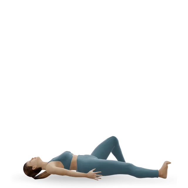
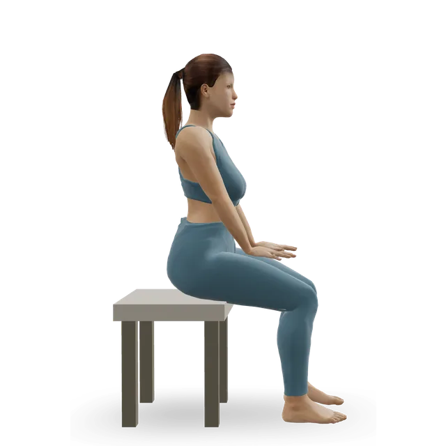
Quad set (static quads) demonstrated on the 3D figure3D demoQuad set (static quads)Beginner · No equipment

10 holds of 5 s, 2 to 3 times a day · Works the last part of straightening

Harder: Straight-leg raise

Straight-leg raise demonstrated on the 3D figure3D demoStraight-leg raiseBeginner · No equipment

2 sets of 10 each leg · In the trial's programme

Easier: Quad set (static quads) · Harder: Leg extension

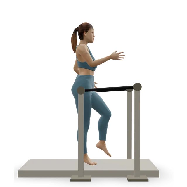
Stationary cycling demonstrated on the 3D figure3D demoStationary cyclingBeginner · Cardio machine

10 to 15 min easy, seat high · In the trial's programme

Harder: Air bike intervals

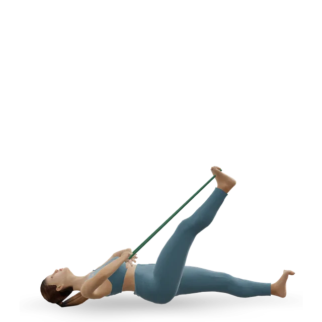
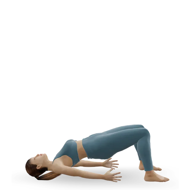
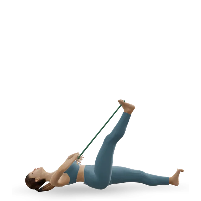
Supine hamstring stretch with a strap demonstrated on the 3D figure3D demoSupine hamstring stretch with a strapBeginner · No equipment

2 to 3 holds of 30 s each leg · Back off if you feel burning or tingling down the leg

Easier: Leg-up hamstring stretch · Harder: Contract-relax hamstring stretch (PNF)

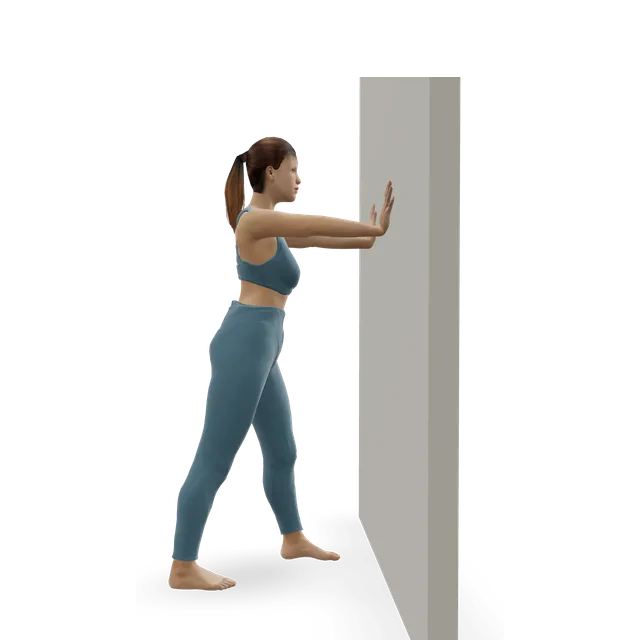
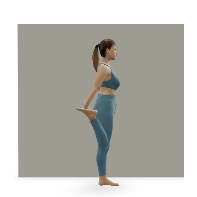
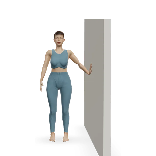
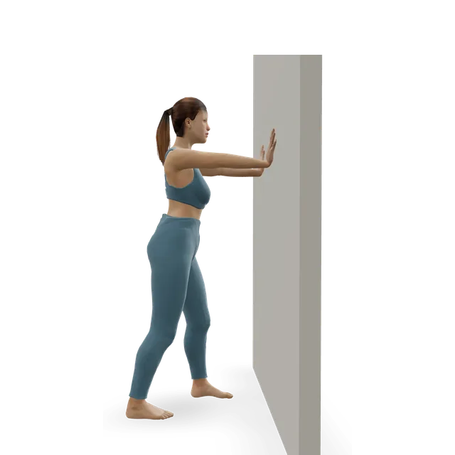
Standing quad stretch demonstrated on the 3D figure3D demoStanding quad stretchBeginner · A wall

2 rounds of 30 to 45 s each side · Only if the knee bend doesn't pinch; hold your trouser leg rather than the ankle if it does

Harder: Couch stretch

Move on when: Stairs and sitting are within the pain rule, and the spot on the inner edge of the kneecap is less tender.

Follow along: a timer, a cue and the next move, one step at a time.
Stage 2

Build: thigh and hip strength in a comfortable range (about weeks 2 to 6)

Strength 2 to 3 days a week, with the feet on the floor or a footplate and a shallow range first, plus the stretches from stage 1.

Leg press demonstrated on the 3D figure3D demoLeg pressBeginner · Strength machine

3 sets of 10 to 12 · Limited depth at first; in the trial's programme

Harder: Bodyweight squat

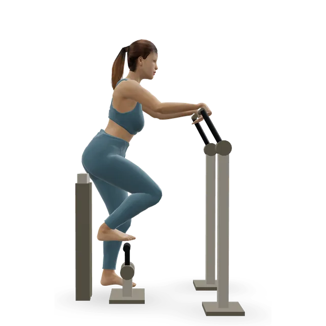
Elliptical trainer demonstrated on the 3D figure3D demoElliptical trainerBeginner · Cardio machine

10 to 20 min at a moderate effort · In the trial's programme

Harder: Stair-stepper machine

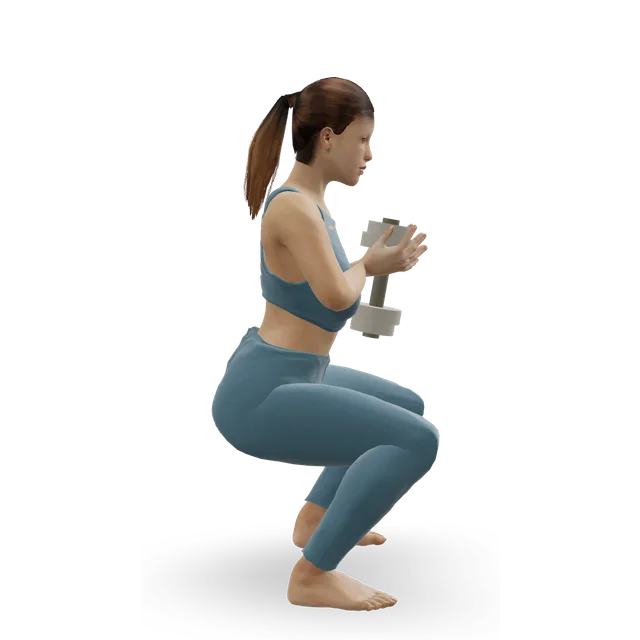
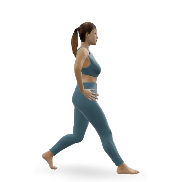
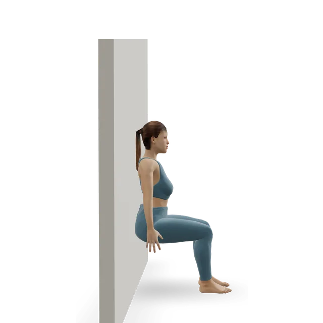
Sit squat (partial squat) demonstrated on the 3D figure3D demoSit squat (partial squat)Beginner · No equipment

3 sets of 10 · To about 45 degrees of knee bend

Harder: Bodyweight squat

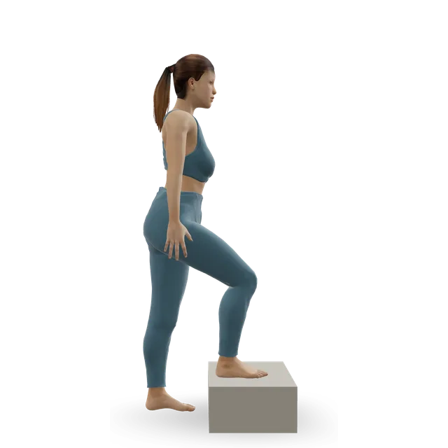
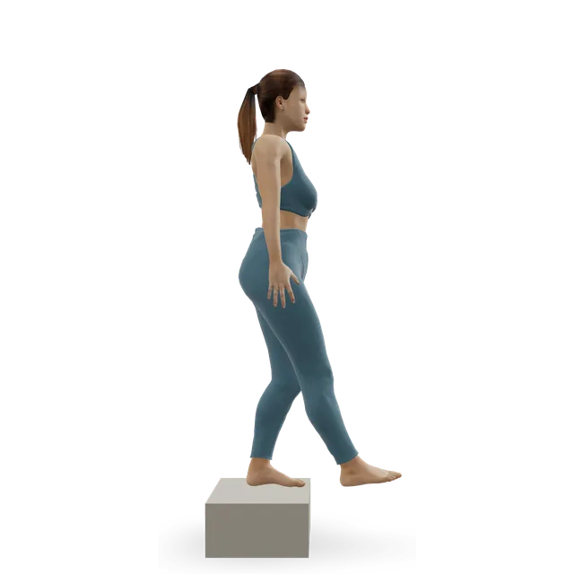
Step-up demonstrated on the 3D figure3D demoStep-upBeginner · A step

3 sets of 8 to 10 each leg · Low step first

Easier: Low step-up · Harder: Dumbbell step-up

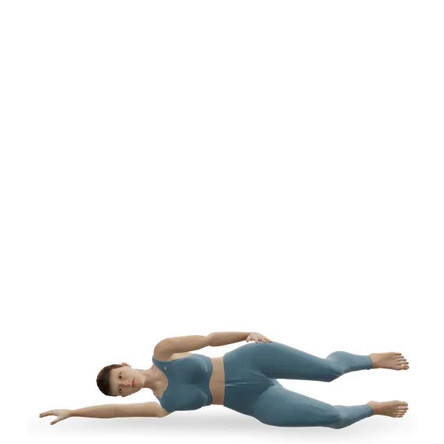
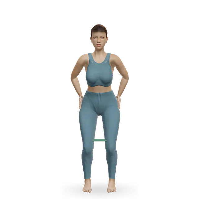
Clamshell demonstrated on the 3D figure3D demoClamshellBeginner · No equipment

3 sets of 12 to 15 each side · Hip strength, as a US professional body advises

Harder: Lateral band walk

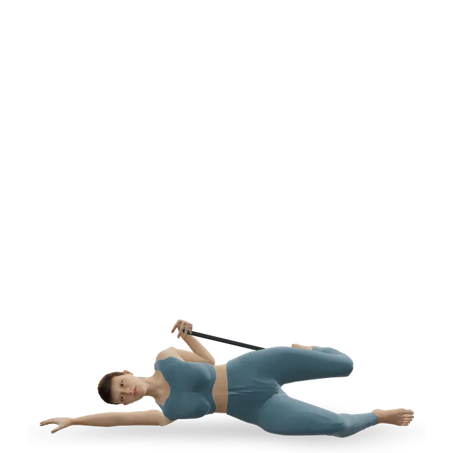
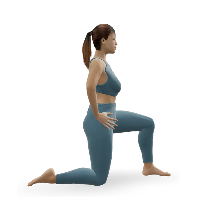
Side-lying quad stretch demonstrated on the 3D figure3D demoSide-lying quad stretchBeginner · No equipment

20 to 30 s, 2 to 4 times each side · Use a belt if the full knee bend pinches

Harder: Face-down quad and hip flexor stretch with a strap

Move on when: Shallow squats, step-ups and the leg press are within the pain rule with no painful catching, and the knee settles by the next morning.

Follow along: a timer, a cue and the next move, one step at a time.
Stage 3

Strengthen and return (about weeks 6 to 12)

Heavier and single-leg work 2 to 3 times a week, then running and sport. Keep the exercises going afterwards: in the Danish trial, people who stopped tended to slip back.

Move on when: Daily life and sport stay within the pain rule. If there's no clear improvement after about 3 months of doing this well, ask about a referral to discuss an injection or surgery.

Follow along: a timer, a cue and the next move, one step at a time.

What to change for now

What to cut back for now

Long sits with the knee bent, deep squats and lunges, jumping and lots of stairs bring the pain on. Stretch the leg out when you sit for a while, use shallow squats, and spread stairs through the day. The trial's programme avoided exercises that straighten the knee against a weight with the foot free, such as the leg extension machine, when they irritated the plica.

Sources 2, 9, 7

Keep it going

In the Danish trial, most people in the physiotherapy group said they stopped after 3 to 6 months, and their scores slipped by 2 years. Two short sessions a week once you feel better is a reasonable way to keep the gains (our suggestion).

Source 7

Positions to swap, and why

Habit or positionWhy it mattersInstead
Long sits with the knee bent, deep squats and lunges, and lots of stairs in a flareThese bring the pain on.Shorter sits with the leg stretched out, shallow squats, and stairs spread through the day.Try: Sit squat (partial squat)
The leg extension machine, or full-range knee extensions, if they catchThe trial's programme avoided exercises like these when they irritated the plica.The leg press and shallow squats, and quad sets for the last part of straightening.Try: Leg press
Pushing through painful catches or snapsPersistent pain with snapping went with conservative care failing.A smaller range, or a different exercise (our suggestion).Try: Quad set (static quads)
Stopping the exercises as soon as it feels betterIn the trial, people who stopped after 3 to 6 months tended to get worse again.Keep two short sessions a week going.Try: Split squat
Asking for an MRI to find the plicaMost knees have one, and MRI is less accurate than examination for plica syndrome.Examination first, with scans to rule out other causes.
Relying on stretching aloneThe programmes in the studies combined strengthening and stretching.Strength work plus stretches.Try: Straight-leg raise

Routines to follow

More moves for this area

Questions people ask

What is a plica?

A fold of the knee's lining left over from its development before birth. Most people have one: 80% of knees at keyhole surgery in a large Japanese study. It only causes trouble when it becomes thickened and irritated.

Is plica syndrome common?

It's an uncommon cause of knee pain: about 2% of people at one knee clinic, and 4% of nearly 2,000 keyhole examinations in another series. It's most often seen in teenagers and young adults.

What does it feel like?

Pain at one spot on the inner front of the knee, often with clicking or snapping as you bend, worse on stairs, squatting and sitting for a long time. Swelling is unusual.

How is it diagnosed? Do I need an MRI?

By examination, after other causes have been ruled out. An MRI is mostly for ruling out other problems: it can show a plica, but most knees have one, and an examination test or ultrasound diagnosed plica syndrome more accurately.

What exercises help?

Strengthening the thigh, especially the last part of straightening, and the hips, with stretches of the hamstrings, calves and quadriceps, for about 3 months. The plan on this page follows the choice of exercises in a Danish trial's physiotherapy programme.

Which exercises should I avoid?

For now, deep squats and lunges, jumping, long sits with the knee bent, and any exercise that makes the plica catch painfully, such as the leg extension machine. Build them back gradually.

How long does it take to get better?

Programmes in the studies ran for about 3 months, and many people improved over that time. If there's no clear improvement after about 3 months of good rehab, ask about a referral.

Should I have the plica removed?

If symptoms persist despite a good programme, removal is a reasonable option. In two small trials it did better than a keyhole examination that left the plica alone and, at 2 years, better than physiotherapy, though only 33 of 48 people were assessed then and many had stopped their exercises. Results were poorer with cartilage damage under the plica.

Does an injection help?

A steroid injection into the plica gave full relief in 73% of 30 people in one series. Reviews suggest it early, as a help to exercise rather than on its own.

Is it the same as runner's knee?

No. Runner's knee is pain around or behind the kneecap; plica syndrome is pain at one spot on its inner edge, often with a click. They're treated in similar ways, and runner's knee has its own guide.

Related exercise guides

Other pain guides

Sources

  1. Schindler 2014 · The 'sneaky plica' revisited: plicae of the knee and their treatment (review, Knee Surg Sports Traumatol Arthrosc)
  2. Lee 2017 · Synovial plica syndrome: a commonly overlooked cause of front-of-knee pain (review, Surg J)
  3. Camanho 2010 · Treating painful plicae of the knee (case series of 63, Clinics)
  4. Nakayama 2011 · How often a medial plica is found: 3,889 knees at keyhole surgery (case series, Arthroscopy)
  5. Hayashi 2013 · Medial plica on MRI in 342 people with knee pain (cross-sectional study, BMC Musculoskelet Disord)
  6. Paczesny 2009 · Diagnosing medial plica syndrome with dynamic ultrasound (diagnostic study, Radiology)
  7. Sauer 2022 · Plica removal vs structured physiotherapy for medial plica syndrome (RCT of 48, Surg J)
  8. Johnson 1993 · Dividing painful plicae vs keyhole examination alone in children and teenagers (RCT of 45 knees, J Bone Joint Surg Am)
  9. APTA ChoosePT · Physical therapy guide to plica syndrome
  10. Nottage 1983 · The medial patellar plica syndrome (case series, AJSM)
  11. Stubbings 2014 · How accurate examination and scans are for medial plica syndrome (meta-analysis, Knee)
  12. Shetty 2007 · Clinical diagnosis of medial plica syndrome (prospective study of 48, J Knee Surg)
  13. Zheng 2026 · How well MRI shows a medial plica (diagnostic study of 156 knees, BMC Musculoskelet Disord)
  14. Dorchak 1991 · Keyhole treatment of a painful plica: long-term follow-up (case series, AJSM)
  15. Amatuzzi 1990 · Conservative treatment of a painful plica (case series of 136, AJSM)
  16. Blanke 2021 · What predicts failure of conservative care in medial plica syndrome (retrospective study of 117, Arch Orthop Trauma Surg)
  17. Gerrard 2018 · Keyhole removal of the medial plica (meta-analysis of 12 studies, Knee Surg Relat Res)
  18. Rovere 1985 · Steroid injection into the plica for medial plica syndrome (case series, AJSM)
  19. Thomeé 1997 · Treating patellofemoral pain in young women with a pain-monitoring model (Phys Ther)
  20. Ageberg 2010 · NEMEX-TJR neuromuscular training for severe hip or knee osteoarthritis, and its pain rule (BMC Musculoskelet Disord)
  21. Smith 2017 · Should exercises be painful in chronic musculoskeletal pain? (meta-analysis, BJSM)
  22. NHS · Knee pain
  23. NHS · Septic arthritis
  24. NHS · DVT (deep vein thrombosis)

Exercise education, not a diagnosis or treatment plan. It is written from the guidelines and studies listed above; review by a clinician is pending. If your pain follows an injury, is severe, is getting worse, or comes with any warning sign listed on this page, get it assessed. Health disclaimer.