Pain guide · Knee pain

Knee OCD (osteochondritis dissecans): a damaged patch of bone and cartilage in children, teenagers and young adults

Knee osteochondritis dissecans (OCD) is damage to a patch of bone and the cartilage over it, usually on the end of the thigh bone. It mostly affects active children and teenagers, more often boys, and can be painless at first; later it causes pain and swelling with sport and, if a piece comes loose, catching or locking. A knee that locks and won't straighten needs checking the same day.

Whether the lesion is stable or unstable decides the treatment, and imaging decides that. In children whose bones are still growing, stable lesions often heal with months away from running, jumping and sport; unstable lesions and loose pieces usually need surgery, so a specialist leads the care. This guide is for young athletes, parents and coaches, and for adults: the signs to act on, what the scans mean, the rest and return times the studies give, and a staged plan with 3D demos, as the specialist allows.

When to get help first

Exercise is for pain that has been checked or is clearly mild and settling.

Get urgent medical help if

  • The knee is locked and you cannot fully straighten it. Get help from NHS 111 now: the NHS lists a knee that locks, gives way or clicks painfully for urgent advice, and in a young person a loose piece of bone and cartilage can do this.
  • Ask for an urgent GP appointment or get help from NHS 111 the same day if your child gets sudden pain in their hip, thigh or knee, or is limping or can't put any weight on 1 leg. Hip problems can sometimes be felt in the thigh or knee.
  • You cannot move the knee or put any weight on it, or it is very painful, badly swollen or has changed shape.
  • A hot, red or swollen knee with sudden severe pain, a very high temperature, or feeling hot, cold or shivery. This can be a joint infection.
  • After surgery: changes in feeling, severe pain or swelling, a change of colour in the leg or foot, oozing from the wound, a high temperature, or feeling hot and cold. Contact your surgical team, or ask for an urgent GP appointment or get help from NHS 111.
  • Same day: in a cast or brace, pain that is getting worse, toes that are numb or burning, a cast that feels too tight, toes that are swollen, blue or white, or a bad smell from under the cast.
  • Throbbing pain and swelling in one calf or thigh, with red or darkened skin or swollen veins: ask for an urgent GP appointment or get help from NHS 111, as it could be a blood clot. Call 999 or go to A&E if you are also short of breath or have chest pain.

See a doctor or physiotherapist if

  • A child, teenager or young adult with knee pain that comes with sport and keeps coming back, or an ache that's hard to pin down: see a GP or physiotherapist. X-rays can find OCD, and early, stable lesions heal best.
  • Swelling after activity, clicking or catching, or a knee that doesn't straighten as far as the other one: get it checked. These can mean the lesion is loosening.
  • Pain or swelling that comes back when running or sport restarts: stop that activity and contact the team.
  • OCD in one knee: ask whether the other knee needs an X-ray. Some children have a lesion in both knees without pain in the second.
  • Any of the NHS reasons to see a GP about a child's leg pain: pain in one leg or one joint, pain in the morning or when walking or taking part in activities, pain bad enough to stop them walking or make them limp, a rash, swelling or unusual bruising on the legs, leg pain with a high temperature, unusual tiredness, or not eating or losing weight.
  • In a brace: sore skin, pressure marks, or a brace that no longer fits as the swelling goes down. Tell your physiotherapist or orthotics team.

Understand it

What it is

Osteochondritis dissecans is a problem of the bone just under the joint cartilage. A patch of bone weakens, the bone and the cartilage over it can crack and loosen, and a piece can come free into the joint as a loose body. In the knee it is usually on the rounded ends of the thigh bone: in one population study, 64% were on the inner (medial) end and 33% on the outer end, and a few on the kneecap or the groove it runs in.

The cause isn't known. Repeated stress from sport, a disturbance in how bone forms under the growth cartilage, a reduced blood supply and genes are all proposed, and in US health records, clotting disorders were linked with it as well as sport. It has nothing to do with obsessive-compulsive disorder.

Sources 1, 2, 3, 4

Who gets it

Mostly active children and teenagers. In a US health system of over a million children, it was diagnosed in 9.5 per 100,000 aged 6 to 19 each year: 15.4 per 100,000 boys and 3.3 per 100,000 girls, more often at 12 to 19 than at 6 to 11, and never under 6. In a US county over 38 years, it peaked at ages 11 to 15. In US records from 2017 to 2024, gymnastics, volleyball and cheerleading carried the highest risk of the sports studied.

Adults get it far less often: 1.21 per 100,000 aged 20 to 45 each year, mostly men.

Sources 3, 5, 4, 6

It can be painless, and in both kneesEmerging evidence

In a Dutch population study, MRI (magnetic resonance imaging) scans of 1,910 13-year-olds found knee OCD in 1 in 174, all boys, and they were no more likely to have knee pain than other children. None of the 7 scanned again at 17 still had a lesion.

In a US population study, 7% of cases were in both knees. Of 80 children with pain in one knee, 15% had a lesion in the other knee too, and two-thirds of those later needed surgery on it.

Sources 7, 3, 8

How it's recognised

Pain and swelling, often brought on by sport, are the usual first signs, and an NHS trust leaflet describes a mild ache that gradually gets worse. Later, a loose piece can make the knee catch, lock or give way. The American Academy of Orthopaedic Surgeons (AAOS) guideline lists the symptoms that should prompt imaging: pain, swelling, locking, catching, popping or giving way, with tenderness, fluid in the knee, lost movement or grating.

Loose pieces have other causes too. A dislocated kneecap can knock off a piece of bone and cartilage, and arthritis can produce loose pieces in older adults; both have their own guides. This one is about OCD.

Sources 1, 9, 10

X-rays first, then MRIStrong evidence

X-rays come first, including a 'tunnel' view that shows the back of the rounded ends of the thigh bone; early or stable lesions can be missed on them. MRI then shows whether the cartilage is intact or cracked, whether fluid has got behind the piece, whether it has come loose, and how big the lesion is. The AAOS guideline supports MRI for this with strong evidence.

Sources 10, 11

Stable or unstable: what the scan can and can't tellEmerging evidence

Stable means the patch is still attached with intact cartilage; unstable means the cartilage has cracked, fluid has got behind the piece, or it has come loose. In adults, the MRI signs of instability are reliable: in 40 patients with an average age of 26, MRI picked out 35 of 36 unstable lesions.

In children the same signs over-call instability. In 36 lesions in children, the signs together caught every unstable lesion but labelled most stable ones as unstable too: only 11% of stable lesions were correctly called stable, against 100% in adults. Keyhole surgery is the final judge when it matters.

Sources 12, 13, 11

What scans can mistake for OCDEmerging evidence

In younger children, the end of the thigh bone hardens unevenly as it grows, especially at the back, and this can look like early OCD. In 38 children, 23 had bone defects suggestive of early OCD; the signs of a normal variant were extra small centres of bone, spiky edges and no swelling in the bone, and 22 of the 23 improved. Comparing with the other knee helps.

In 7 pain-free brothers and sisters of children with OCD, these variants were common and occasionally progressed to OCD, once needing surgery.

Sources 14, 15, 11

How it usually goesEmerging evidence

About half to three-quarters of stable lesions in children heal without surgery, depending on how studies define healing. In 47 stable lesions in children treated with a cast, then a brace and no running, jumping or sport, two-thirds were healing at 6 months. In a multicentre study of 185 knees, 77% avoided surgery; with strict criteria (complete healing on MRI and a full return to sport), 38% of 64 healed. Across 13 studies, success without surgery ranged from about 40% to 88%.

Older children do less well without surgery: 12 to 19 year olds were 7.4 times as likely as 6 to 11 year olds to need an operation. Larger lesions, and swelling, locking, catching or giving way at the start, made healing less likely. Across Europe, results were better in younger patients, and in a small older series, 3 of 5 knees with open growth plates did well without surgery against 1 of 9 with closed ones.

Sources 16, 17, 18, 19, 20, 21, 22
What helps, and how strong the evidence is

Stable lesions in growing children: months of protection, often enoughEmerging evidence

Treatment means avoiding activities that repeatedly load or squash the joint (running, jumping, squatting and sport), sometimes with crutches, a brace or a short time in a cast. How long varies: often 3 to 6 months, and up to 10 to 18 months in an NHS trust leaflet, with the specialist following healing on X-rays or MRI. One review advises at least 3 months away from high-impact activity, and running again only once pain has gone and an X-ray shows healing; full healing on X-ray often takes more than a year.

About half to three-quarters of stable lesions in children heal this way, depending on how studies define healing. These are cohorts and case series, not trials.

Sources 11, 1, 9, 16, 17, 18, 19

No method of protection has been shown to be bestEmerging evidence

The AAOS guideline couldn't recommend for or against any specific non-surgical treatment, such as casting, bracing, an unloader brace, bone stimulators or rest from activity alone, because no prospective study had compared them. Nor could it recommend for or against physiotherapy during non-surgical care.

A cast helps children stick to the rest but stiffens the knee and wastes the thigh, while a brace allows movement. In 185 knees with stable lesions on the inner end of the thigh bone, an unloader brace didn't reduce the need for surgery.

Sources 10, 17, 11

When a stable lesion doesn't heal: drillingEmerging evidence

If a stable lesion hasn't healed after months of protection, usually at least 3 to 6, surgeons may drill small holes into it to encourage healing. In a trial of 91 children comparing two ways of drilling, results and healing were similar at 2 years. In 139 knees, full movement was back at about 13 weeks, 95% were back to daily activities by 3 months, and 96% healed on X-ray; larger lesions were more likely to need another operation.

Most young athletes returned to sport after drilling in the studies, and it takes months.

Sources 11, 23, 24, 19

Unstable or loose lesions: surgery is usually offeredEmerging evidence

The AAOS guideline says children and adults with an unstable or displaced lesion that causes symptoms should be offered surgery, based on limited evidence. The options are fixing the piece back with screws, pins or stitches, sometimes with drilling or a bone graft, or, if it can't be saved, replacing the damaged surface with a plug of bone and cartilage from elsewhere in the knee, donor tissue or grown cartilage cells.

Fixed pieces healed in 72% to 76% of knees in long-term studies, as well with closed growth plates as with open ones. Removing the piece alone led to more arthritis: 70% by 30 years, against 51% when the piece was kept or restored. Most young athletes returned to sport after surgery in the studies, and it takes months.

Sources 10, 11, 1, 25, 26, 27

The long-term outlookEmerging evidence

Most children do well. In 95 children followed for 14 years on average, 14% still had knee pain, 6% had arthritis with symptoms and 3% had had a knee replacement, at an average age of 52.

After non-surgical care at any age, arthritis reached 5% at 5 years, 10% at 10 and 30% at 35 years, and was more likely with diagnosis as an adult, a body mass index (BMI) over 25, or a lesion on the kneecap.

Sources 28, 29, 27

How much pain is OK while you exercise?

No pain in the knee joint, no swelling and no catchingEmerging evidence

This rule is our suggestion, the same as our elbow OCD guide's. No study has tested a pain rule for knee OCD. If the specialist or physiotherapist has given you a programme, follow theirs.

Running, jumping and other weight-bearing work: no pain in the knee joint during, straight after or the next morning. Strength exercises: no pain in the joint; a working ache in the thigh or hip muscles that settles within the hour is fine.

Catching, locking, giving way, new swelling, or a knee that doesn't straighten as far as before are never part of the rule: stop and contact the specialist. Symptoms that come back with running or sport mean stopping that activity and contacting the team.

It's stricter than our adult knee rule, because the joint surface of a growing bone is healing, and lesions can be painless until they're advanced.

Sources 7, 10, 1, 11

The exercise plan: 4 stages

Start at the first stage. Each move opens a full guide with a 3D demo.

Stage 1

Protect the joint surface, keep the rest of you fit (often 3 to 6 months; the specialist sets the time)

No running, jumping, deep squatting or sport: one review advises at least 3 months away from high-impact activity. Use crutches, a brace or a cast exactly as instructed. Keep the thigh working and the knee moving within the limits you're given, and keep fit with the upper body and trunk, and with swimming or cycling only if the specialist allows. Children and teenagers can still aim for the NHS's average of 60 minutes of activity a day using what's allowed.

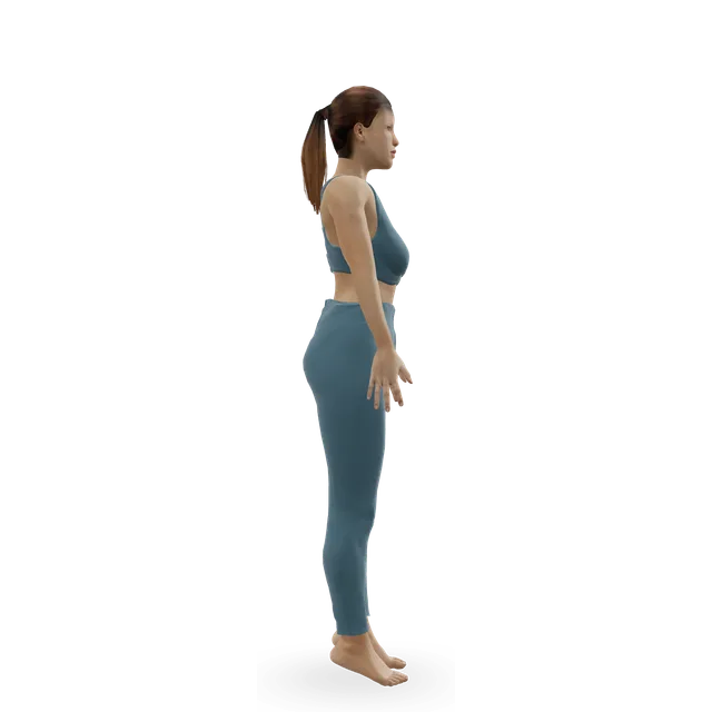
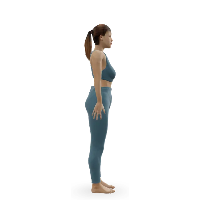
Quad set (static quads) demonstrated on the 3D figure3D demoQuad set (static quads)Beginner · No equipment

10 holds of 5 s, 2 to 3 times a day · The knee hub's dose. In a cast, tighten the thigh inside it only if the team agrees

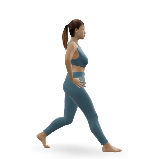
Harder: Straight-leg raise

Straight-leg raise demonstrated on the 3D figure3D demoStraight-leg raiseBeginner · No equipment

Lift about 15 cm, hold 5 s, 10 times, 2 to 4 times a day · The repetitions are an NHS trust leaflet's (given after surgery); how often is editorial

Easier: Quad set (static quads) · Harder: Leg extension

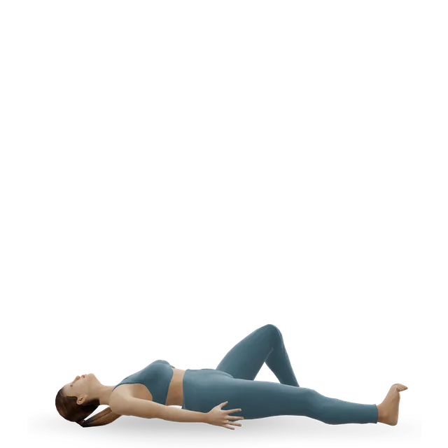
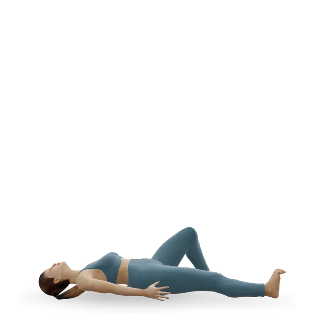
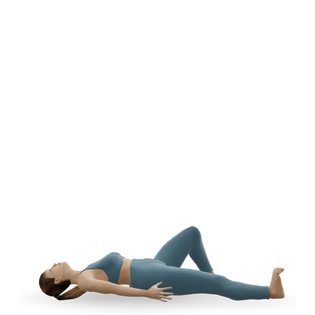
Supine heel slide demonstrated on the 3D figure3D demoSupine heel slideBeginner · No equipment

Bend as far as is comfortable, hold 5 s, 10 times · The NHS trust leaflet's dose. Only if the knee is allowed to bend; stay inside the range your team allows

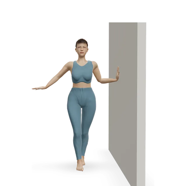
Harder: Hip windshield wipers

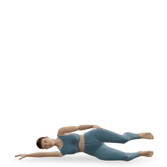
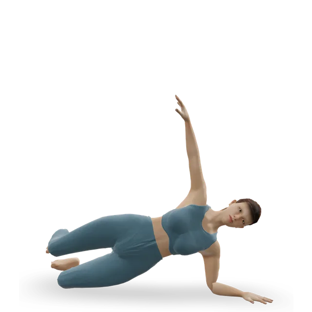
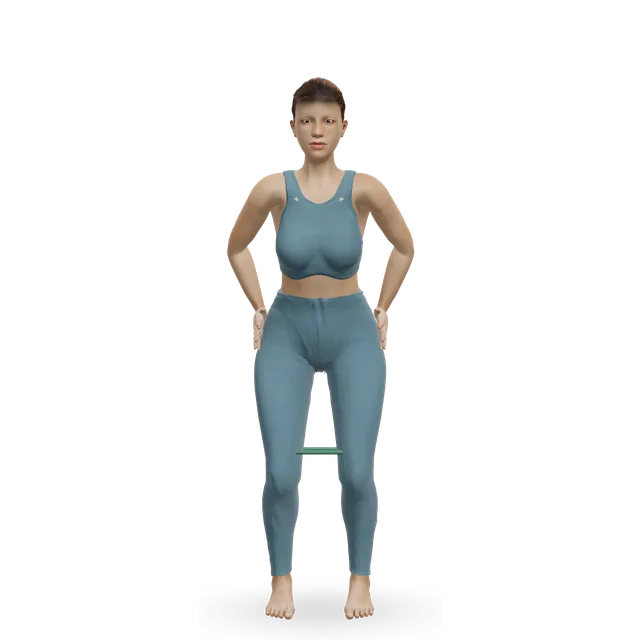
Clamshell demonstrated on the 3D figure3D demoClamshellBeginner · No equipment

2 sets of 12 each side · Editorial dose

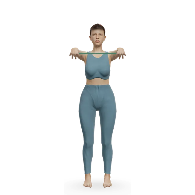
Harder: Lateral band walk

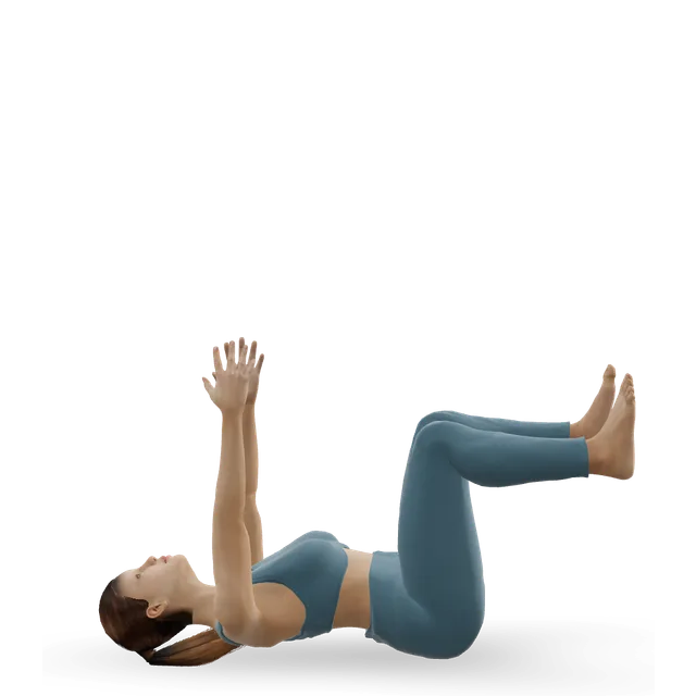
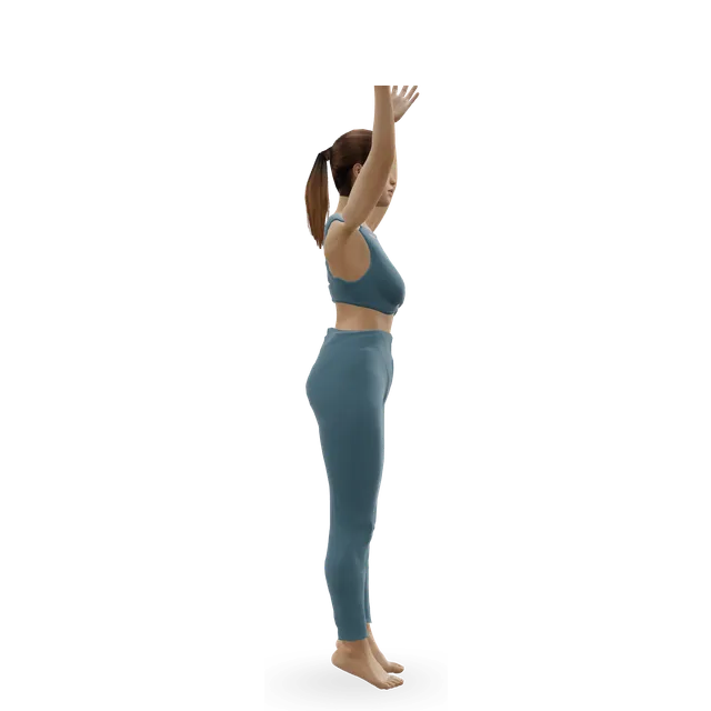
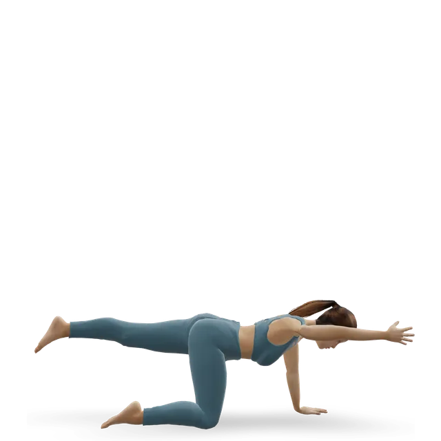
Dead bug demonstrated on the 3D figure3D demoDead bugBeginner · No equipment

2 sets of 6 to 8 each side, slowly · Our elbow OCD guide's editorial dose

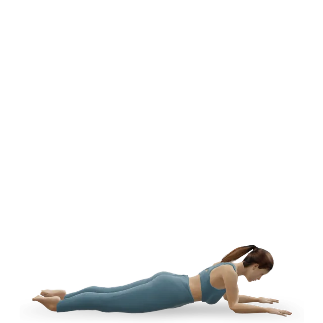
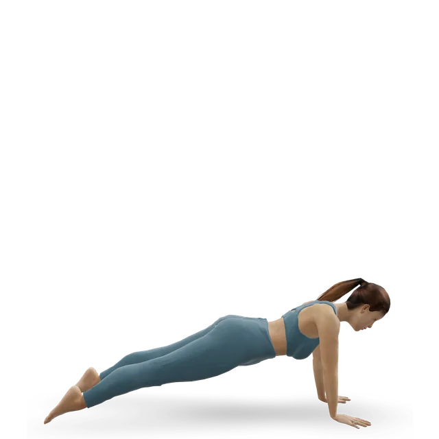
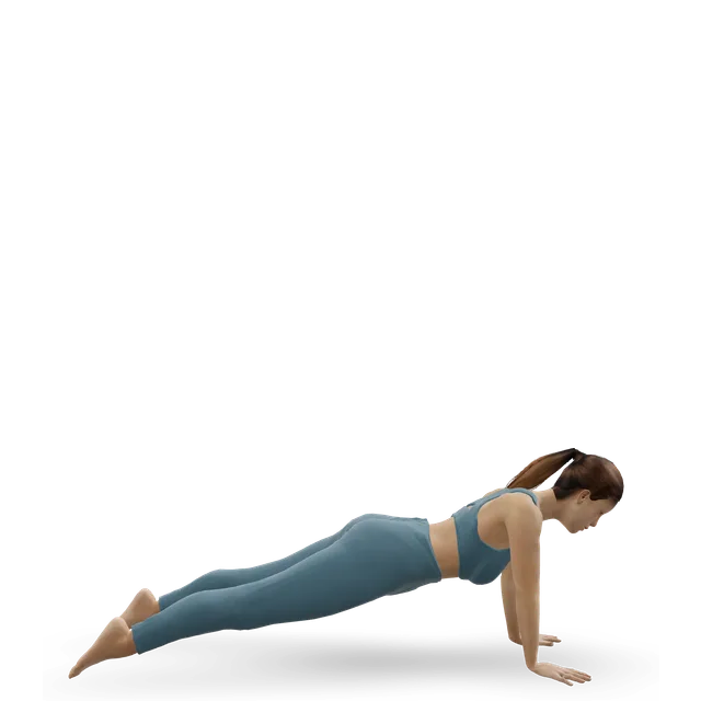
Harder: Forearm plank

Move on when: Daily life and walking are pain-free, the knee bends and straightens fully without swelling, and the specialist sees healing and clears strengthening.

Follow along: a timer, a cue and the next move, one step at a time.
Stage 2

Strength without impact (once the specialist clears it)

2 to 3 strength sessions a week, with low-impact fitness such as cycling or swimming as allowed. Still no running or jumping. Doses are our knee guides' or editorial.

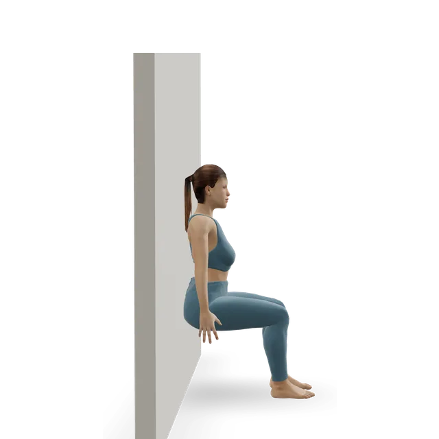
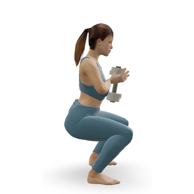
Wall sit demonstrated on the 3D figure3D demoWall sitBeginner · A wall

4 holds of 20 to 45 s, shallow · The knee hub's dose

Easier: Straight-leg raise · Harder: Spanish squat (band squat hold)

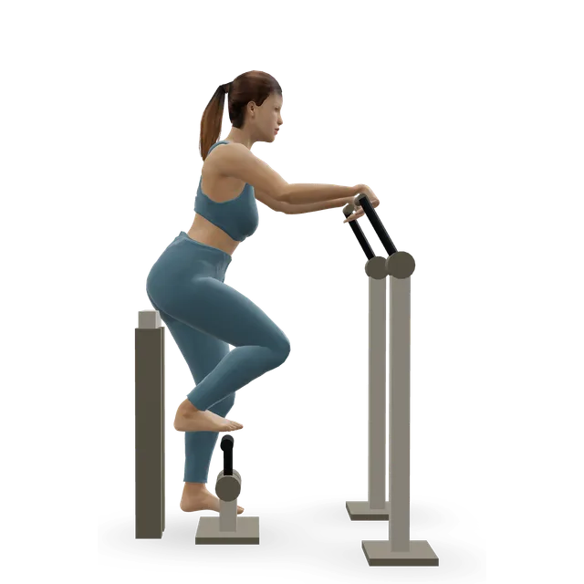
Leg press demonstrated on the 3D figure3D demoLeg pressBeginner · Strength machine

3 sets of 10 to 12 · Limited depth at first. Editorial dose

Harder: Bodyweight squat

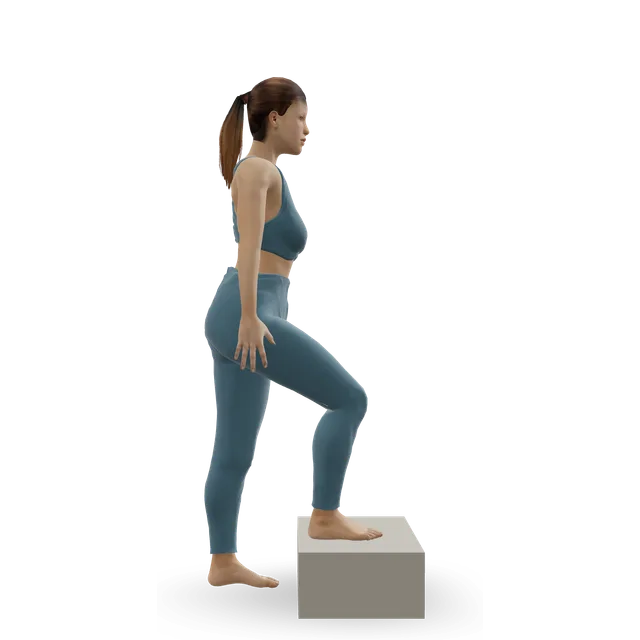
Step-up demonstrated on the 3D figure3D demoStep-upBeginner · A step

3 sets of 8 to 10 each leg, low step · The knee hub's dose

Easier: Low step-up · Harder: Dumbbell step-up

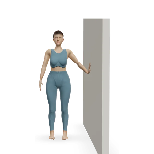
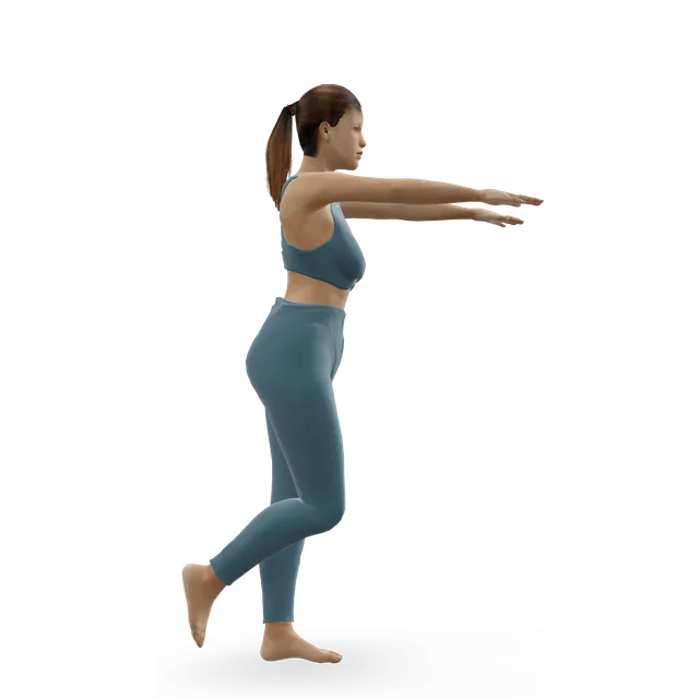
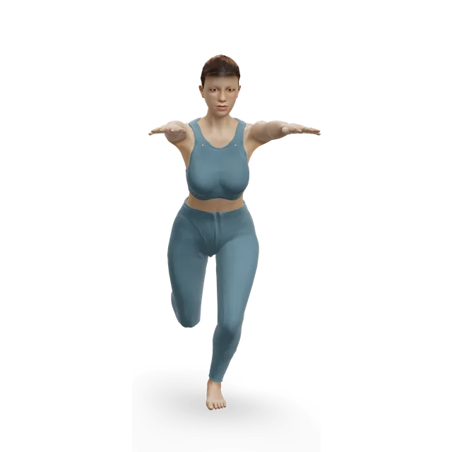
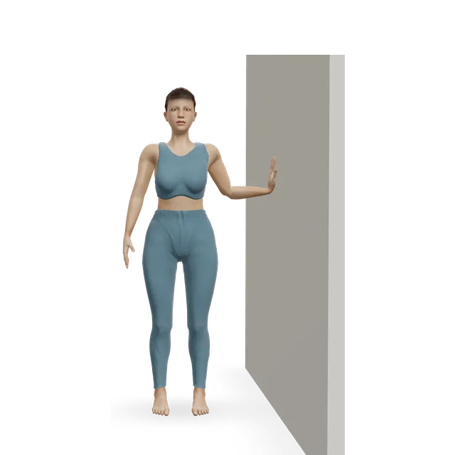
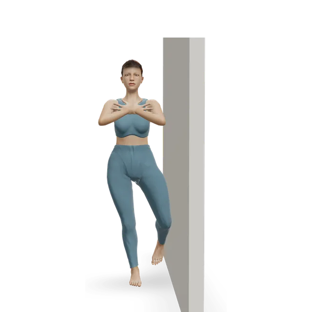
Single-leg stand demonstrated on the 3D figure3D demoSingle-leg standBeginner · A wall

3 holds of 30 s each leg · The knee hub's dose

Easier: Tandem stance and heel-to-toe walk · Harder: Single-leg reach matrix

Stationary cycling demonstrated on the 3D figure3D demoStationary cyclingBeginner · Cardio machine

15 to 30 min, building gradually · Once allowed. Editorial dose

Harder: Air bike intervals

Move on when: At least 3 months off high-impact activity, no symptoms, at least partial healing on X-ray, and the specialist clears running (one centre's criteria).

Follow along: a timer, a cue and the next move, one step at a time.
Stage 3

Running and landing (as the specialist allows)

The back-to-running steps in the notes below the plan, every other day at first, with single-leg strength and landing practice. Doses are editorial.

Move on when: About 6 weeks of running without symptoms, further healing on the follow-up X-ray, and the specialist clears sport.

Follow along: a timer, a cue and the next move, one step at a time.
Stage 4

Back to sport, step by step (as the specialist allows)

Sport drills, then training, then matches. Do the knee control warm-up before every session, keep strength work going twice a week, and keep the follow-up X-rays going until the lesion has fully healed. Doses are editorial.

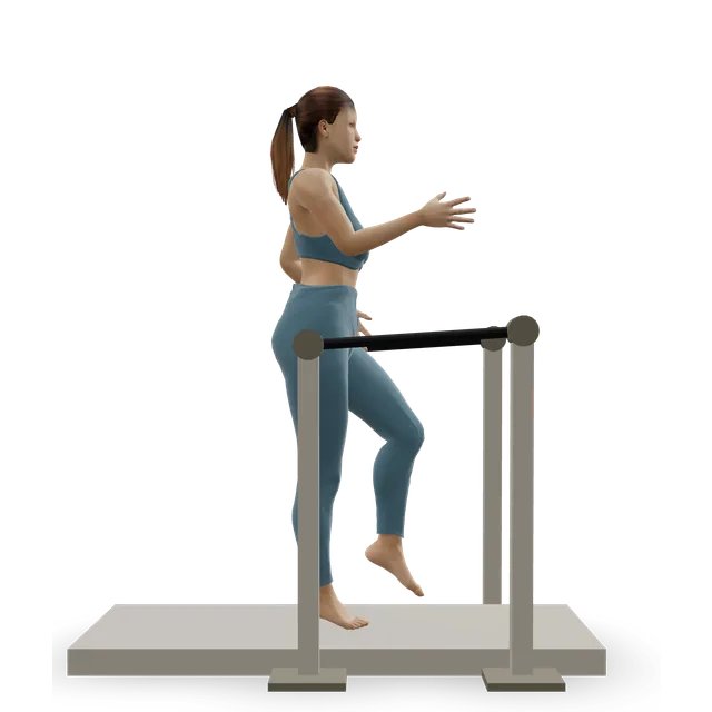
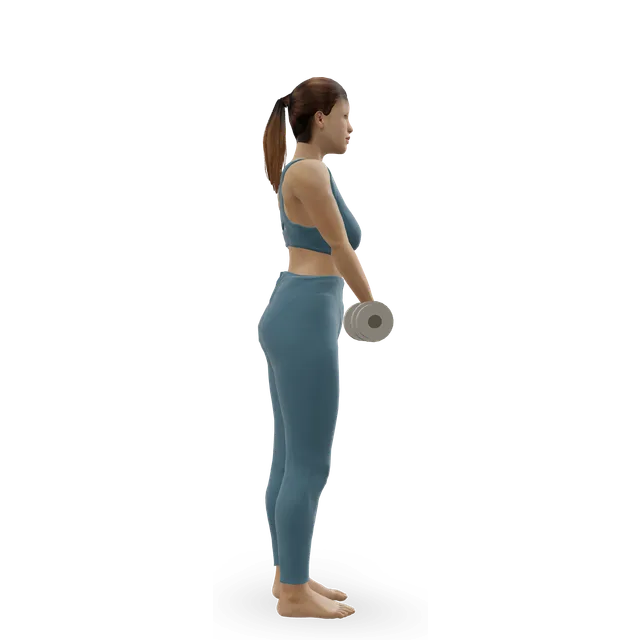
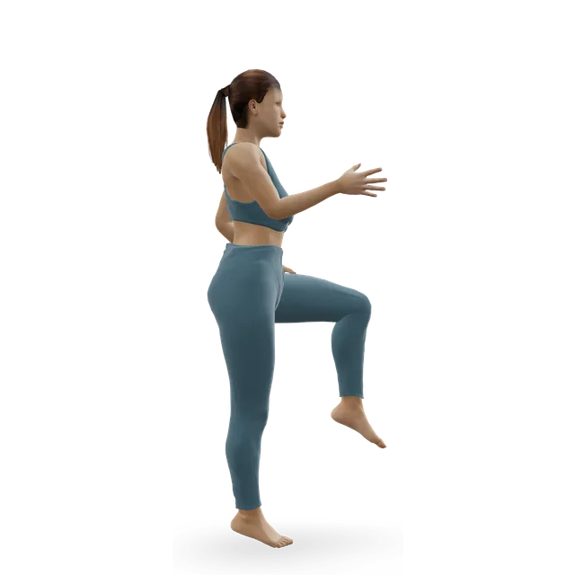
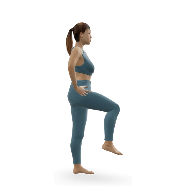
Single-leg hop and stick demonstrated on the 3D figure3D demoSingle-leg hop and stickIntermediate · No equipment

3 sets of 5 each leg · No hopping if there's any bone pain, as the move page says

Easier: Standing broad jump with stuck landing · Harder: Alternate-leg bounding

Move on when: Full training causes no pain, swelling, catching or locking for 2 weeks, with follow-up imaging as the specialist advises.

Follow along: a timer, a cue and the next move, one step at a time.

What to change for now

Back to running: written steps (one centre's protocol)

No running programme has been tested for knee OCD. These steps follow one US centre's criteria, reported in a 2026 review. If your child's specialist has a programme, use theirs.

  • 1. Start only when the specialist has cleared it: at least 3 months off high-impact activity, no symptoms, and at least partial healing on X-ray.
  • 2. Run on the flat, guided by a physiotherapist: walk-jog intervals, building time before speed (the walk-jog detail is our suggestion).
  • 3. Any return of pain, swelling, catching or locking means stop running and contact the team.
  • 4. Expect an X-ray about 6 weeks after you start running.
  • 5. If there are still no symptoms and the X-ray shows further healing, the specialist can clear a return to competitive sport.
  • 6. If the lesion hasn't fully healed by then, expect follow-up about every 6 months until it has.

Source 11

For parents and coaches

Knee pain that keeps coming back with sport, swelling after training, or a knee that catches needs checking rather than playing through. Tell the clinician about any pain in the other knee, because some children have a lesion in both. While the knee is protected, keep your child active with what's allowed, towards the NHS's average of 60 minutes of activity a day.

Sources 1, 8, 30

Keep the follow-up appointments

Healing is judged on X-rays or MRI, not on how the knee feels: pain can settle long before the bone heals, and full healing on X-ray often takes more than a year. Even after healing and clearance, 7 of 25 children in one centre later needed surgery, so keep going to follow-up until the specialist discharges you.

Sources 11, 31

After surgery (as your team allows)

Your surgeon's protocol comes first. After drilling a stable lesion, full movement was back at about 13 weeks and daily activities at about 3 months in one large series. After fixing an unstable piece, expect crutches with little or no weight for about 6 weeks and then months of physiotherapy. OrthoInfo, the AAOS patient site, describes a gradual return to sport from about 4 to 5 months, and one NHS trust leaflet says no impact until X-rays show healing (3 to 9 months). These are typical times, not deadlines.

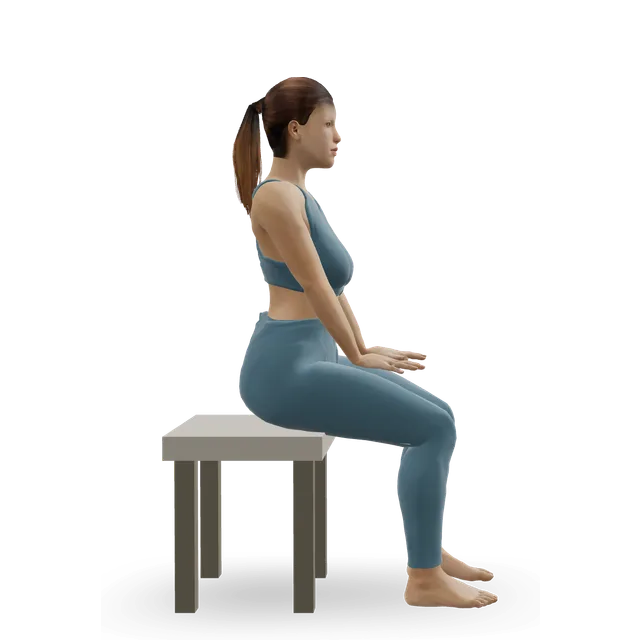
That leaflet's first exercises after surgery, about 4 times a day, are bending the knee with the heel sliding (hold 5 s, 10 times), letting the knee straighten with the heel on a stool (hold 10 s, 10 times), a straight-leg raise (hold 5 s, 10 times) and straightening the knee while seated, without weights until allowed.

Sources 24, 1, 9

Positions to swap, and why

Habit or positionWhy it mattersInstead
Playing or training through knee pain or swelling during treatmentRest from loading is the treatment, and symptoms that come back with impact mean stopping and contacting the team.Stop the activity and tell the specialist.
Jumping, running or sport before clearanceOne review advises at least 3 months away from high-impact activity, until pain has gone and an X-ray shows healing.Upper body and trunk work, and swimming or cycling if allowed.Try: Dead bug
Deep squat holds and kneeling stretches while the knee is protectedSquatting is among the joint-squashing loads to avoid.Shallow wall holds once strength work is cleared.Try: Wall sit
Coming out of the cast or brace, or putting weight through the leg, before you're toldThe team's plan protects the healing joint surface.Follow the instructions exactly, and ask if anything rubs.
Judging healing by how it feelsPain can settle long before the bone heals; full healing on X-ray often takes more than a year.Keep the follow-up appointments and X-rays.
Expecting a special brace to prevent surgeryAn unloader brace made no difference to the need for surgery in 185 knees.Follow the activity limits; wear a brace if the specialist uses one.
Ignoring the other kneeOf 80 children with pain in one knee, 15% had a lesion in the other.Mention any pain in the other knee, and ask about an X-ray.
Going back to sport by the calendarThe return depends on symptoms and healing on X-ray.The back-to-running steps, then sport step by step.Try: Single-leg hop and stick

Routines to follow

More moves for this area

Questions people ask

What is knee OCD?

Osteochondritis dissecans: a patch of bone just under the joint cartilage, usually on the end of the thigh bone, weakens and can crack, loosen or come free. It has nothing to do with obsessive-compulsive disorder.

Who gets it?

Mostly active children and teenagers, boys about 4 to 5 times as often as girls, and teenagers more often than younger children. Adults get it too, but much less often.

What are the symptoms?

Knee pain and swelling with activity, often vague at first. Later, the knee can catch, lock or give way if a piece comes loose. Many early lesions cause no pain at all.

Does my child need an MRI?

Usually, once an X-ray has shown a lesion: an MRI shows whether the cartilage is intact and whether a piece has come loose, which guides treatment. X-rays come first, and doctors may X-ray the other knee too.

What do stable and unstable mean?

A stable lesion is still attached with intact cartilage and can heal. An unstable one has cracked cartilage, fluid behind it or a loose piece, and usually needs surgery. In children, MRI can make a lesion look less stable than it is, so the specialist weighs the scan with the examination.

How long does my child need off sport?

For a stable lesion, months: often 3 to 6, and sometimes longer, with one NHS trust leaflet saying 10 to 18 months. Running restarts only once the pain has gone and an X-ray shows healing, and the specialist follows healing on X-rays or MRI.

Will my child need surgery?

Not usually at first, if the lesion is stable and the growth plates are open: about half to three-quarters of stable lesions heal without it, depending on how studies define healing. Teenagers near the end of growth, larger lesions, and unstable or loose pieces are more likely to need an operation.

What does the operation involve?

For a stable lesion that hasn't healed, drilling small holes to help it heal. For an unstable piece, fixing it back or, if it can't be saved, replacing the damaged surface with a plug of bone and cartilage. Keeping or restoring the surface leads to less arthritis later than removing the piece.

When can my child play again after surgery?

Most young athletes returned to sport after surgery in the studies, but it takes months, and longer after fixing an unstable piece than after drilling. The team decides, from strength and X-rays.

Can it be in both knees?

Yes. In one study, 15% of children with pain in one knee had a lesion in the other, so doctors often X-ray both.

Will it cause arthritis later?

Most children do well: in one group followed for 14 years, 6% developed arthritis with symptoms. The risk is higher with diagnosis as an adult, a BMI over 25, a lesion on the kneecap, or removal of the loose piece.

Can adults get knee OCD?

Yes, though it's rarer and less likely to heal without surgery. In adults, the MRI signs of an unstable lesion are more reliable than in children.

Related exercise guides

Other pain guides

Sources

  1. AAOS OrthoInfo · Osteochondritis dissecans
  2. Chau 2021 · Osteochondritis dissecans: epidemiology, causes, management and outcomes (review, J Bone Joint Surg Am)
  3. Kessler 2014 · Knee OCD in children and teenagers in a US health system (population study, AJSM)
  4. Magoline 2026 · Medical and sporting risk factors for OCD in children (matched cohort, OJSM)
  5. Pareek 2017 · Knee OCD with symptoms in a US county over 38 years (population study, Osteoarthritis Cartilage)
  6. Weiss 2018 · How often adults develop OCD (population study, AJSM)
  7. Kemmeren 2026 · Knee OCD in 13-year-olds in the general population, and at 17 (population cohort, J ISAKOS)
  8. Yellin 2023 · OCD in both knees in children with symptoms in one (cohort, J Child Orthop)
  9. Liverpool University Hospitals NHS · Osteochondritis dissecans (OCD) (patient leaflet, 2020; review date 2023)
  10. AAOS 2023 · Diagnosis and treatment of osteochondritis dissecans (clinical practice guideline)
  11. Rosenberg 2026 · Management and outcomes of knee OCD in children and teenagers (review, Curr Rev Musculoskelet Med)
  12. De Smet 1996 · MRI signs of an unstable OCD lesion, checked at surgery (diagnostic study, Skeletal Radiol)
  13. Kijowski 2008 · MRI signs of instability in children's and adults' knee OCD (diagnostic study, Radiology)
  14. Gebarski 2005 · Early OCD or normal bone growth in children's knees? (imaging study, Pediatr Radiol)
  15. Kajabi 2026 · Bone growth variants that progressed to OCD in pain-free siblings (MRI cohort, Skeletal Radiol)
  16. Wall 2008 · How often stable knee OCD heals without surgery in children (prospective cohort, J Bone Joint Surg Am)
  17. ROCK Group 2026 · An unloader brace for stable knee OCD in 185 knees (multicentre cohort, AJSM)
  18. Johnstone 2024 · Which stable knee OCD lesions heal without surgery (cohort analysis, OJSM)
  19. Muchintala 2025 · Return to sport after stable knee OCD in adolescents (systematic review, AJSM)
  20. Weiss 2016 · How often children and teenagers with OCD need surgery (population study, OJSM)
  21. Hefti 1999 · OCD in 509 knees across Europe (multicentre study, J Pediatr Orthop B)
  22. De Smet 1997 · Untreated knee OCD: predicting the outcome from X-rays and MRI (case series, Skeletal Radiol)
  23. Heyworth 2023 · Two ways of drilling stable knee OCD in 91 children (RCT, AJSM)
  24. Baghdadi 2022 · Drilling stable knee OCD in 139 knees (case series, Arthrosc Sports Med Rehabil)
  25. Wu 2018 · Fixing unstable knee OCD with open or closed growth plates (cohort, AJSM)
  26. Husen 2023 · Fixing unstable knee OCD: results after 11 years (cohort, AJSM)
  27. Sanders 2017 · Arthritis after removing or restoring an OCD fragment (population cohort, AJSM)
  28. Hevesi 2020 · Knee OCD in children: pain and arthritis 14 years on (population cohort, Cartilage)
  29. Sanders 2017 · Arthritis after non-surgical care of knee OCD (population cohort, OJSM)
  30. NHS · Physical activity guidelines for children and young people
  31. Metz 2022 · Later surgery after healed, stable knee OCD in children (retrospective study, J Pediatr Orthop)
  32. NHS · Knee pain
  33. NHS · Hip pain in children (irritable hip)
  34. NHS · Growing pains
  35. NHS · DVT (deep vein thrombosis)

Exercise education, not a diagnosis or treatment plan. It is written from the guidelines and studies listed above; review by a clinician is pending. If your pain follows an injury, is severe, is getting worse, or comes with any warning sign listed on this page, get it assessed. Health disclaimer.