What it is
The capitellum is the rounded outer part of the lower end of the upper arm bone, where the head of the radius presses against it. In a growing child its bone forms gradually and its blood supply is limited. Repeated squashing and shearing damage the bone under the cartilage, which softens, cracks and can separate, taking cartilage with it. A piece that comes free can float in the joint as a loose body.
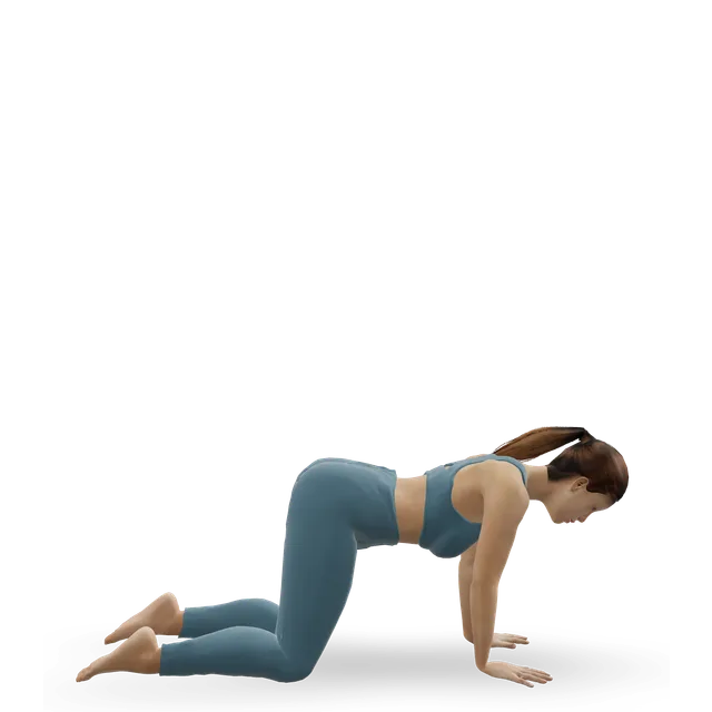
Throwing squashes the outer elbow as the arm comes forward. Gymnastics loads it through a straight elbow in handstands, vault and tumbling, so gymnasts' lesions sit further back on the capitellum than throwers' (28° against 57.6° from the bone's long axis).
Sources 1, 2, 3, 4, 5, 6
Who gets it
Young athletes aged about 10 to 17: boys who throw, and gymnasts, who are often girls. In one US children's hospital's group of 154, 39% were gymnasts and 28.5% played baseball or softball. In a US population, it was diagnosed in 6.0 per 100,000 people aged 10 to 24 each year, and in 15.3 per 100,000 boys aged 10 to 15; 89% did sport, and 90% of those did overhead sports.
Ultrasound screening of Japanese baseball players found it in 1.4% of 6 to 12 year olds, 2.1% of 10 to 12 year olds and 3.4% of 12 to 18 year olds, and 1.8% of 6 to 11 year olds developed it within a year. In 31 healthy high-level gymnasts, 15 of 62 elbows showed changes in the capitellum on ultrasound, often in both arms, and about 10% of gymnasts with OCD have it in both elbows.
Sources 2, 7, 8, 9, 10, 11, 12, 13, 3
How it's recognised
Pain on the outer elbow with throwing, lifting or weight on the hands, often for months before anyone looks at it; losing some straightening; and later swelling, clicking, catching or locking. The examination checks tenderness on the outer elbow, movement compared with the other arm, and catching.
Many lesions don't hurt at first. Of the children found by screening in three Japanese programmes, 34.4%, 34.8% and 68% had ever had elbow pain. Inner elbow pain in a young thrower is usually Little League elbow, which has its own guide; the two can happen together, so outer elbow pain needs checking even when the inner side hurts most.
Sources 1, 3, 2, 14, 9, 12, 10, 6
Stable or unstable: imaging decidesEmerging evidence
X-rays come first, including a view with the elbow bent to 45° for throwers and a straighter view for gymnasts. But they miss early lesions: routine X-rays showed OCD on first reading in 7 of 15 confirmed cases, and loose bodies in 3 of 7. In another group, X-rays found 27% and magnetic resonance imaging (MRI) 40% of the loose bodies later seen at surgery.
MRI decides stability. Experts agree that intact cartilage, no fluid behind the lesion, unbroken bone and no displaced piece suggest a stable lesion; broken cartilage or bone, fluid beneath the cartilage, a displaced piece and loose bodies suggest an unstable one, which is unlikely to heal without surgery. In an older classification still in use, stable lesions had an open growth plate, early changes and good movement, and healed with rest; unstable lesions had a closed growth plate, a broken-up fragment or 20° or more of lost movement, and did better with surgery.
In Japan, ultrasound screening finds lesions early: 65.8% of players flagged by ultrasound were confirmed to have OCD, against 1.9% of those flagged by an examination alone.
Sources 15, 5, 16, 17, 18, 19, 9
What scans can mistake for OCDEmerging evidence
In baseball players under 10, irregular areas on the capitellum on ultrasound (3.4% of 321) had all gone 2 years later while the children kept throwing: a normal stage of bone growth. On MRI, a normal notch at the back of the capitellum appeared in 14 of 22 volunteers without symptoms. And 24% of healthy high-level gymnasts' elbows showed capitellum changes on ultrasound, linked with tenderness but not with how well the elbow worked.
Sources 20, 21, 13
How it usually goesEmerging evidence
Stable lesions heal with rest, slowly: 90.5% of early lesions healed in one group, but healing on X-ray took 14.9 months on average, and 16 of 17 children with an open growth plate healed in another. Unstable lesions rarely heal without surgery: 1 of 9 advanced lesions healed, and non-surgical care failed in 70% of a US group, especially with a loose body.
After surgery, most return to their sport (76% to 81%), usually at about 6 months, and about 1 in 9 needs another operation. Long term, 54% of 24 young people in an older series treated with 6 months of rest had elbow pain with daily activities 5 years later, and in a US group followed for over 10 years, elbows treated surgically had better function and fewer catching or locking symptoms (9% against 50%).
Sources 15, 22, 17, 7, 23, 24, 25
Panner's diseaseEmerging evidence
Panner's disease affects the whole capitellum in children under about 10, usually boys (90% of 30 reported cases), usually in the dominant arm. On X-ray the capitellum looks smaller and broken up, but there is no loose piece and no cartilage damage, and it remodels. Pain, swelling and losing about 20° of straightening are typical.
It settles without surgery, with rest from stressful elbow use, but healing takes months. In 7 children (average age 10, 4 gymnasts and 3 baseball players), overhead activity and loading the arm were restricted for an average of 7 months, healing was complete at 12 months, and they were back to sport at 15 months, all with well-remodelled capitella. An age of 12 to 17, a small separate lesion, cartilage damage, loose bodies and catching point to OCD instead.
Sources 4, 26, 2, 27