Pain guide · Foot pain

Accessory navicular syndrome: a painful extra bone on the inner side of the foot, in teenagers and adults

An accessory navicular is an extra bone, or an extra piece of bone, on the inner side of the foot, where the tendon that holds up the arch attaches. Across 39 studies, about 1 in 6 people had one, and most never know. In some, often active teenagers around the growth spurt or after a sprain, the bump becomes sore, gets rubbed by shoes and aches with sport: accessory navicular syndrome.

Treatment starts without surgery: less of the sport that hurts, wider shoes, padding or insoles, and calf and arch strength. The evidence for each is thin and no exercise trial exists, so the plan borrows from trials in the tendon that attaches there. If months of good care don't help, a specialist may discuss removing the bone, which helps most people who have it. This guide is for teenagers, parents and adults: the warning signs, how it's diagnosed, what each treatment can and can't do, and a staged plan with 3D demos back to sport.

When to get help first

Exercise is for pain that has been checked or is clearly mild and settling.

Get urgent medical help if

  • Call 999 or go to A&E: after an injury, you heard a crack, the foot or ankle has changed shape or points at an odd angle, a bone is sticking out, there is a bad cut over it, you're in severe pain, or the toes look blue or white, feel numb or are cold to touch. These can mean a broken bone. Don't drive yourself.
  • Same day (urgent GP appointment or NHS 111): after an injury, you can't put weight on it or walk more than a few steps, the pain is very bad or getting worse, there is a lot of swelling or bruising or it is getting worse, or it feels very stiff. If you think it's broken, don't treat it yourself.
  • Pain, swelling, redness and warmth over a bone, or a very high temperature with feeling generally unwell: ask for an urgent GP appointment or contact NHS 111. It could be a bone infection. Take your child to a GP if they don't want to use a leg and seem irritable: young children don't always get a high temperature with a bone infection.
  • Same day: a sudden, very painful, hot, swollen, red joint, often the big toe, especially with a high temperature, feeling shivery or being unable to eat. It may be gout, but it could be a joint infection (septic arthritis), which is urgent.
  • After surgery: redness, swelling, oozing or a smell from the wound, or a fever. Contact your surgical team the same day, or NHS 111 out of hours.
  • Same day: in a cast or boot, pain that is getting worse, toes that are numb or burning, a cast that feels too tight, toes that are swollen, blue or white, or a bad smell from under the cast.
  • Same day (GP or NHS 111): throbbing pain and swelling in one calf, with red or darker skin. It could be a blood clot (DVT). Watch for it after an ankle injury or time in a cast or boot.
  • Call 999: calf or leg pain or swelling with breathlessness or chest pain. It could be a clot that has reached the lungs.

See a doctor or physiotherapist if

  • A painful bump on the inner side of the foot that keeps coming back with sport: see a GP or physiotherapist once, to confirm what it is. An X-ray shows the extra bone.
  • One spot on a foot bone that hurts with every step and later at night: a possible stress fracture.
  • Leg pain in the morning, pain bad enough to stop your child walking, a rash, swelling or unusual bruising, a high temperature, unusual tiredness, or not eating or losing weight: see a GP. A limp that carries on away from sport needs checking too.
  • Bone pain that is worse at night, or a lump or swelling that is growing or isn't the familiar bony bump: see a GP.
  • No better after about 6 weeks of rest from the painful sport, shoe changes and insoles, or pain that stops your child enjoying sport: see a GP or physiotherapist again. If it hasn't settled after several months, a specialist opinion may help.
  • Pain behind and below the inner ankle bone, an arch that is getting flatter on one side, or difficulty rising onto tiptoe on that foot: see a GP or physiotherapist. In adults it can be the tibialis posterior tendon itself, which our posterior tibial tendon guide covers.
  • Flat feet that are painful, stiff, weak or numb, that you didn't have before, or that affect only one foot.
  • Pain that isn't better after 2 weeks of looking after it yourself, is getting worse, keeps coming back, or stops you doing normal things.
  • Tingling or loss of feeling in the foot, or a foot that feels weak.
  • Any foot pain if you have diabetes: foot problems can be more serious.

Understand it

What it is

The navicular is the boat-shaped bone on the inner side of the midfoot. The tibialis posterior tendon, which helps hold up the arch, attaches to the bump on its inner side. An accessory navicular is an extra bone, or an extra piece of bone, at that bump. It forms when a separate growth centre in the bone doesn't fuse with the rest, and it tends to run in families.

There are three types. Type 1 is a small, round bone inside the tendon. Type 2 is a larger piece joined to the navicular by a bridge of cartilage or fibrous tissue, which the tendon's pull or a twist can strain: in operated feet, the bridge showed tiny cracks and signs of repair. Type 3 has fused to the navicular, leaving a large bump that shoes rub. Type 2 causes most of the pain: 72.7% of 226 painful feet in one US children's hospital, and over 70% of painful cases in a review.

In 27 feet with an accessory navicular and flat feet, MRI (magnetic resonance imaging) showed the tendon attaching straight into the extra bone, with little or no slip carrying on under the foot, in 20. That is why the extra bone, tibialis posterior strength and the arch are discussed together.

Sources 1, 2, 3, 4, 5, 6

How common it is: most never hurtStrong evidence

A 2024 review of 39 studies (11,015 people) found an accessory navicular in 17.5% of people, about 1 in 6, and half of them had one in both feet. Men and women were affected alike. It was commonest in East Asian populations (38.4%) and least common in North Americans (8.0%), with Europeans in between (12.1%). NHS leaflets give 4% to 14%, and single studies range from about 8% of foot and ankle X-rays in Turkey to about half of adults in a Japanese population study, depending on the population and how it was looked for.

Most never cause trouble. In 875 Japanese schoolchildren checked with ultrasound, 20.8% of feet with an accessory navicular (55 of 264) had ever been painful, most often at 10 to 11 in girls and 12 to 13 in boys. In people X-rayed for foot pain, the extra bone was blamed for the pain in 10.6% of those who had one. So an extra bone on an X-ray isn't a diagnosis on its own: a tender bump that matches the story is.

Sources 2, 3, 7, 8, 9, 10

Who gets pain

Mostly active children and teenagers, usually between about 10 and 15, around the growth spurt, when sport loads the bridge harder. Clinic series are mostly girls (78% of 169 children in one, 72% of 71 patients in another), although the school study found boys and girls equally likely to have pain.

It often starts after a sprain, more training or new or tight shoes: 31% of the 169 children linked it to an injury, and in a Korean surgical series, 74% of athletes' feet and 39% of non-athletes' had a history of injury. Professional footballers had more accessory naviculars than other men (35.9% of feet against 24%).

Adults get it too. An NHS foot surgery service notes that pain may only start in adulthood, perhaps after a trip or fall. In older adults it may play a part in tibialis posterior tendon problems, and in a Japanese population study, having a type 2 bone in both feet went with more falls, though walking speed and balance tests were no different.

Sources 9, 4, 12, 13, 14, 11, 8

How it's recognised, and what scans do and don't meanEmerging evidence

A bony bump on the inner side of the midfoot, just in front of and below the inner ankle bone, tender to press, sometimes red or swollen after sport, and rubbed by shoes, football boots, ski boots or skates. It aches with walking, running and jumping, and symptoms often come and go with activity. A clinician presses on the bump itself: tenderness on the bone, rather than along the length of the tendon, points to the accessory navicular. An X-ray confirms it, and an angled (oblique) view shows it best.

MRI is kept for pain that doesn't settle, or before an injection or surgery. Painful type 2 bones show swelling inside the bone (marrow oedema) in the extra bone and the navicular: in all 17 operated feet in one series, and in all 27 athletes and 16 of 20 non-athletes in another. But in 1,988 adults having an ankle MRI for other reasons, 21% of type 1 and 2 bones showed the same swelling. Swelling fits a painful accessory navicular; it doesn't prove the bump is the cause.

Sources 3, 4, 5, 15, 13, 16

How it usually goesEmerging evidence

Children's pain often settles as the foot matures, but the evidence is small. In one small Japanese school study, 15 of 21 painful feet had settled 2 years later; treatment wasn't recorded, so this isn't a general rate. Over those 2 years, about a third of the extra bones in 11 to 13 year olds fused to the navicular, though none of the painful ones did. In 19 US children X-rayed every year, 8 fused, at about 12.5 years in girls and 14 in boys.

In two US children's hospitals, about 3 in 10 children with a painful accessory navicular went on to surgery (30% of 169, and 28.8% of a random sample of 299), at a median of about 5 months in one. In the first, 28% had complete relief with non-surgical care, after an average of 8 months, and about 4 in 10 partial relief. Athletes settled less often: in a Korean series, at least 6 months of non-surgical care helped 17 of 50 non-athletes but only 2 of 29 athletes.

Sources 17, 18, 4, 19, 13

Flat feet, and pains that aren't an accessory navicular

The extra bone doesn't cause flat feet, as far as studies show: arches were the same in 49 people with one and 179 without. But the two often go together (17.6% to 21.5% of the 169 children had flat feet on X-ray measures), and a flatter foot made surgery more likely in one series. A flat foot that doesn't hurt needs nothing, as our flat feet guide explains.

Pain behind and below the inner ankle bone, often in women over 40, with the arch dropping on one side, is more often the tibialis posterior tendon itself. Pain on the top of the midfoot, at one spot, with every step and later at night, can be a stress fracture. A soft, smooth lump on the top of the foot is more often a ganglion cyst. Each has its own guide.

Sources 20, 4, 12, 21, 3
What helps, and how strong the evidence is

Rest from the sport that hurts, and shoes that don't press: sensible first steps, untestedEmerging evidence

NHS and specialist sources agree on the first steps: rest from the activities that bring it on, wider and more comfortable shoes that don't press on the bump, anti-inflammatory painkillers for a flare, and for a bad flare, a below-knee walking boot or a cast for about 2 weeks. An NHS foot surgery service adds that a supportive trainer helps. These steps are low-risk, but no study has compared them.

Rest from the sport that hurts doesn't mean rest from everything: cycling and swimming usually keep fitness going (our suggestion, as in our Sever's disease guide).

Sources 3, 4, 1, 22, 11

Insoles and orthoses: reasonable to try, not provenMixed evidence

An NHS leaflet says custom insoles take pressure off the area. In one US children's hospital, insoles alone were tried in 40 children and only 2 went on to surgery, against 34% of those put in a boot or cast, but treatment wasn't randomised, so milder cases probably got insoles.

Only one trial has compared insoles: in 54 professional athletes, custom orthoses worn for 12 months eased pain more than standard insoles (pain fell from about 8.5 out of 10 to under 2, against about 4). But the trial was registered only after it ended, the athletes knew which insoles they had, and it was small. In 27 children with flexible flat feet, pain and arch measures improved over 4 years of custom orthoses, with no comparison group. Insoles are reasonable to try; custom insoles haven't been shown to beat standard ones.

Sources 3, 4, 23, 24

Borrowed: strength work from trials in the tibialis posterior tendonEmerging evidence

No study has tested exercise for an accessory navicular. A review suggests toe-gripping exercises to strengthen tibialis posterior, and an NHS leaflet includes physiotherapy to strengthen the foot, ankle and calf after surgery. The plan borrows from trials in adults with tibialis posterior tendon problems, as our posterior tibial tendon guide does.

In 36 adults with early tendon problems, insoles and stretching helped, and adding progressive strength work helped more. In another 36 adults, adding home strengthening made little difference: both groups improved. A review of 3 trials (93 people) found moderate benefits for strengthening alongside stretching and insoles. These were adults with a different problem, so the doses are borrowed starting points, and children start gentler.

Sources 1, 25, 26, 27, 28

A steroid injection: a specialist's option, rarely usedMixed evidence

An NHS children's orthopaedic service offers an ultrasound-guided steroid injection by a consultant radiologist if MRI confirms inflammation and non-surgical care has failed. A US surgeon writes that one is very rarely needed, and only 3% of 71 patients in one series had one. No study reports how well it works.

Sources 3, 22, 12

Surgery when months of good care fail: most people improveEmerging evidence

Surgery is considered if several months of non-surgical care haven't helped; a review says 6 months. Children who had surgery had a median of about 5 months, or an average of nearly 12 months, of non-surgical care first. The usual operation removes the extra bone through a small cut over the bump. The commonest version, the Kidner procedure (52% of 625 operations in a 2025 review), also re-attaches the tendon to the navicular; simple removal leaves it. Fusing the bridge is another option, which one review prefers for large bones.

Most people improve. In 298 children and teenagers, 94% had less pain and 93% returned to their main sport. In 32 operated feet, 87.5% had good or excellent results; in 25 feet, all were relieved of pain; in 14 adult feet followed for over 8 years, 13 were pain-free; and athletes did as well as non-athletes in 42 feet. An NHS children's service quotes about 90% symptom-free.

Simple removal and the Kidner procedure did equally well in a comparison of 50 feet in children with flat feet (86% and 82% satisfied), and reviews find both work. But after simple removal, 8 of 17 people with flat feet struggled to rise onto tiptoe on one leg, one reason surgeons often re-attach the tendon. These are case series and one comparison, not trials against non-surgical care.

Sources 3, 1, 19, 4, 29, 30, 31, 5, 32, 33, 28, 34, 35, 21

Surgery: the risks and the recoveryEmerging evidence

Complications occurred in about 11% after Kidner-type operations and 25% after fusion in the 2025 review. An NHS foot surgery service lists infection (2 in 100), slow skin healing (8 in 100), long-lasting swelling and thick or sensitive scars (4 in 100 each), the tendon pulling off the bone and blood clots, and says about 20% of people still have some arch pain afterwards.

NHS leaflets describe the recovery. Children: the foot raised for 72 hours in a half-plaster with a heel-walking shoe, a clinic check at about 2 weeks for new insoles and physiotherapy, running usually at 4 to 6 weeks, contact sport at 8 to 12 weeks, and pain-free activity for most by 3 to 4 months. Adults whose tendon is re-attached: 6 to 8 weeks in a cast without putting weight on the foot, full weight at about 10 weeks, sport considered from about 4 months, and the full benefit by about 6 months. Your surgical team sets your timeline.

Sources 29, 11, 28

How much pain is OK while you exercise?

Children: no more than 3 out of 10 in sport (borrowed from Sever's disease). Strength work and adults: the foot and ankle ruleEmerging evidence

Children and teenagers in sport: keep pain during activity to 3 out of 10 or less, where 0 is no pain and 10 is severe pain. A little discomfort during the exercises is fine if it settles after stopping. This rule comes from an NHS children's hospital booklet for Sever's disease (heel pain in growing children). No accessory navicular leaflet gives a rule, so it is borrowed.

Strength work, and adults: discomfort up to about 5 out of 10 that settles within an hour, with no more swelling or stiffness the next morning, is fine. If it's worse the next day, do a little less, not nothing. The 5 out of 10 figure matches our other foot guides and borrows the pain-monitoring rule from an Achilles tendon trial, in which carrying on with activity under the rule did no harm. It hasn't been tested here.

Rubbing is different: pain from a shoe, boot or skate pressing on the bump means changing the shoe or padding, not pushing through. The exceptions: one spot on top of the midfoot that hurts with every step should be pain-free (a possible stress fracture), a hot, red, swollen bump needs checking, and burning, tingling or numbness means stop that movement. After surgery, your team's limits come first.

Sources 36, 37

The exercise plan: 3 stages

Start at the first stage. Each move opens a full guide with a 3D demo.

Stage 1

Settle: take the pressure off, keep moving (about weeks 0 to 2)

Cut back the running, jumping and kicking that hurts, not all activity: cycle, swim or train the upper body instead. Wear wider, softer shoes, pad around the bump and use insoles if a clinician has given them (see the notes below). Ice for 10 to 15 minutes after activity if it helps. If a clinician has put the foot in a boot or cast, do only what they allow. Doses are editorial unless marked.

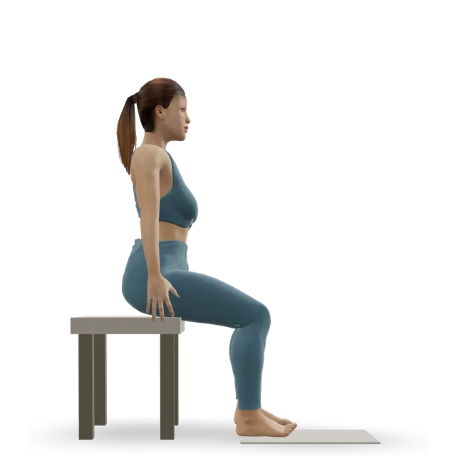
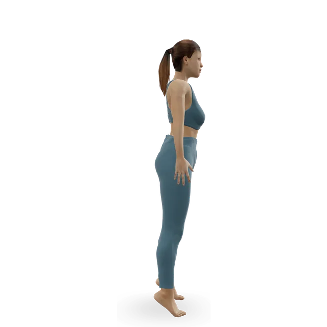
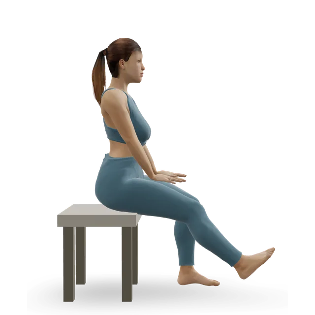
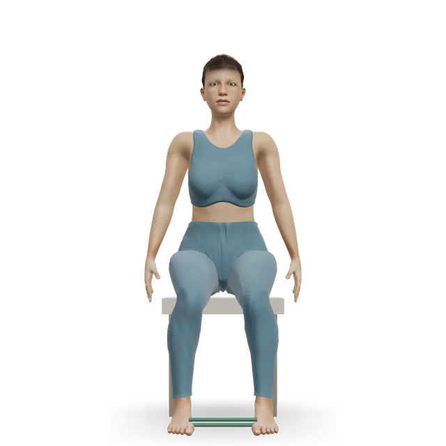
Seated bent-knee calf raise (soleus raise) demonstrated on the 3D figure3D demoSeated bent-knee calf raise (soleus raise)Beginner · A bench or box

2 sets of 15 · A gentle start. Editorial, as in our Sever's disease and posterior tibial tendon guides

Harder: Slow calf raise

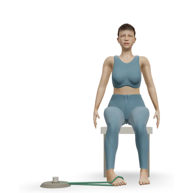
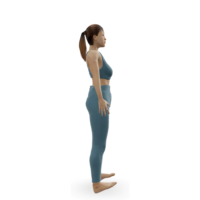
Band ankle inversion demonstrated on the 3D figure3D demoBand ankle inversionBeginner · A band + A bench or box

2 sets of 15 with a light band, once a day · Slow and pain-free: a lighter start before the trial dose in stage 2. Editorial

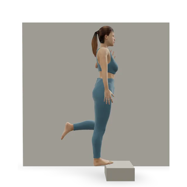
Harder: Single-leg calf raise from a step

Towel toe curl demonstrated on the 3D figure3D demoTowel toe curlBeginner · Other / check instructions + A bench or box

2 sets of 10 · A review suggests toe-gripping exercises for this tendon. Editorial dose

Easier: Big toe press (seated toe-flexor hold) · Harder: Short foot + toe splay

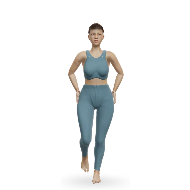
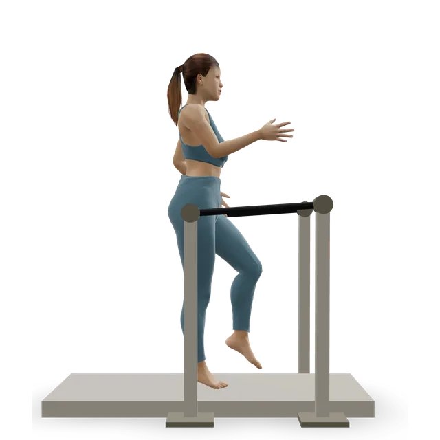
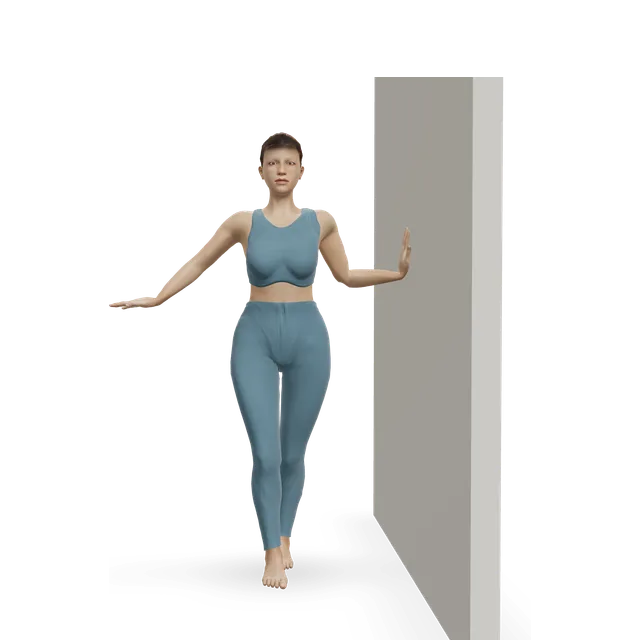
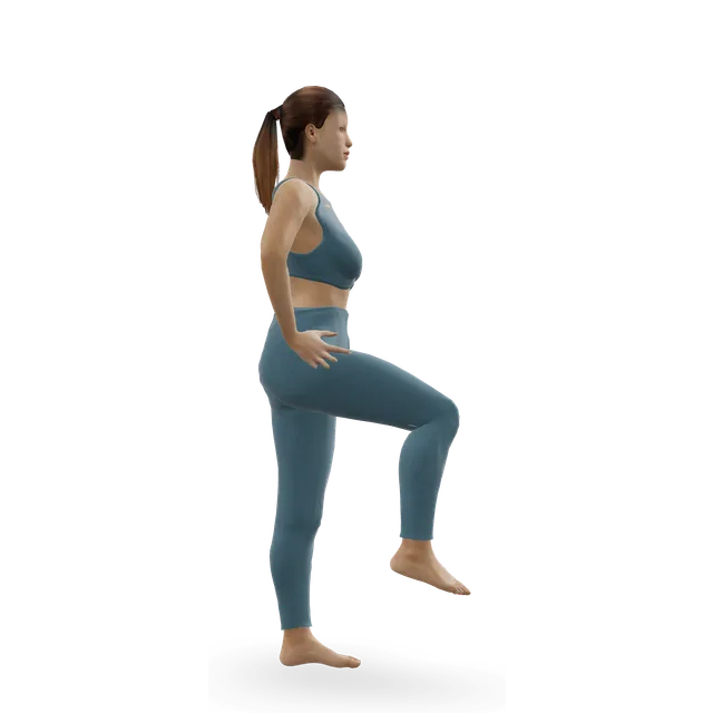
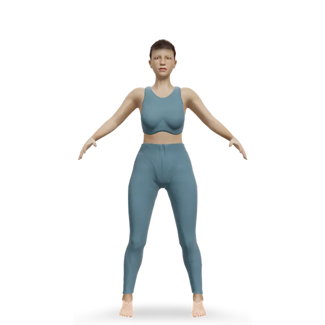
Single-leg stand demonstrated on the 3D figure3D demoSingle-leg standBeginner · A wall

3 holds of 20 to 30 s on each leg, near a support · The move page's dose

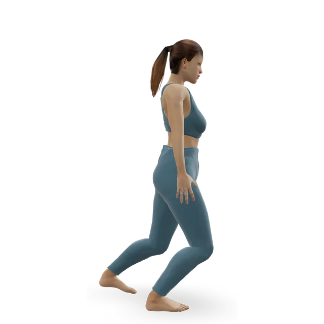
Easier: Tandem stance and heel-to-toe walk · Harder: Single-leg reach matrix

Stationary cycling demonstrated on the 3D figure3D demoStationary cyclingBeginner · Cardio machine

15 to 30 min, easy · Fitness while the foot settles. Editorial

Easier: Recumbent exercise bike · Harder: Air bike intervals

Move on when: Walking and school PE (or work) are comfortable in normal shoes, the bump isn't sore with everyday walking, and the foot is no worse the next morning.

Follow along: a timer, a cue and the next move, one step at a time.
Stage 2

Build: arch and calf strength (about weeks 2 to 8)

Strength work most days, as in the tendon trials this stage borrows from. Keep sport at a level that stays within the pain rule. Children use the gentler doses marked.

Band ankle inversion demonstrated on the 3D figure3D demoBand ankle inversionBeginner · A band + A bench or box

3 sets of 15, twice a day, with a band heavy enough that the last few are hard · Take 2 to 3 s to return. The dose from an adult tibialis posterior trial, as in our posterior tibial tendon guide (borrowed); children once a day (editorial)

Harder: Single-leg calf raise from a step

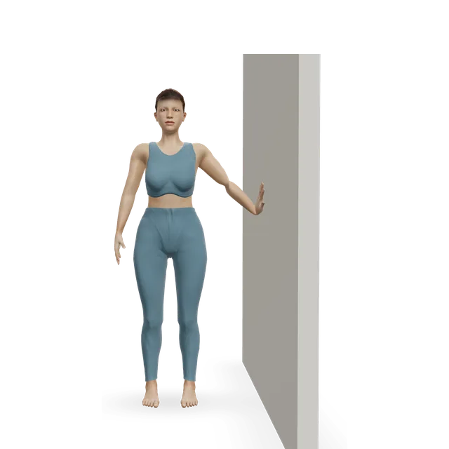
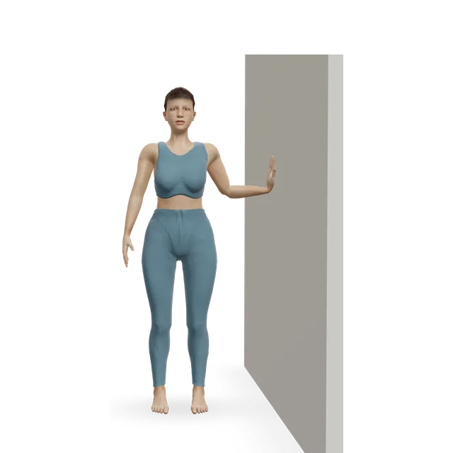
Slow calf raise demonstrated on the 3D figure3D demoSlow calf raiseBeginner · A wall

2 sets of 10, building to 3 sets of 30, twice a day · Another adult tendon trial's dose (borrowed). Children: about 1 minute at a time on both feet, as an NHS Sever's disease booklet suggests

Easier: Seated bent-knee calf raise (soleus raise) · Harder: Standing dumbbell calf raise

Tibialis raise demonstrated on the 3D figure3D demoTibialis raiseBeginner · No equipment

2 sets of 15 to 20 · The move page's dose

Harder: Single-leg tibialis raise

Single-leg stand demonstrated on the 3D figure3D demoSingle-leg standBeginner · A wall

3 holds of 30 s, then with your eyes closed or on a cushion · An editorial step up from the move page's dose

Easier: Tandem stance and heel-to-toe walk · Harder: Single-leg reach matrix

Single-leg reach matrix demonstrated on the 3D figure3D demoSingle-leg reach matrixIntermediate · No equipment

3 rounds of 5 reaches on each leg · Shallow reaches first. The move page's dose

Easier: Standing crossed-leg side reach · Harder: Single-leg Romanian deadlift

Move on when: Single-leg heel raises on the floor are close to the other side without pain at the bump. For children, the NHS Sever's disease booklet's target is a good guide: single-leg heel raises in a row of just under double their age (about 20 at age 11), and hopping, skipping and jumping quietly without pain.

Follow along: a timer, a cue and the next move, one step at a time.
Stage 3

Back to running, jumping and sport (from about weeks 6 to 8)

Add back one sport session a week. For children, the NHS Sever's disease booklet's limits are sensible (borrowed): no more than 4 high-intensity sessions a week, not on 2 days in a row, and weekly hours of organised sport no more than the child's age. Adults: build running back over 2 to 4 weeks (editorial).

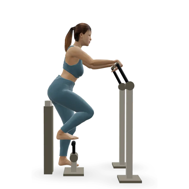
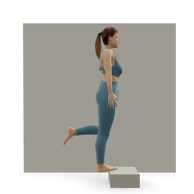
Single-leg calf raise from a step demonstrated on the 3D figure3D demoSingle-leg calf raise from a stepIntermediate · A step + A wall

Children: build to just under double their age in a row. Adults: 3 sets of 10 to 15 · Floor first, then off a step. The NHS booklet's target (borrowed) and the move page's dose

Easier: Slow calf raise · Harder: Single-leg pogo hops

Ankle bounces (mini pogo) demonstrated on the 3D figure3D demoAnkle bounces (mini pogo)Beginner · No equipment

2 sets of 20, with quiet landings · The NHS booklet's hop check, as in our Sever's guide; adults can build to the move page's 4 sets of 20

Harder: Pogo hops

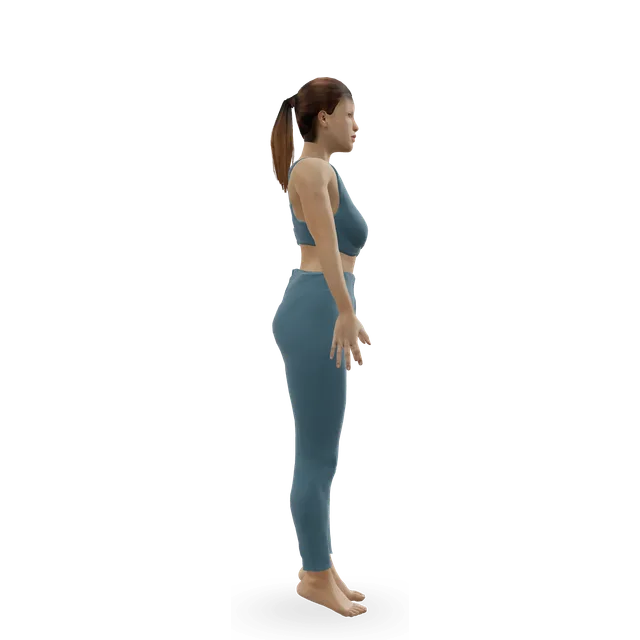
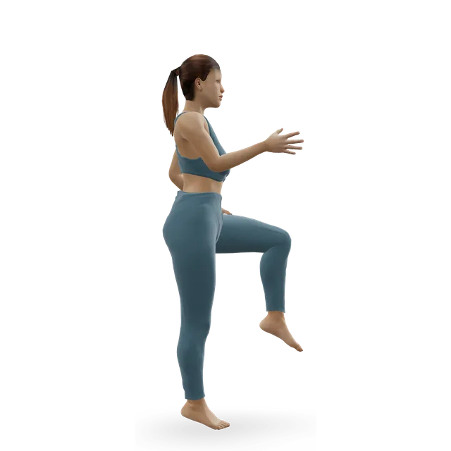
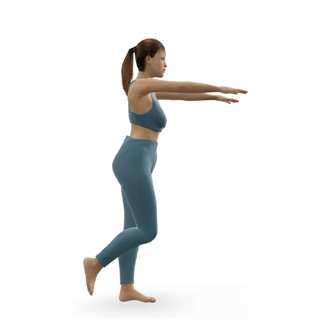
Skipping demonstrated on the 3D figure3D demoSkippingBeginner · No equipment

3 runs of 20 m · The booklet's skip check; adults can build to the move page's 6 runs

Easier: Ankle bounces (mini pogo) · Harder: Alternate-leg bounding

Pogo hops demonstrated on the 3D figure3D demoPogo hopsIntermediate · No equipment

2 sets of 10 to 20 · The booklet's jump check; adults can build to the move page's 5 sets of 15

Easier: Ankle bounces (mini pogo) · Harder: Single-leg pogo hops

Single-leg hop and stick demonstrated on the 3D figure3D demoSingle-leg hop and stickIntermediate · No equipment

3 sets of 5 in each direction on each leg · Small hops in one direction first. The move page's dose

Easier: Standing broad jump with stuck landing · Harder: Alternate-leg bounding

Treadmill jogging demonstrated on the 3D figure3D demoTreadmill joggingIntermediate · Cardio machine

Gentle running, then training · Walking, swimming and cycling first, then gentle running: the NHS booklet's order

Easier: Treadmill walking · Harder: Sprint intervals

Move on when: Full training stays within the pain rule and settles soon after. If it flares in a growth spurt, drop back a stage. If the bump still hurts after several months of doing this well, ask about a specialist opinion.

Follow along: a timer, a cue and the next move, one step at a time.

What to change for now

Shoes, padding and insoles

Wider shoes are the first step in one children's hospital, an NHS service says a supportive trainer helps, and an NHS leaflet says custom insoles take pressure off the area. The details below are our suggestions.

  • 1. Choose wider, softer shoes with room on the inner side of the midfoot, and supportive trainers for walking.
  • 2. For football boots, ski boots and skates, ask a shop or podiatrist to stretch or pad the area over the bump.
  • 3. A ring-shaped pad around the bump, not over it, can stop rubbing.
  • 4. Use insoles or arch supports if a clinician advises them. Custom insoles take pressure off the area, but they haven't been shown to beat standard ones.
  • 5. Give each change 2 to 4 weeks before you judge it.

Sources 4, 11, 3, 23

Back to sport for children: an NHS booklet's checks for Sever's disease (borrowed)

These checks come from an NHS children's hospital booklet for Sever's disease, another common foot pain in active 10 to 15 year olds. No accessory navicular leaflet gives return-to-sport steps, so they are borrowed. If your child's physiotherapist has a plan, follow theirs.

  • 1. Your child can do single-leg heel raises in a row on each leg, building towards just under double their age (about 20 at age 11).
  • 2. They can hop, skip and jump quietly without pain.
  • 3. Restart one activity or training session a week.
  • 4. Keep pain during sport at 3 out of 10 or less, settling soon after.
  • 5. No more than 4 high-intensity sessions a week, not on 2 days in a row, and weekly hours of organised sport no more than their age.

Source 36

After surgery (as your team allows)

Your surgical team's plan comes first. An NHS children's service describes keeping the foot raised for 72 hours, a half-plaster and heel-walking shoe, crutches if needed, a clinic check at about 2 weeks for new insoles and physiotherapy, running usually at 4 to 6 weeks and contact sport at 8 to 12 weeks.

An NHS foot surgery service that re-attaches the tendon in adults describes about 6 to 8 weeks in a cast without putting weight on the foot, partial weight in a rigid-soled trainer at 8 weeks, full weight at about 10 weeks, driving once you can do an emergency stop without pain, and a gradual return to sport from about 4 months. Once your team agrees, the plan above picks up at stage 2.

Sources 28, 11

Positions to swap, and why

Habit or positionWhy it mattersInstead
Tight or narrow shoes, boots or skates that press on the bumpRubbing irritates the bump; wider shoes are the first step in one children's hospital.Wider, softer shoes, a ring-shaped pad, or boots stretched over the bump.
Playing through pain above 3 out of 10 in sport (children)The limit an NHS booklet uses for Sever's disease, borrowed here.Cut back running, jumping and kicking for now, and cycle or swim instead.Try: Stationary cycling
Hard sessions on 2 days in a row, or more weekly hours of organised sport than the child's ageThe NHS Sever's booklet's weekly limits for growing athletes, borrowed.No more than 4 high-intensity sessions a week, not on 2 days in a row.
Pogo hops and sprinting straight after a flarePush-off and landing load the cartilage bridge most (our reasoning).Stage 2 strength first, then small, quiet ankle bounces.Try: Ankle bounces (mini pogo)
Barefoot sprinting and jumping while it's soreLess support under the arch (our suggestion, as in our Sever's guide).Supportive trainers, with insoles if you have them.
Pressing or massaging the bump hardIt presses on the irritated bridge (our reasoning).Ice after activity, and padding around the bump.

Routines to follow

More moves for this area

Questions people ask

What is an accessory navicular?

An extra bone, or extra piece of bone, on the inner side of the foot next to the navicular, where the tibialis posterior tendon attaches. It forms when a growth centre doesn't fuse with the main bone, and it runs in families. Most people with one never have pain.

How common is it?

Common: across 39 studies, about 1 in 6 people had one, often in both feet, and it was commoner in East Asian populations. Only a minority ever hurt: about 1 in 5 in a study of schoolchildren.

Why has my child's foot started hurting now?

Pain usually starts between about 10 and 15, around the growth spurt, when sport loads the cartilage bridge between the extra bone and the navicular. A sprain, more training or new or tight shoes often set it off.

Does my child need an X-ray or an MRI scan?

A clinician can usually feel the bump, and an X-ray confirms it. An MRI scan is kept for pain that doesn't settle or before an injection or surgery. Swelling inside the bone on MRI fits a painful accessory navicular, but it is also seen in people without pain.

Will my child grow out of it?

Often, though the evidence is small. In one Japanese school study, 15 of 21 painful feet had settled 2 years later, but treatment wasn't recorded. In two US children's hospitals, about 7 in 10 children avoided surgery.

Can my child keep playing sport?

Usually, with less of the running and jumping that hurts. Keep pain during sport to 3 out of 10 or less, a rule borrowed from an NHS Sever's disease booklet, and swim or cycle while it's sore. A short spell in a boot may be used for a bad flare.

What shoes and insoles help?

Wider, softer shoes that don't press on the bump come first, and insoles can take pressure off it. One small trial in professional athletes favoured custom orthoses over standard insoles, but it had serious limits, so custom insoles aren't proven to be better.

What exercises help?

No exercise has been tested for an accessory navicular itself. The plan borrows band exercises that turn the foot in and slow heel raises from trials in adults with tibialis posterior tendon problems, where they reduced pain and disability. Children start with gentler doses.

Does it cause flat feet?

Studies haven't shown that: arches were the same in people with and without one. Flat feet and an accessory navicular often go together, though, and a flatter foot made surgery more likely in one series.

When is surgery considered?

When several months of good non-surgical care haven't helped. The usual operation removes the extra bone and often re-attaches the tendon (the Kidner procedure). In 298 young people, 94% had less pain and 93% returned to their sport, though about 1 in 10 have a complication.

How long is recovery after surgery?

Your surgical team sets the timeline. For children, an NHS service gives running at 4 to 6 weeks, contact sport at 8 to 12 weeks, and pain-free activity for most by 3 to 4 months. Adults whose tendon is re-attached may spend 6 to 8 weeks in a cast, with sport from about 4 months.

Can adults get accessory navicular syndrome?

Yes. Pain can start in adulthood, sometimes after a trip or fall, and in older adults it may play a part in tibialis posterior tendon problems. In one series, adults aged 16 to 64 did well after surgery.

Related exercise guides

Other pain guides

Sources

  1. Xiang 2023 · Accessory navicular in children (review, World J Clin Cases)
  2. Stolarz 2024 · How common the accessory navicular is: 39 studies, 11,015 people (meta-analysis, Surg Radiol Anat)
  3. Royal Berkshire NHS · Accessory navicular (extra foot bone) (children's orthopaedic leaflet, 2025)
  4. Wynn 2019 · Non-surgical treatment of a painful accessory navicular in 169 children (case series, Iowa Orthop J)
  5. Grogan 1989 · The painful accessory navicular: 25 operated feet (case series, Foot Ankle)
  6. Kiter 1999 · The tibialis posterior tendon in 27 feet with an accessory navicular and flat feet (MRI study, Acta Orthop Scand)
  7. Candan 2022 · Extra bones of the foot and ankle on 1,651 X-rays (radiographic study, Foot Ankle Orthop)
  8. Maenohara 2025 · The accessory navicular and physical function in 1,995 adults (population cohort, Foot Ankle Spec)
  9. Sasaki 2025 · The accessory navicular and pain in 875 Japanese children (ultrasound study, PLoS One)
  10. Kalbouneh 2017 · The accessory navicular on the X-rays of 1,240 people with foot pain (radiographic study, Clin Anat)
  11. Kent Community Health NHS · Accessory navicular operation (podiatric surgery patient guide, 2025)
  12. Knapik 2019 · What predicts surgery for a painful accessory navicular: 71 patients (case series, J Child Orthop)
  13. Jegal 2016 · Accessory navicular syndrome in athletes and non-athletes (case series, Foot Ankle Int)
  14. Kinoshita 2024 · Extra foot bones in elite male footballers (radiographic study, Int Orthop)
  15. Choi 2004 · MRI of 17 painful type 2 accessory naviculars, checked at surgery (case series, Korean J Radiol)
  16. Akdemir 2026 · The accessory navicular on 1,988 adult ankle MRI scans (cross-sectional study, Clin Pract)
  17. Sasaki 2025 · The accessory navicular over 2 years in 11 to 13 year olds (cohort study, PLoS One)
  18. Knapik 2016 · The accessory navicular on yearly X-rays in children (longitudinal study, J Child Orthop)
  19. Nguyen 2024 · When non-surgical care fails for an accessory navicular in children (case-cohort study, J Pediatr Orthop)
  20. Sullivan 1979 · The accessory navicular and flat feet (comparative study, Clin Orthop Relat Res)
  21. Kiter 2000 · Simple removal of a painful accessory navicular with flat feet: 17 patients (case series, J Orthop Sci)
  22. Parekh · Ortho-pinion: accessory navicular (opinion column, AAOS OrthoInfo)
  23. Pu 2025 · Custom orthoses against standard insoles in 54 professional athletes (randomised trial, registered after it ended, BMC Musculoskelet Disord)
  24. Shu 2024 · Four years of custom orthoses in 27 children (cohort without a comparison group, Indian J Orthop)
  25. Kulig 2009 · Orthoses and resistance exercise for early tibialis posterior tendon problems: 36 adults (randomised trial, Phys Ther)
  26. Houck 2015 · Adding strengthening to orthoses and stretching for stage II tibialis posterior tendon dysfunction (randomised trial, Foot Ankle Int)
  27. Ross 2018 · Exercise for tibialis posterior tendon dysfunction: 3 trials, 93 people (systematic review, BMJ Open Sport Exerc Med)
  28. Royal Berkshire NHS · Accessory navicular surgery (children's orthopaedic leaflet, 2025)
  29. Samsonov 2025 · Surgery for the accessory navicular: 23 studies, 625 patients (systematic review, Foot Ankle Spec)
  30. Leonard 2010 · Adolescent accessory navicular (review, Foot Ankle Clin)
  31. Pretell-Mazzini 2014 · Surgery for a painful accessory navicular in children: 32 feet (case series, Am J Orthop)
  32. Kopp 2004 · Surgery for a painful accessory navicular in adults: 14 feet (case series, Foot Ankle Int)
  33. Njoku Austin 2025 · The Kidner procedure in athletes and non-athletes (case series, Foot Ankle Orthop)
  34. Cha 2013 · Simple removal against the Kidner procedure in 50 feet (prospective comparative study, Foot Ankle Int)
  35. Wariach 2022 · The Kidner procedure against simple removal (systematic review, Curr Rev Musculoskelet Med)
  36. NHS Lothian · Sever's disease (Royal Hospital for Children and Young People booklet, 2023)
  37. Silbernagel 2007 · Carrying on with sport under a pain-monitoring rule in Achilles tendon pain (randomised trial, AJSM)
  38. NHS · Broken ankle
  39. NHS · Sprains and strains
  40. NHS · Osteomyelitis
  41. NHS · Septic arthritis
  42. NHS · Gout
  43. NHS · Pain in the top of the foot
  44. NHS · Flat feet
  45. NHS · Symptoms of bone cancer

Exercise education, not a diagnosis or treatment plan. It is written from the guidelines and studies listed above; review by a clinician is pending. If your pain follows an injury, is severe, is getting worse, or comes with any warning sign listed on this page, get it assessed. Health disclaimer.