Pain guide · Knee pain

Broken kneecap (patella fracture): brace or surgery, and the exercises that follow

A broken kneecap (patella fracture) usually follows a fall straight onto the knee or a blow to it. If the pieces haven't moved apart and you can lift your straight leg, it's usually treated in a splint or brace that keeps the knee straight, and you can often walk on it as comfort allows. If the pieces have separated, or the knee won't straighten, an operation usually holds them together.

Either way, the first weeks are spent with the knee straight, bending comes back in steps your team sets, and full strength takes months: weakness in the thigh is common years later. This guide explains the signs, what each treatment means for exercise, and a staged plan from the splint to kneeling, stairs and sport, with a 3D demo of each move. Your fracture clinic's plan comes first.

When to get help first

Exercise is for pain that has been checked or is clearly mild and settling.

Get urgent medical help if

  • Go to A&E: you've injured your knee and it has changed shape, becomes very swollen very quickly, you felt a pop or snap, bone is sticking out, or it's bleeding heavily. Call 999 if you can't get to A&E because the pain stops you moving your leg. Don't drive yourself. While you wait, keep the knee still and supported with padding, and don't stand or walk on it.
  • Same day (NHS 111): you've injured your knee and it's very painful, swollen or bruised, or you can't stand up or move your knee. After a fall onto the front of the knee, not being able to straighten it or lift the straight leg also needs a same-day check.
  • You hurt your knee and cannot take 4 steps, or you are 55 or over and hurt it in a fall: get it checked the same day for a broken bone.
  • A child or teenager: after a jump, landing or fall, a pop or sudden severe pain below the kneecap, and the knee will not straighten or take weight. Get it checked the same day: the bony bump below the kneecap can break away instead of the tendon tearing, or a sleeve of bone and cartilage can pull off the lower kneecap.
  • Same day (NHS 111), while you're recovering: the pain gets worse; a very high temperature, or feeling hot, cold or shivery; the leg, foot or toes feel numb or burning, or look swollen, blue or white; the cast, splint or brace rubs or feels too tight or too loose; or there's a bad smell or it feels wet under the cast.
  • After surgery: changes in feeling, severe pain or swelling, a change of colour in the leg or foot, oozing from the wound, a high temperature, or feeling hot and cold. Contact your surgical team, or ask for an urgent GP appointment or get help from NHS 111.
  • During recovery: a new pop, suddenly being unable to lift the straight leg, or the kneecap changing shape. Contact your team the same day, because the fixation may have failed.
  • Throbbing pain and swelling in one calf or thigh, with red or darkened skin or swollen veins: ask for an urgent GP appointment or get help from NHS 111, as it could be a blood clot. Call 999 or go to A&E if you are also short of breath or have chest pain.
  • Leg pain or swelling with breathlessness or chest pain: call 999 or go to A&E, and do not drive yourself. It can be a blood clot that has reached the lungs.
  • A hot, red or swollen knee with sudden severe pain, a very high temperature, or feeling hot, cold or shivery. This can be a joint infection.

See a doctor or physiotherapist if

  • In a brace: skin redness that doesn't fade after 15 to 20 minutes, a marked increase in swelling, sores or blisters, or pain, irritation or rubbing. Contact the physiotherapy team as soon as possible.
  • Pins and needles or numbness down the leg, or pain anywhere other than the knee: tell the fracture clinic or your physiotherapist.
  • Months after the injury, the knee still sags when you lift the straight leg, or bending isn't coming back as your team expects: ask for a review.
  • Metalwork that rubs, or hurts when you kneel: ask your team about removing it.
  • Front-of-knee pain, stiffness or swelling that lasts or builds months later: see your GP or physiotherapist. It can be wear behind the kneecap.

Understand it

What it is

The kneecap (patella) is a small bone at the front of the knee. It protects the joint and links the thigh muscles to the shin bone, and its back is covered in smooth cartilage.

A break can be stable (the pieces in contact or a millimetre or two apart, usually staying in place as they heal), displaced (the pieces separated, often with a step in the joint surface), comminuted (in several pieces, which may be stable or not) or open (through the skin, which needs urgent surgery and antibiotics). US orthopaedic surgeons (the American Academy of Orthopaedic Surgeons) say that in most kneecap fractures the pieces move out of place.

Source 1

How it happens, and whoEmerging evidence

Usually a fall straight onto the knee or a sharp blow, such as the knee hitting the dashboard in a crash; sometimes a sudden, violent contraction of the thigh muscle pulls the kneecap apart.

In a population study of 756 kneecap fractures over 10 years, about 13 people per 100,000 broke one each year, the average age was 54 and 56% were women. Rates were highest in women aged 60 to 80 (about 36 per 100,000 a year) and, among males, in those aged 10 to 19 (about 15 per 100,000). In German national data, women were about 1.5 times as likely as men to have one.

Sources 1, 2, 3, 4

How it's recognised

Pain and swelling at the front of the knee, bruising, being unable to straighten the knee or keep it straight while lifting the leg (a straight-leg raise), and often being unable to stand or walk. The edges of a displaced break can often be felt, and blood in the joint can make the knee tensely swollen.

NICE (the National Institute for Health and Care Excellence) asks clinicians to use the Ottawa knee rules to decide who needs an X-ray; tenderness of the kneecap alone is one of them. X-rays from several angles show the break, and a CT (computed tomography) scan helps plan surgery for breaks in several pieces.

Sources 1, 2, 5

The straight-leg raise question

Whether you can straighten the knee and lift the straight leg tells the team whether the thigh muscles can still straighten the knee across the break. Health New Zealand puts it plainly: if the break is stable and you can fully straighten and raise your leg, you may be treated in a brace; if it's unstable, you might need surgery.

Being able to lift the leg doesn't rule out a break: an undisplaced break often allows it, which is why the X-ray is still needed.

Sources 2, 1

What an X-ray does and doesn't mean

Some people are born with a kneecap in two or more pieces that never joined (a bipartite patella), which can look like a fracture on an X-ray. Because it's often in both knees, the doctor may X-ray the other knee to compare.

Source 1

How it usually goesEmerging evidence

Without surgery, the bone takes about 6 weeks to heal, sometimes 6 to 8 weeks or longer, with some pain and swelling for 3 to 6 months. After surgery, most people are back to their normal activities in 3 to 6 months, longer after severe breaks.

Severe arthritis is uncommon, but milder wear behind the kneecap, lasting thigh weakness, some loss of bending and straightening, and long-term front-of-knee pain are common. In 8,726 Swedish adults, function was measurably worse a year after the fracture than before it, more so in people aged 65 or under, women, and people with more complex or operated breaks. Operated breaks are usually the more displaced ones, so this doesn't show that surgery itself made people worse, and only 27% answered the one-year questionnaire.

Sources 6, 2, 1, 7

Children and teenagersEmerging evidence

In males, kneecap fractures are most common at age 10 to 19. In children of about 7 to 17, a landing or a direct blow, usually in sport, can also tear a sleeve of cartilage and bone off the lower tip of the kneecap (a sleeve fracture).

Of 90 such children and teenagers at one hospital (average age 10.7, 77% boys), 71% were treated without surgery, in a brace locked straight, a cast or a knee splint; those who couldn't straighten the knee, or whose pieces were further apart, had surgery, and results were similar in the two groups.

Sources 3, 8

What helps, and how strong the evidence is

When a brace is enough and when surgery is neededStrong evidence

Health New Zealand and US orthopaedic surgeons agree: a stable break, with the knee able to straighten and lift, is usually treated in a splint, cast or brace holding the knee straight; a displaced break, or a knee that can't straighten, usually needs an operation. Two-part breaks across the middle are usually fixed with wires or screws (a tension band); small fragments at one end may be removed and the tendon reattached; removing the whole kneecap is rare.

A Cochrane review found 11 small trials (564 adults, all at high risk of bias), every one comparing one operation with another, with very low-quality evidence. No trial has compared surgery with a brace, so the choice rests on the break and the person. An older series is reassuring for stable breaks: of 40 treated without surgery, 80% were pain-free and 90% had full movement about 2.5 years later. In Germany, the share of kneecap fractures treated with surgery rose from about half in 2006 to three-quarters in 2020.

Sources 2, 1, 9, 10, 4

Older people with displaced breaks: a judgement callMixed evidence

In 18 people with breaks at least 1 cm apart, 12 of them limited by other illnesses, who walked and bent the knee as pain allowed without surgery, no one had severe pain and 3 had significant limits at 2 years, though the author noted that surgery can do better. In 99 people aged 65 and over, fixation brought more complications than in younger people: 2% infection, 4% of breaks not joining and 11% further operations, and less straightening at 6 months. These are small, old or single-centre studies, so the choice is one to talk through with your surgeon.

Sources 11, 12

The first weeks: NHS practiceStrong evidence

As your fracture clinic allows. Without surgery, NHS fracture clinics fit a splint that keeps the knee straight, often a 'cricket pad' splint with the kneecap in its central hole. You wear it under or over clothes, taking it off only to wash or use a cold pack, with the knee kept straight. You may put weight through the leg as comfort allows, with or without crutches, and a fracture clinic review at about 2 weeks decides how long the splint stays on. The first exercises, 3 times a day with or without the splint, are pointing the foot up and down 10 times and tightening the thigh to push the knee straighter, held for 10 seconds, 7 to 10 times. US surgeons note that some breaks allow no weight for 6 to 8 weeks.

After surgery, US orthopaedic surgeons expect full weight in a splint or brace locked straight, with the knee kept straight for 2 to 4 weeks; Health New Zealand describes a brace for 4 to 6 weeks and no driving while you wear it; and one NHS trust's hinged brace stays on, day and night, until the team removes it, between 6 weeks and 3 months after surgery. Hospitals differ, so your own plan comes first.

Sources 13, 6, 14, 1, 2, 15

Bending again, after surgery: one US protocol and NHS leafletsEmerging evidence

As your fracture clinic allows. We found no trial of a rehabilitation plan after a broken kneecap, and a Cochrane review found no trials of non-surgical treatment. Timings after surgery vary between hospitals, and UK practice may differ from the one US hospital protocol given here.

That protocol, after fixation: no bending for 2 weeks, then 0 to 30 degrees to week 4, 0 to 60 to week 6, 0 to 90 to week 8 and full range after that. The brace stays locked straight for walking for 6 weeks, weight builds from partial to as much as is comfortable over the first 6 weeks, and straight-leg raises start at 2 weeks if the knee doesn't sag. It adds a static bike at 6 weeks, an elliptical trainer at 8, lunges at 12, straight-line jogging at 16 if a functional test is passed, and sport at 5 to 6 months once a return-to-sport test is over 90% of the other leg.

An NHS knee fracture leaflet gives knee bends, knee straightening and thigh exercises, each held for 2 to 3 seconds, 10 times, 3 times a day, in short sessions of 5 to 10 minutes.

Sources 9, 16, 17

Why strength work matters long after the bone healsEmerging evidence

Three small cohorts found lasting weakness after surgery. In 40 people a mean 6.5 years on, the injured thigh was 26% weaker in a static test and 29% to 31% less powerful, 1 in 5 couldn't fully straighten the knee by themselves, and knee scores for sport averaged 45 out of 100. In 30 people in the year after surgery, strength was down 41%, power 47% and endurance 34%, and 80% had front-of-knee pain in daily activities. In 58 people a median 5 years after surgery for complex breaks, the deficit averaged 36%, and bigger deficits went with more pain and worse function.

These studies can't show that more rehabilitation would close the gap, but they show the gap is common, and that it's worth measuring and training.

Sources 18, 19, 20

The metalworkEmerging evidence

Across 14 studies (5,659 people), 29.6% had their metalwork removed because it caused symptoms; the fixation failed in 5.2%, infection followed in 3.1% and the bone failed to join in 1.7%. In smaller cohorts, removal reached 37% to 52%. Health New Zealand says wires are likely to be taken out, after at least 3 months, and painful screws not usually before 12 months, when the bone has fully healed.

Sources 21, 19, 18, 20, 2

Back to daily life, kneeling and sportEmerging evidence

Most people are back to normal activities in 3 to 6 months. An NHS knee fracture leaflet expects normal daily activities in 6 to 12 weeks, apart from high-impact tasks such as running, and no sport for at least 12 weeks; an NHS discharge leaflet for kneecap fractures says no contact sport for 8 to 12 weeks, as your consultant advises. Driving waits until you're out of the brace, walking comfortably and able to do an emergency stop without pain.

Kneeling is often uncomfortable for months. No study gives a kneeling timeline, so ours (on a cushion, once the wound has healed and your team agrees) is a suggestion. After surgery, the US protocol's sport at 5 to 6 months applies to its own patients; your fracture clinic decides yours.

Sources 1, 17, 22, 14, 2, 16
How much pain is OK while you exercise?

Settled within 30 minutes, no worse the next morning, and never a popEmerging evidence

An NHS knee fracture leaflet's rule: it's normal to feel aching, discomfort or stretching after exercise. If pain lasts more than about 30 minutes or gets stronger, do the exercise less forcefully or less often, and talk to your physiotherapist if that doesn't help. Use painkillers and heat or ice before exercising, and short, frequent sessions (5 to 10 minutes, 4 times a day) rather than one long one.

Once the knee is free to bend, the rule from our other knee guides can be borrowed: rate the pain 0 to 10; 0 to 2 is safe, up to 5 acceptable, above 5 too much, aiming for 0 to 3 most of the time, and back to your usual level by the next morning. A knee that swells after a session has done too much (our suggestion).

The team's limits come first: never push past the bend you're allowed or take the splint off early. A new pop, suddenly being unable to lift the straight leg, or a change in the shape of the kneecap isn't part of any pain rule: contact your team the same day.

Sources 17, 13, 16

The exercise plan: 4 stages

Start at the first stage. Each move opens a full guide with a 3D demo.

Stage 1

Protect it: the knee straight in the splint or brace (typically the first 2 to 6 weeks, as your fracture clinic allows)

As your fracture clinic allows. Wear the splint or brace exactly as instructed; take it off only to wash, keeping the knee straight, or for a cold pack, then put it straight back on. Without surgery you may walk on the leg as comfort allows, with or without crutches; after surgery, many teams allow full weight in a brace locked straight, though some limit weight at first. Rest with the leg supported and the knee straight, raised for the first 24 to 72 hours, with a wrapped cold pack for up to 15 minutes every few hours. A fracture clinic review at about 2 weeks decides the next step. Exercises 3 times a day, with or without the splint.

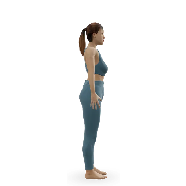
Ankle circles demonstrated on the 3D figure3D demoAnkle circlesBeginner · No equipment

Point your foot up and down 10 times, 3 times a day, then circle it · An NHS fracture clinic leaflet's dose (up and down); the circles are editorial

Harder: Ankle CARs (controlled articular rotations)

Quad set (static quads) demonstrated on the 3D figure3D demoQuad set (static quads)Beginner · No equipment

With the leg straight and supported, tighten the thigh to push the knee straighter: hold 10 s, 7 to 10 times, 3 times a day · An NHS fracture clinic leaflet's dose; an older NHS leaflet holds for 5 s

Harder: Straight-leg raise

Straight-leg raise demonstrated on the 3D figure3D demoStraight-leg raiseBeginner · No equipment

In the splint or brace, lift the leg about 5 cm and hold 5 to 10 s, 5 to 10 times · An NHS brace leaflet's dose, once your team agrees

Easier: Quad set (static quads) · Harder: Leg extension

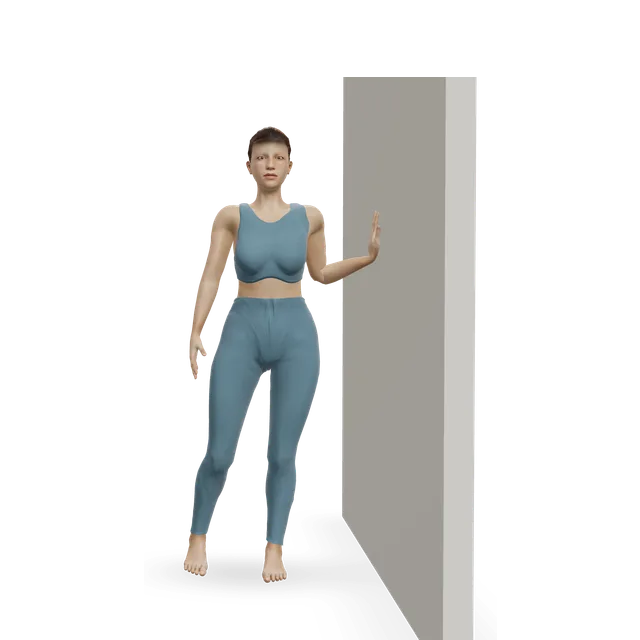
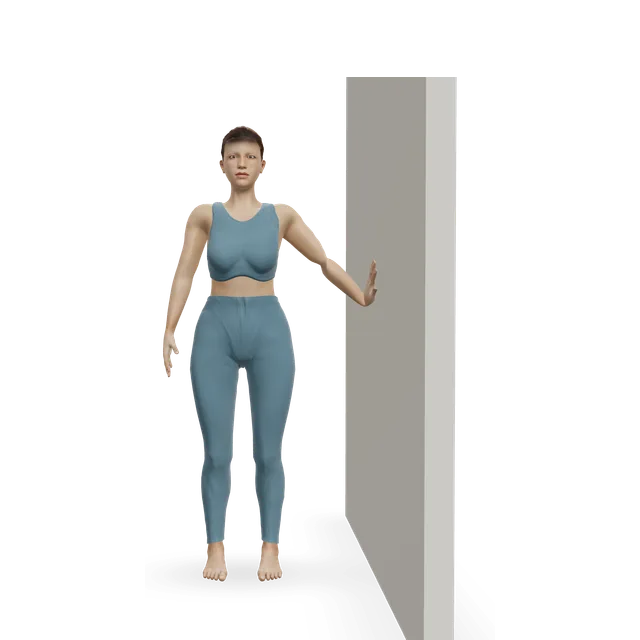
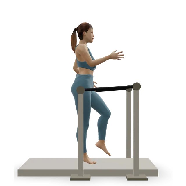
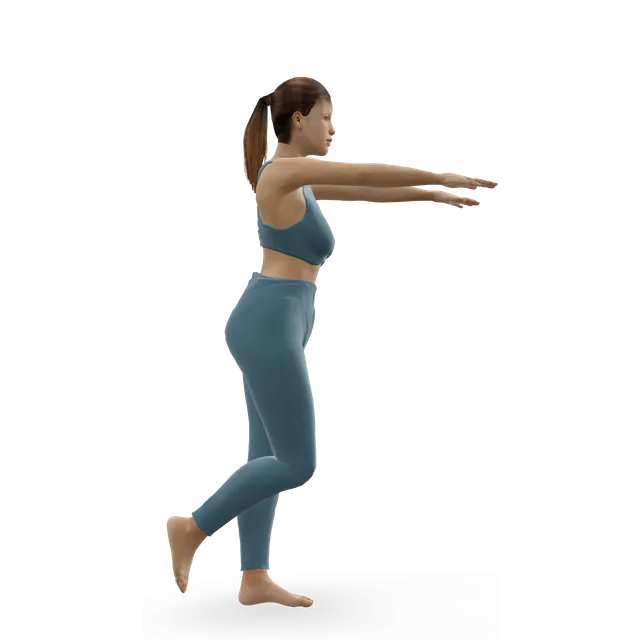
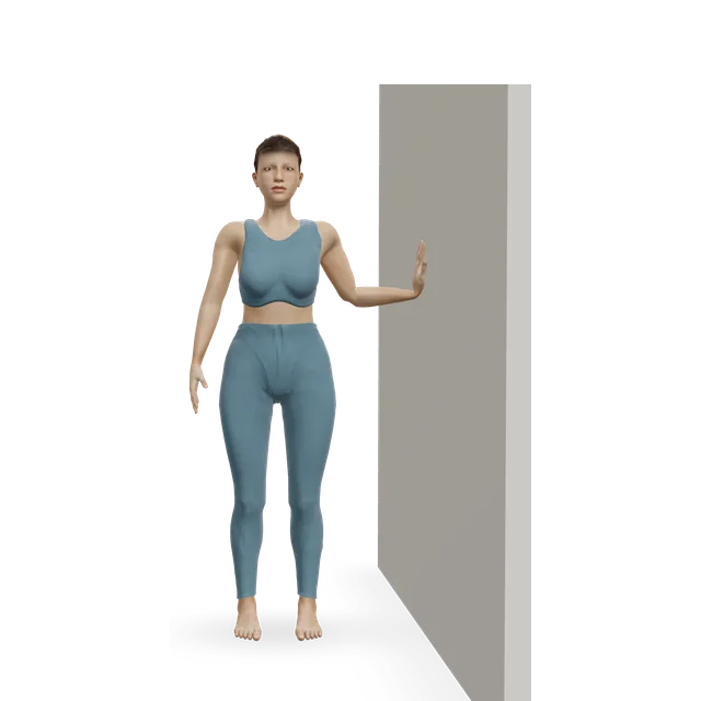
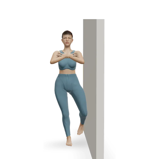
Wall side leg lift (standing side kick at the wall) demonstrated on the 3D figure3D demoWall side leg lift (standing side kick at the wall)Beginner · A wall

Splint on, holding a support: lift the leg out to the side, up for a slow count of 3 and down for 5, up to 10 each leg · The Otago strength and balance programme's dose

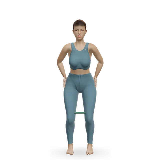
Harder: Lateral band walk, band at the feet

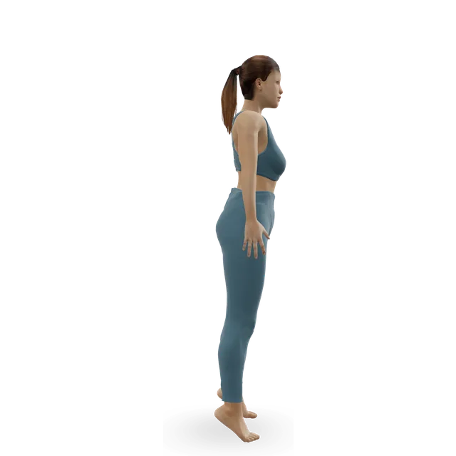
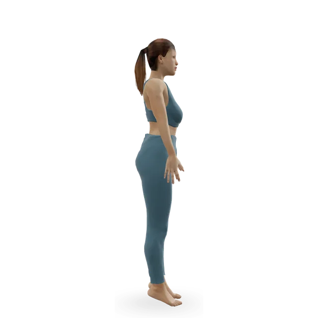
Slow calf raise demonstrated on the 3D figure3D demoSlow calf raiseBeginner · A wall

Splint on, holding a worktop: up for a count of 3 and down for 5, 10 to 20 times · The Otago programme's dose

Easier: Seated bent-knee calf raise (soleus raise) · Harder: Standing dumbbell calf raise

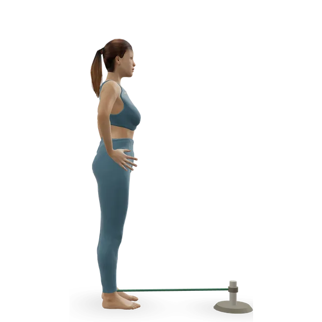
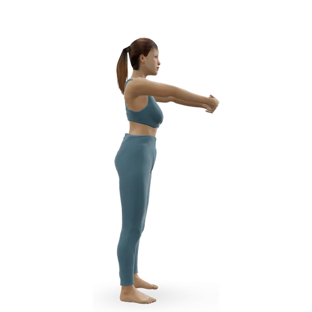
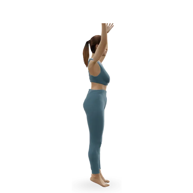
Standing band hip extension demonstrated on the 3D figure3D demoStanding band hip extensionBeginner · A band + An anchor point

No band at first: holding a support, splint on, take the straight leg back without leaning forward, 10 times each side · Editorial dose. Stop where the low back starts to arch

Harder: Back extension on a 45-degree bench

Move on when: Your team frees the knee to bend, often at 2 to 6 weeks, and you can lift the straight leg without the knee sagging.

Follow along: a timer, a cue and the next move, one step at a time.
Stage 2

Bending again and walking normally (typically weeks 2 to 8, as your fracture clinic allows)

As your fracture clinic allows. Bend the knee only within the range you're given: in one US hospital protocol after surgery, 0 to 30 degrees from week 2, 0 to 60 from week 4, 0 to 90 from week 6 and full range after week 8, with the brace locked straight for walking until week 6; UK practice may differ, and without surgery your fracture clinic sets the range. Wean off the brace and crutches as allowed, and walk normally. Short, frequent sessions suit better than one long one. Add the inner-range thigh exercise below if your team includes it.

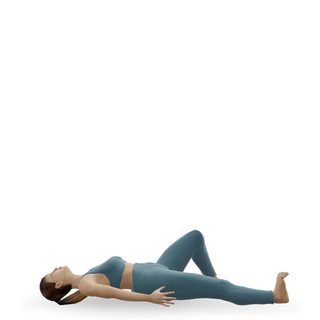
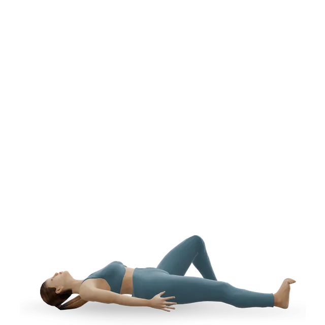
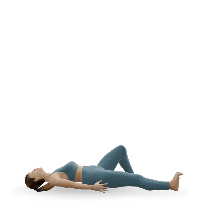
Supine heel slide demonstrated on the 3D figure3D demoSupine heel slideBeginner · No equipment

Slide the heel towards you within your allowed range, hold 2 to 3 s: 10 times, 3 times a day · An NHS knee fracture leaflet's dose. Stay inside the range your team allows

Harder: Hip windshield wipers

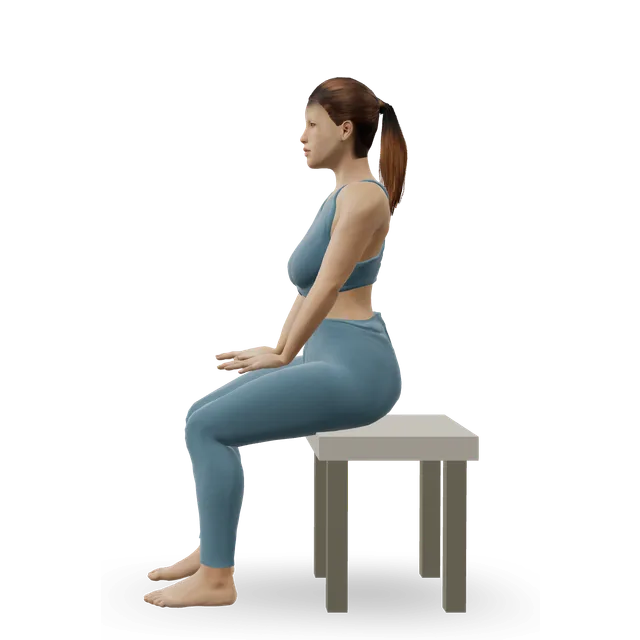
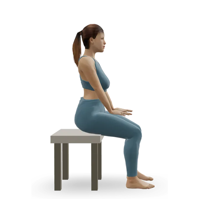
Seated knee extension (chair) demonstrated on the 3D figure3D demoSeated knee extension (chair)Beginner · A bench or box + Something with weight

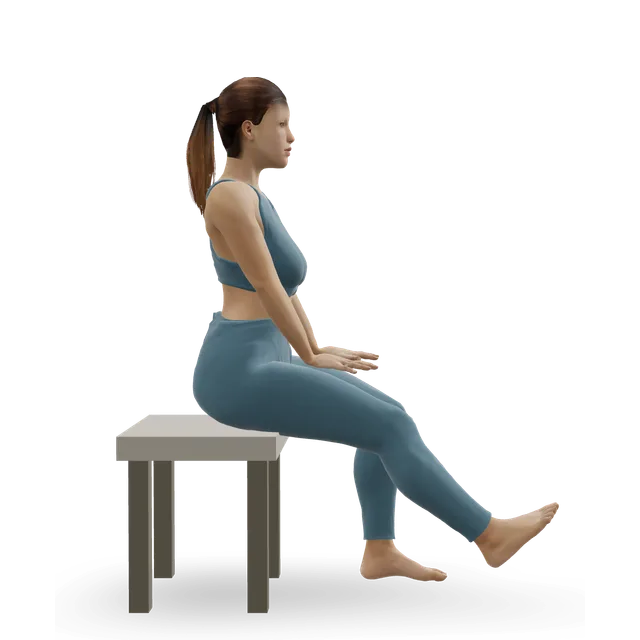
Sitting, straighten the knee, hold 2 to 3 s and lower with control: 10 times, 3 times a day, no weight yet · An NHS knee fracture leaflet's dose

Easier: Quad set (static quads) · Harder: Leg extension

Stationary cycling demonstrated on the 3D figure3D demoStationary cyclingBeginner · Cardio machine

10 to 15 minutes, no or low resistance, once the knee bends far enough to pedal round · Editorial dose; one US hospital protocol starts the bike at 6 weeks after surgery

Easier: Recumbent exercise bike · Harder: Air bike intervals

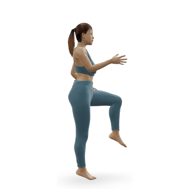
Single-leg stand demonstrated on the 3D figure3D demoSingle-leg standBeginner · A wall

Fingertips on a wall: hold 5 to 10 s, 3 times each side; build to 30 s · The first dose is the NHS balance exercises'; the build is editorial. Once full weight is allowed

Easier: Tandem stance and heel-to-toe walk · Harder: Single-leg reach matrix

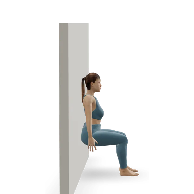
Sit squat (partial squat) demonstrated on the 3D figure3D demoSit squat (partial squat)Beginner · No equipment

2 sets of 10, shallow, within your allowed bend · Editorial dose

Harder: Bodyweight squat

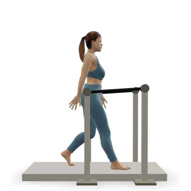
Treadmill walking demonstrated on the 3D figure3D demoTreadmill walkingBeginner · Cardio machine

Walking out of the brace, building from 10 minutes · Editorial dose. Walking outside counts

Harder: Treadmill jogging

Move on when: The knee bends to about 90 degrees or more, you walk without the brace or crutches and without a limp, and you can lift the straight leg with no sag.

Follow along: a timer, a cue and the next move, one step at a time.
Stage 3

Strength (typically months 2 to 4, as your team allows)

As your team allows. Strength on two legs and then one, with balance work, 2 to 3 sessions a week, because lasting thigh weakness is common. In the US protocol, an elliptical trainer joins at about 8 weeks and lunges at about 12 after surgery. Doses are editorial unless a source is named.

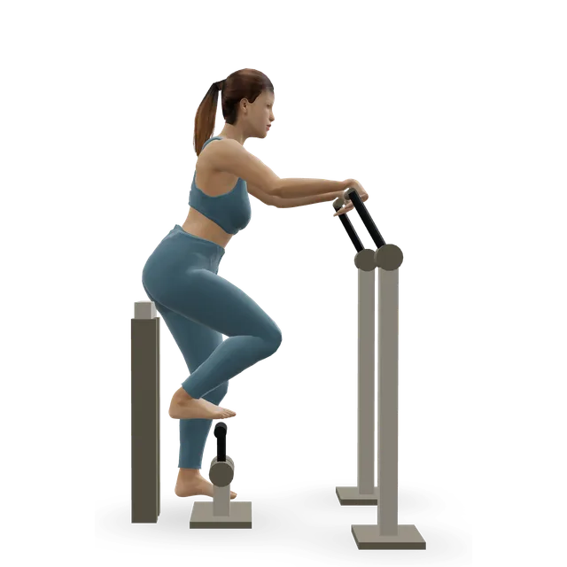
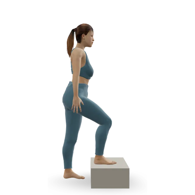
Step-up demonstrated on the 3D figure3D demoStep-upBeginner · A step

3 sets of 8 to 10 each leg · Editorial dose

Easier: Low step-up · Harder: Dumbbell step-up

Leg press demonstrated on the 3D figure3D demoLeg pressBeginner · Strength machine

3 sets of 10 to 12, within the depth your team allows · Editorial dose

Harder: Bodyweight squat

Seated knee extension (chair) demonstrated on the 3D figure3D demoSeated knee extension (chair)Beginner · A bench or box + Something with weight

With a light ankle weight or band: 3 sets of 10 to 15, 2 to 3 days a week · The move page's dose, once your team agrees to resisted straightening. Work in the bent-knee half if the full range hurts

Easier: Quad set (static quads) · Harder: Leg extension

Split squat demonstrated on the 3D figure3D demoSplit squatIntermediate · No equipment

3 sets of 8 each leg, from about 12 weeks after surgery · Editorial dose; the timing is the US protocol's for lunges. Pad the back knee

Easier: Bodyweight squat · Harder: Reverse lunge

Elliptical trainer demonstrated on the 3D figure3D demoElliptical trainerBeginner · Cardio machine

10 to 20 minutes at a moderate effort · The move page's dose; from about 8 weeks after surgery in the US protocol

Harder: Stair-stepper machine

Move on when: The knee bends fully or nearly, walking and stairs are normal, it doesn't swell after sessions, and your team agrees.

Follow along: a timer, a cue and the next move, one step at a time.
Stage 4

Kneeling, impact and sport (from about month 4, as your team allows)

As your team allows. NHS leaflets keep sport out for at least 12 weeks, and contact sport out for 8 to 12 weeks after a stable break, as your consultant advises. After surgery, the US protocol allows straight-line jogging from 16 weeks if a functional test shows good movement and symmetry, jumping from two feet to one at 16 to 20 weeks, and sport at 5 to 6 months once a return-to-sport test is over 90% of the other leg. Start kneeling on a cushion once the wound has healed and your team agrees (our suggestion).

Get up and down from the floor demonstrated on the 3D figure3D demoGet up and down from the floorBeginner · No equipment

5 to 8 slow cycles down and up, kneeling on a cushion, changing the leading leg · The move page's dose; here, the first real test of kneeling

Easier: Sit to stand from a chair · Harder: Bodyweight get-up

Treadmill jogging demonstrated on the 3D figure3D demoTreadmill joggingIntermediate · Cardio machine

The NHS walk-jog programme, every other day, once 30 minutes of fast walking is pain-free · An Oxford NHS return-to-running leaflet, used here as a guide; the weeks are in the notes below

Easier: Treadmill walking · Harder: Sprint intervals

Pogo hops demonstrated on the 3D figure3D demoPogo hopsIntermediate · No equipment

Short sets of 10 to 20, two-footed · Editorial dose

Easier: Ankle bounces (mini pogo) · Harder: Single-leg pogo hops

Snapdown to athletic landing demonstrated on the 3D figure3D demoSnapdown to athletic landingBeginner · No equipment

2 to 3 sets of 5, freezing each landing · Editorial dose

Harder: Box drop landing

Single-leg hop and stick demonstrated on the 3D figure3D demoSingle-leg hop and stickIntermediate · No equipment

3 sets of 5 each leg, small hops first · Editorial dose. Only once your team clears you

Easier: Standing broad jump with stuck landing · Harder: Alternate-leg bounding

Shuttle runs demonstrated on the 3D figure3D demoShuttle runsAdvanced · No equipment

Short sets, with wide, unhurried turns first · Editorial dose

Easier: Treadmill jogging

Move on when: Your team clears you. Keep two strength sessions a week afterwards (our suggestion).

Follow along: a timer, a cue and the next move, one step at a time.

What to change for now

Typical timings, which your fracture clinic may change

These come from NHS leaflets, Health New Zealand, US surgeons and one US hospital protocol, and they differ. Your own clinic's plan comes first.

  • Without surgery: a straight-leg splint until the fracture clinic review at about 2 weeks, then as advised, walking as comfort allows.
  • After surgery: the knee kept straight for 2 to 4 weeks (US surgeons) or a brace for 4 to 6 weeks (Health New Zealand); one NHS trust's hinged brace stays on until it's removed, 6 weeks to 3 months after surgery.
  • Bending after surgery, in one US hospital protocol: none for 2 weeks, 30 degrees by 4 weeks, 60 by 6, 90 by 8, then full. UK practice may differ.
  • Driving: not in the brace.
  • Sport: not for at least 12 weeks in NHS leaflets; 5 to 6 months after surgery in the US protocol.

Sources 13, 6, 1, 2, 15, 16, 17

Splint and brace care

Wear it as instructed. Take it off only to wash or use a cold pack, keeping the knee straight, and put it straight back on; with a hinged brace, don't take it off at all, even at night, unless your physiotherapist says so. Keep the brace and padding dry, tighten the straps as they loosen, and check the skin: redness that doesn't fade after 15 to 20 minutes, sores, blisters or rubbing need the physiotherapy team.

Sources 13, 15

Inner-range thigh tightening (written steps)

An NHS brace leaflet's exercise, for when your team allows some bend. It has no 3D demo.

  • 1. Only within the range your team allows, and only once they've shown you.
  • 2. Lie or sit with the leg resting over a rolled-up towel or pillow under the knee.
  • 3. Pull your toes towards you, then lift your foot until the knee is straight, keeping the back of the knee on the towel.
  • 4. Hold for 5 to 10 seconds, then lower slowly.
  • 5. Repeat 5 to 10 times (Royal Berkshire NHS) or 10 times (King's College Hospital NHS), 3 times a day.

Source 15

Stairs on crutches (written steps)

The usual NHS technique. Use stairs one step at a time until your team says otherwise.

  • 1. Use a stair rail or banister if you can, with your crutch or stick in the other hand.
  • 2. Going up: the good leg first, then the injured leg, then the crutch or stick.
  • 3. Going down: the crutch or stick first, then the injured leg, then the good leg.
  • 4. If you have weight limits, your physiotherapist will teach you the technique before you go home.

Sources 17, 15

The walk-jog programme (written steps)

From an Oxford NHS leaflet written for coming back from any injury lay-off. We use it here as a guide, once your team clears you for running.

  • 1. Start once 30 minutes of fast walking is pain-free, and your team is happy for you to run.
  • 2. Week 1: jog 1 minute, walk 1 minute, for 30 minutes. Do each week's session every other day.
  • 3. Week 2: jog 2 minutes, walk 1 minute, for 30 minutes.
  • 4. Week 3: jog 4 minutes, walk 1 minute, for 30 minutes.
  • 5. Week 4: jog 9 minutes, walk 1 minute, for 30 minutes.
  • 6. Week 5: jog 15 minutes, walk 1 minute.
  • 7. Week 6: jog 30 minutes without stopping.
  • 8. Move up a level after a week without pain, and down a level if it hurts. From week 7, add no more than 10% a week.

Source 24

Blood clots

NICE says blood-thinning injections may be considered while the leg is held straight in a splint or brace, if the risk of a clot outweighs the risk of bleeding, and that stopping them can be considered if this goes on beyond 42 days. Your team decides whether you need them.

Source 23

Positions to swap, and why

Habit or positionWhy it mattersInstead
Taking off or unlocking the splint or brace before you're toldIt protects the break; NHS leaflets say off only to wash, with the knee kept straight.Wear it as instructed, and ask the team if it rubs.
Resting with a pillow under the kneeNHS leaflets keep the leg straight early on.Support under the heel, the knee straight, and thigh tightening.Try: Quad set (static quads)
Deep squats or kneeling before your team agreesAfter surgery, bending comes back in steps over 8 weeks or more in one US hospital protocol.Heel slides within your allowed range.Try: Supine heel slide
The leg extension machine or ankle weights earlyResisted straightening is added later, once your team agrees.Thigh tightening and straight-leg raises.Try: Quad set (static quads)
Going upstairs leading with the injured legThe NHS crutch technique: the good leg goes up first and the injured leg comes down first.A rail in one hand and a crutch in the other, good leg first going up.
Running or jumping before you're clearedNHS leaflets keep sport out for at least 12 weeks; one US hospital protocol waits about 16 weeks after surgery for jogging.A static bike, then an elliptical trainer.Try: Stationary cycling
Kneeling straight onto the scar or the wiresFront-of-knee pain and metalwork that rubs are common.A cushion or kneeling pad; ask about removing the metalwork if it persists.
Stopping strength work when physiotherapy endsThigh weakness of a quarter or more is common years later in small studies.Two strength sessions a week.Try: Leg press
Driving in the braceHealth New Zealand says you can't.Wait until the brace is off and you can do an emergency stop without pain.
SmokingIt affects how well bones heal.NHS stop-smoking support.

Routines to follow

More moves for this area

Questions people ask

How do I know if I've broken my kneecap?

Pain and swelling at the front of the knee after a fall onto it or a blow, bruising, and difficulty straightening the knee, lifting the straight leg or walking. It needs an X-ray. Go to A&E if the knee has changed shape, swells very quickly or bone is showing.

Can you walk with a broken kneecap?

Often yes, in a splint or brace that keeps the knee straight: NHS fracture clinics let you put weight through the leg as comfort allows after a stable break. After surgery, most people walk with full weight in a brace locked straight, though some hospitals limit weight at first. Follow your fracture clinic's plan.

Do I need surgery for a broken kneecap?

Not usually if the break is stable and you can fully straighten and lift the leg: a splint or brace is usually enough. If the pieces have separated, or the knee can't straighten, an operation usually holds them together. No trial has compared the two directly.

How long does a broken kneecap take to heal?

About 6 weeks for the bone, sometimes 6 to 8 weeks or longer, with some pain and swelling for 3 to 6 months. Most people are back to normal activities in 3 to 6 months.

How long will I wear a splint or brace?

Without surgery, until your fracture clinic review at about 2 weeks, and then as advised. After surgery, the knee is usually kept straight for 2 to 6 weeks, and one NHS trust's hinged brace stays on for 6 weeks to 3 months. It's your fracture clinic's call.

When can I bend my knee?

When your team says. In one US hospital protocol after surgery, bending starts at 2 weeks and builds to 90 degrees by 8 weeks; UK hospitals may differ. Without surgery, your fracture clinic decides at your review.

Will the wires need to come out?

About 3 in 10 people have the metalwork removed because it causes symptoms. Health New Zealand says wires are likely to be taken out after at least 3 months, and screws only if they're painful, usually not before 12 months.

When can I kneel?

When the wound has healed and your team agrees, starting on a cushion. Front-of-knee pain is common for a long time, so a kneeling pad helps; ask about removing the metalwork if it's the cause.

When can I drive?

Not while you're in a brace. Once you're out of it, can walk comfortably and can do an emergency stop without pain, and your insurer agrees.

When can I play sport again?

NHS leaflets keep sport out for at least 12 weeks, and contact sport out for 8 to 12 weeks after a stable break. After surgery, one US hospital protocol allows jogging from about 16 weeks and sport at 5 to 6 months, once strength tests are passed. Your fracture clinic's plan comes first.

Will my knee ever feel normal?

Most people get back to daily life, but thigh weakness of a quarter or more, and some front-of-knee pain, are common years later in small studies. Keeping up strength work is sensible, and a physiotherapist can measure the difference between your legs.

My child can't straighten their knee after a jump: is it a fracture?

It could be a sleeve fracture, where a piece of cartilage and bone pulls off the lower tip of the kneecap, or a tendon injury. Get it checked the same day.

Related exercise guides

Other pain guides

Sources

  1. AAOS OrthoInfo · Patellar (kneecap) fractures
  2. Health New Zealand Te Whatu Ora · Broken kneecap (fractured patella)
  3. Larsen 2016 · Incidence and epidemiology of patellar fractures (population study of 756, Orthopedics)
  4. Rau 2023 · Patella fractures and their surgery in Germany, 2006 to 2020 (national data, BMC Musculoskelet Disord)
  5. NICE NG38 · Fractures (non-complex): assessment and management (2016)
  6. York and Scarborough Teaching Hospitals NHS · Fracture of the patella (virtual fracture clinic letter)
  7. Schmidt 2025 · Patient-reported outcomes a year after patella fractures in the Swedish Fracture Register (observational study of 8,726, Bone Jt Open)
  8. Yellin 2024 · Patellar sleeve injuries in 90 children and teenagers (case series, J Child Orthop)
  9. Sayum Filho 2021 · Interventions for treating fractures of the patella in adults (Cochrane review)
  10. Braun 1993 · Non-surgical treatment of stable patellar fractures (case series of 40, Clin Orthop Relat Res)
  11. Pritchett 1997 · Non-surgical treatment of widely displaced patella fractures (case series of 18, Am J Knee Surg)
  12. Contractor 2025 · Outcomes after patella fracture repair in people aged 65 and over (cohort of 248, J Knee Surg)
  13. East Sussex Healthcare NHS · Undisplaced patella fracture (emergency department shared care plan, 2025)
  14. Whittington Health NHS · Undisplaced patella fracture (virtual fracture clinic leaflet, 2022, now past its review date)
  15. Royal Berkshire NHS · Hinged knee brace information and exercises (physiotherapy leaflet, 2025)
  16. University of Kansas Health System · Patellar fracture fixation: rehabilitation protocol (US hospital protocol)
  17. West Hertfordshire Teaching Hospitals NHS · Knee fracture (outpatient physiotherapy leaflet, 2025)
  18. LeBrun 2012 · Function a mean 6.5 years after surgery for patella fractures (cohort of 40, J Orthop Trauma)
  19. Lazaro 2013 · Function in the year after surgery for patella fractures (prospective cohort of 30, J Bone Joint Surg Am)
  20. Dalaslan 2026 · Thigh strength a median 5 years after surgery for patella fractures (cohort of 58, Arch Orthop Trauma Surg)
  21. Vesterager 2024 · Complications after surgery for patella fractures (meta-analysis of 14 studies, Eur J Trauma Emerg Surg)
  22. Sherwood Forest Hospitals NHS · Discharge advice: patella (knee cap) fracture (leaflet, 2023, now past its review date)
  23. NICE NG89 · Venous thromboembolism in over 16s: reducing the risk of hospital-acquired deep vein thrombosis or pulmonary embolism (2018, updated 2019)
  24. Oxford University Hospitals NHS · Return to Running Programme (OxSport patient leaflet, 2024)
  25. NHS · Dislocated kneecap
  26. NHS · Knee pain
  27. NHS · Broken leg
  28. NHS · DVT (deep vein thrombosis)

Exercise education, not a diagnosis or treatment plan. It is written from the guidelines and studies listed above; review by a clinician is pending. If your pain follows an injury, is severe, is getting worse, or comes with any warning sign listed on this page, get it assessed. Health disclaimer.