Pain guide · Foot pain

Tarsal coalition: a stiff, painful flat foot and ankles that keep spraining, and what helps

A tarsal coalition is a bar of bone, cartilage or tough tissue joining two of the bones at the back or middle of the foot. It forms before birth, is in both feet in about half of people who have one, and often never causes trouble. But as the bar hardens in late childhood and the teens, it can leave the foot stiff and flat, aching below the ankle with sport, and the ankle spraining again and again. It needs an X-ray, and often a computed tomography (CT) or magnetic resonance imaging (MRI) scan, and a specialist manages it.

This guide is for teenagers, parents and adults. It covers the signs to get checked, what scans can and can't show, what rest, insoles, boots and casts can do, when surgery helps and how removing the bar compares with fusing the joint, recovery times, and a staged plan with 3D demos for strength, balance and getting back to sport, as your team allows.

When to get help first

Exercise is for pain that has been checked or is clearly mild and settling.

Get urgent medical help if

  • Call 999 or go to A&E: after an injury, you heard a crack, the foot or ankle has changed shape or points at an odd angle, a bone is sticking out, there is a bad cut over it, you're in severe pain, or the toes look blue or white, feel numb or are cold to touch. These can mean a broken bone. Don't drive yourself.
  • Contact NHS 111: after an injury, severe ankle pain, feeling faint, dizzy or sick from the pain, a snap, grinding or popping noise at the time of the injury, the ankle or foot at an odd angle, or not being able to walk. These might be signs of a broken ankle or an infection.
  • Same day (urgent GP appointment or NHS 111): after an injury, you can't put weight on it or walk more than a few steps, the pain is very bad or getting worse, there is a lot of swelling or bruising or it is getting worse, or it feels very stiff. If you think it's broken, don't treat it yourself.
  • A child who has hurt their foot or may have broken a toe: don't treat it yourself; take them to an urgent treatment centre or A&E.
  • Same day: a sudden, very painful, hot, swollen, red joint, often the big toe, especially with a high temperature, feeling shivery or being unable to eat. It may be gout, but it could be a joint infection (septic arthritis), which is urgent.
  • Same day: in a cast or boot, pain that is getting worse, toes that are numb or burning, a cast that feels too tight, toes that are swollen, blue or white, or a bad smell from under the cast.
  • After surgery: redness, swelling, oozing or a smell from the wound, or a fever. Contact your surgical team the same day, or NHS 111 out of hours.
  • Same day (GP or NHS 111): throbbing pain and swelling in one calf, with red or darker skin. It could be a blood clot (DVT). Watch for it after an ankle injury or time in a cast or boot.
  • Call 999: calf or leg pain or swelling with breathlessness or chest pain. It could be a clot that has reached the lungs.

See a doctor or physiotherapist if

  • In a teenager or young adult, a stiff, painful flat foot with repeated sprains: see a GP. It can be a tarsal coalition, where two foot bones are joined by a bar of bone or tissue.
  • Flat feet that are painful, stiff, weak or numb, that you didn't have before, or that affect only one foot.
  • A flat foot whose arch doesn't come back when your child stands on tiptoe, a flat foot on one side only, or a flat foot with a tight calf: see a GP. Children's orthopaedic surgeons call a one-sided flat foot a red flag that needs further assessment.
  • Spasm or tightness of the muscles on the outside of the lower leg that pulls the foot outwards, with pain at the back of the foot: see a GP or physiotherapist. It's a protective response to pain, and a coalition is one cause.
  • An ankle that keeps giving way months after a sprain.
  • Pain that isn't better after 2 weeks of looking after it yourself, is getting worse, keeps coming back, or stops you doing normal things.
  • One spot on a foot bone that hurts with every step and later at night: a possible stress fracture.
  • Diagnosed with a coalition and still in pain after about 6 months of rest, insoles, a boot or cast and physiotherapy, or pain stops you walking comfortably or playing sport: ask your specialist about surgery.
  • After surgery: pain or stiffness that is getting worse rather than better weeks after the operation, the foot flattening or tilting further, or the old pain coming back months later: contact your team. The bar can grow back in about 1 in 10 feet.

Understand it

What a tarsal coalition is

The tarsal bones at the back and middle of the foot (the heel bone, the talus, the navicular and the cuboid) normally separate before birth. In a coalition, two or more stay joined by a bridge, or bar, of bone, cartilage or fibrous tissue. It's thought to be inherited, from bones that don't fully separate in the womb.

Two kinds make up over 90%: calcaneonavicular, joining the heel bone to the navicular, and talocalcaneal, joining the talus to the heel bone at the joint below the ankle. One NHS leaflet gives 48% and 44%.

Sources 1, 2, 3, 4

How common it is, and in both feetMixed evidence

Estimates range from 0.03% to 13% of people, depending on how carefully it's looked for. Professional and NHS sources give about 1 in 100, under 1% or 3% to 5%. Scans find more: 6.0% of 839 people having CT scans of both feet in one Korean hospital, and about 11% of people having an ankle MRI scan, a 2026 review says. A US population study notes that older estimates were up to 2% and newer ones 11% to 13%.

About half of people with a coalition have one in both feet (56.5% on the CT scans), though often only one foot hurts. It runs in families: one NHS leaflet says relatives are affected 38% of the time.

Sources 5, 1, 2, 3, 6, 7, 8

Who gets symptoms, and whenEmerging evidence

Many coalitions never cause symptoms and are never found: the authors of one study put pain at about a quarter of cases, and one NHS children's hospital says around 75% of children with a coalition never need treatment. As a child grows, the bar hardens into bone and the foot stiffens, usually between 8 and 16. Pain typically starts in the second decade of life or later, often after an injury such as a sprain.

In a US county over 52 years, painful coalitions were newly diagnosed in 3.5 per 100,000 children a year (calcaneonavicular 1.9, talocalcaneal 1.2), most often at 10 to 14 in both boys and girls. Talocalcaneal coalitions came to attention later, at 13.9 years on average, than calcaneonavicular ones, at 12.7.

Sources 1, 2, 9, 6, 4, 7, 3

The signs

A flat foot that stays flat when the teenager rises onto tiptoe (in a flexible flat foot, the arch comes back); pain below the ankle, around the middle or back half of the foot, worse with sport, sometimes with a limp; and ankle sprains that keep happening, because the stiff foot makes the ankle roll more, with difficulty on uneven ground.

The back of the foot moves less when turning the sole in and out, and in 20% to 30% the muscles on the outside of the leg go into spasm, a protective response to pain, one NHS leaflet says. Pain can also come from the tendons and ligaments around the stiff joints, and later from arthritis in them.

Sources 1, 2, 3, 10, 4

How it's diagnosed, and what a scan finding meansEmerging evidence

The British Society for Children's Orthopaedic Surgery (BSCOS) says whether the arch comes back on tiptoe separates flexible from stiff flat feet; that foot pain, stiffness or a tight calf should prompt a referral for further assessment; that a flat foot on one side only is a red flag; and that X-rays, including an oblique view, should be considered for a painful flat foot.

Many calcaneonavicular bars show on X-rays, but talocalcaneal bars are best seen on CT or MRI. The American Academy of Orthopaedic Surgeons (AAOS) calls CT the gold standard, and MRI shows bars of cartilage or fibrous tissue that don't show on X-rays or CT. In 86 children with a suspected talocalcaneal coalition, the X-ray 'C sign' picked up 54% and the 'talar beak' 11% of those confirmed on CT, while ultrasound picked up 96%.

Because coalitions are common and often painless, finding one on a scan doesn't by itself mean it's the cause of the pain: the story and the examination have to fit (our reasoning from the scan studies).

Sources 10, 4, 1, 11, 5, 8

How it usually goesMixed evidence

Many coalitions are not a problem for years, if at all, one NHS leaflet says. Once a coalition is painful, the stiffness itself doesn't go away, and over time the lack of movement can lead to arthritis in the neighbouring joints.

How many painful coalitions need surgery depends on the study. In a clinic series of 50 painful coalitions in children treated without surgery first, surgery wasn't needed in 79% of calcaneonavicular and 62% of talocalcaneal coalitions. In a US population study, 74% of painful calcaneonavicular and 56% of talocalcaneal coalitions had surgery, against 11% of other types. One NHS children's hospital says up to half of the children who need treatment avoid surgery. The settings and follow-up differ, so there's no single answer.

Sources 3, 1, 6, 12, 7

Look-alikes

A painless, flexible flat foot is normal and has its own guide. Other causes of pain around the midfoot and below the ankle also have their own guides: an accessory navicular (a painful bump on the inner midfoot), tibialis posterior tendon problems with a dropping arch (in adults), pain in the hollow in front of the outer ankle bone after sprains (sinus tarsi syndrome, which a coalition can cause), and an ankle that keeps giving way (chronic ankle instability).

Sources 13, 10, 4, 2
What helps, and how strong the evidence is

Non-surgical care first, for at least 6 monthsStrong evidence

A 2019 review says the first treatment should always be non-surgical, for at least 6 months, and that there is consensus that surgery is offered when that fails. The options NHS hospitals and AAOS describe:

  • A break from high-impact activity, typically 3 to 6 weeks (AAOS), or limiting high-impact sport for a few weeks (an NHS hospital).
  • Insoles: arch supports, heel cups and wedges, or custom insoles; an ankle boot that limits joint movement, or a rocker-bottom shoe.
  • A walking boot or a below-knee cast: weight-bearing, for 4 to 6 weeks in one NHS service.
  • Anti-inflammatory painkillers, and a steroid injection for some.
  • Physiotherapy for stretching and strengthening.
  • Where the muscles on the outside of the leg are in spasm, one NHS podiatric surgery service may numb the nerve behind the knee to relax them and see whether the foot can be corrected for a cast or insoles.

Sources 4, 1, 3, 2, 6, 8

How well non-surgical care works: two studies, different answersMixed evidence

In a clinic series of 50 painful coalitions in children (average age 11.4), 53% of 81 non-surgical treatment courses relieved the pain, and casting, a walking boot and supportive measures did equally well. In 47 feet with a talocalcaneal coalition treated with manipulation under anaesthetic, 5 weeks in a cast and then insoles, 55% had a satisfactory result and 15% went on to surgery; the authors put published success at 25% to 68%.

But in a US population study, most painful calcaneonavicular (74%) and talocalcaneal (56%) coalitions ended up having surgery. These come from different settings and follow-up times. Non-surgical care is always tried first, it helps some teenagers for good, and many still need an operation. No trial has compared non-surgical treatments.

Sources 12, 9, 7

Exercise: untested for coalitions, borrowed from ankle instabilityEmerging evidence

No study has tested exercise for a coalition. NHS hospitals include physiotherapy for stretching and strengthening in non-surgical care, and a stiff foot makes the ankle roll more and sprain.

So the plan borrows from ankle instability trials, labelled as borrowed. In a trial of 39 people with unstable ankles, 6 weeks of either balance training or band and heel-raise training, 20 minutes 3 times a week, improved strength, balance and side hopping. In 522 athletes aged 12 to 70, an 8-week home balance programme after a sprain cut repeat sprains from 33% to 22% over a year. These were not people with coalitions, and band work stays within the movement the foot has (our reasoning).

Sources 2, 6, 1, 14, 15

Surgery: removing the barEmerging evidence

Surgery is offered when pain and limits persist despite good non-surgical care. The bar is removed and fat, muscle or another material is placed in the gap to stop it reforming. AAOS says removal preserves normal foot motion and relieves symptoms in most people who don't have arthritis, and a review calls open removal the treatment of choice, with sound evidence of good long-term results.

Calcaneonavicular bars: across 11 studies (274 patients, 394 feet, average age 12.5, followed 2 to 23 years), removal was successful in 82.9%. The bar grew back in 52 of 380 feet, and material placed in the gap reduced regrowth.

Talocalcaneal bars: removal does best when the bar covers less than half of the joint surface, the heel isn't tilted outwards more than about 16°, and there's no arthritis. But one NHS leaflet notes that some surgeons remove the bar first whatever its size, and in 24 young people followed into their twenties, calcaneonavicular and talocalcaneal removals did equally well, with good results even with larger bars and more tilted heels. A heel that stays tilted outwards may need realigning at the same time or later.

Sources 4, 1, 2, 3, 16, 8, 9, 17

What happens after removalEmerging evidence

Of 304 people who had a coalition removed (average age 24), 8.6% had another removal and 5.3% a fusion over about 9 years, and more than 85% needed no further operation. In 165 children's feet, 10.9% had a complication (6 infections, 6 bars that grew back and 6 other further operations), more often with older age, obesity or a tilted heel.

One NHS hospital quotes regrowth in about 10%, and ongoing symptoms more often after talocalcaneal removal and in older patients. In 78 people (97 removals), getting back to the activity they wanted took 18 weeks on average.

Sources 18, 19, 2, 20

Fusion, and removal against fusionMixed evidence

Fusing the affected joints is kept for large or multiple coalitions, arthritis, severe deformity or a removal that has failed. It reliably relieves pain at the cost of the joint's movement. No trial has compared removal with fusion: reviews agree removal comes first in young people without arthritis, and that fusion gives more predictable results with advanced arthritis or a bar covering more than half of the joint.

One NHS service notes that trying removal first won't harm the chances of a later fusion. In a population study, doing a fusion at the same time as the first removal made a later fusion more likely. And surgery won't give a normal foot: the foot stays stiffer than a normal foot, an NHS hospital says.

Sources 4, 8, 3, 2, 18

How much pain is OK while you exercise?

Teenagers: no more than 3 out of 10 (borrowed from Sever's disease); adults: the site's foot and ankle rule

No coalition source gives a pain rule, so these are borrowed. For teenagers, from an NHS children's hospital booklet for Sever's disease, another growing foot problem, as our Freiberg's and Iselin's guides do: keep pain during activity to 3 out of 10 or less, where 0 is no pain and 10 is the worst. A little discomfort is fine if it settles quickly after you stop.

For adults, the site's foot and ankle rule (editorial): discomfort up to about 5 out of 10 that settles within an hour, with no more swelling or stiffness the next morning. If it's worse the next day, do a little less, not nothing.

For everyone: work within the movement the foot has, and don't force turning the sole in or out, because those joints are joined (our reasoning). Spasm of the muscles on the outside of the leg is a protective response to pain: if it starts, stop that exercise and let the foot settle. A feeling of the ankle giving way is never part of the rule: drop back, and use a brace or tape for that activity. In a cast or boot, or after surgery, do only what your team allows.

Sources 21, 3

The exercise plan: 3 stages

Start at the first stage. Each move opens a full guide with a 3D demo.

Stage 1

Settle the foot (the first weeks, in a cast or boot if your team uses one)

A break from high-impact sport, typically for 3 to 6 weeks; supportive shoes, with insoles or arch supports if advised; and a walking boot or a weight-bearing below-knee cast for 4 to 6 weeks if it's very painful, as your team allows. Keep fit without the activities that hurt. In a cast, leave out the ankle circles and calf raises until it's off.

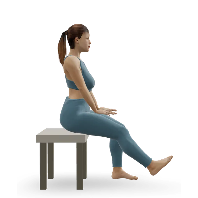
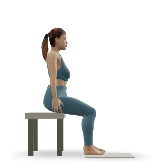
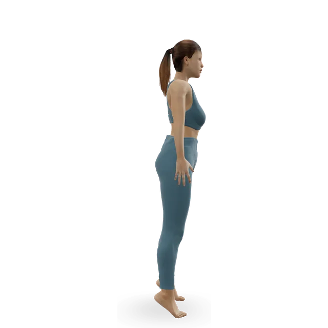
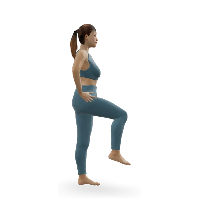
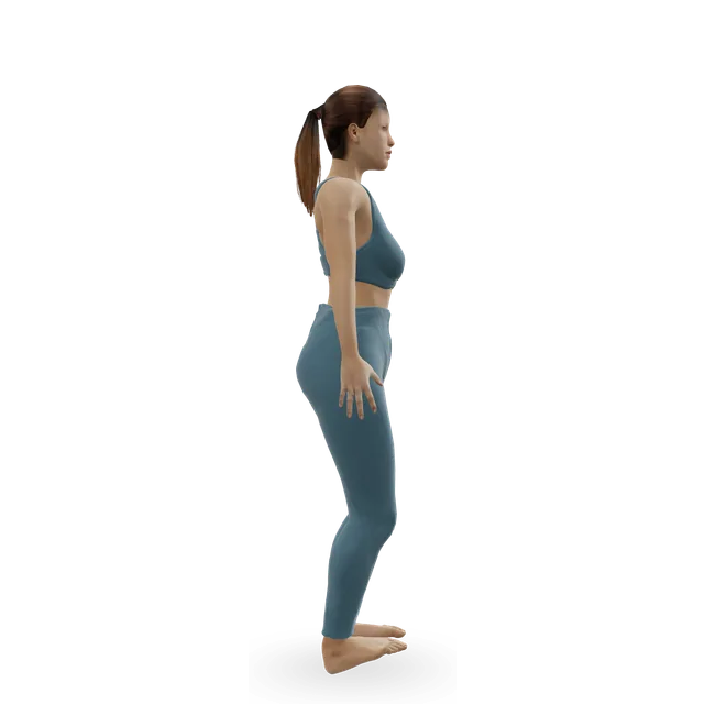
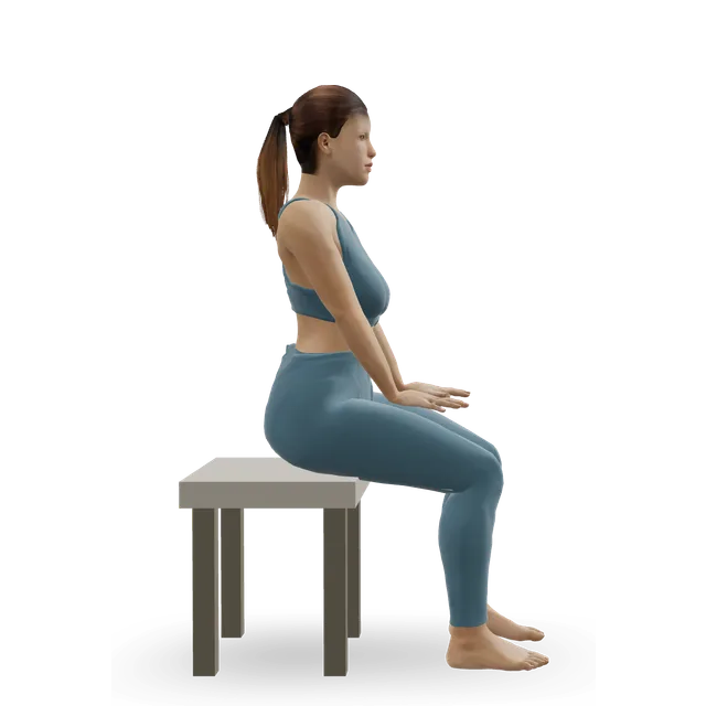
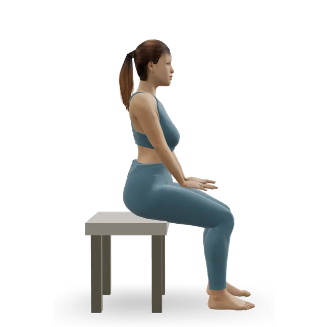
Seated bent-knee calf raise (soleus raise) demonstrated on the 3D figure3D demoSeated bent-knee calf raise (soleus raise)Beginner · A bench or box

2 to 3 sets of 15 · Editorial dose, as in our sinus tarsi syndrome guide

Harder: Slow calf raise

Ankle circles demonstrated on the 3D figure3D demoAnkle circlesBeginner · No equipment

10 slow circles each way, within the movement the foot has · The move page's dose; not forcing the stiff joint is editorial

Harder: Ankle CARs (controlled articular rotations)

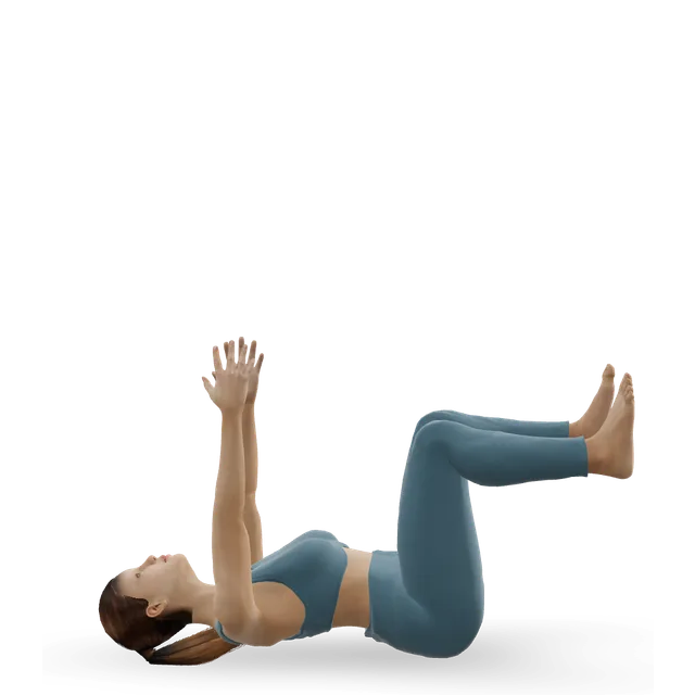
Dead bug demonstrated on the 3D figure3D demoDead bugBeginner · No equipment

2 sets of 6 to 8 on each side, slowly · Editorial dose, as in our guide to osteochondritis dissecans of the knee

Easier: Dead bug heel taps · Harder: Forearm plank

Stationary cycling demonstrated on the 3D figure3D demoStationary cyclingBeginner · Cardio machine

15 to 30 min, easy · Editorial dose, as in our sinus tarsi syndrome guide

Easier: Recumbent exercise bike · Harder: Air bike intervals

Move on when: Everyday walking in your shoes (with insoles if given) is comfortable, pain in daily life is no more than 3 out of 10 for a teenager, and any cast or boot is off.

Follow along: a timer, a cue and the next move, one step at a time.
Stage 2

Strength and balance (borrowed from an ankle instability trial)

Three sessions a week of about 20 minutes, in supportive trainers, borrowed from a trial in people with unstable ankles. Work within the movement the foot has, and don't force the sole in or out (our reasoning).

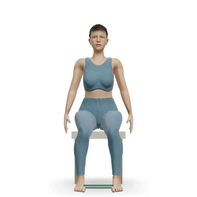
Band ankle eversion demonstrated on the 3D figure3D demoBand ankle eversionBeginner · A band + A bench or box

3 sets of 10 at 3 to 5 s a rep, then 4 sets and 3 sets of 10 in alternate weeks · A heavier band every 2 weeks: the Hall 2018 trial's progression, borrowed. Stop short of any pinch or muscle spasm (editorial)

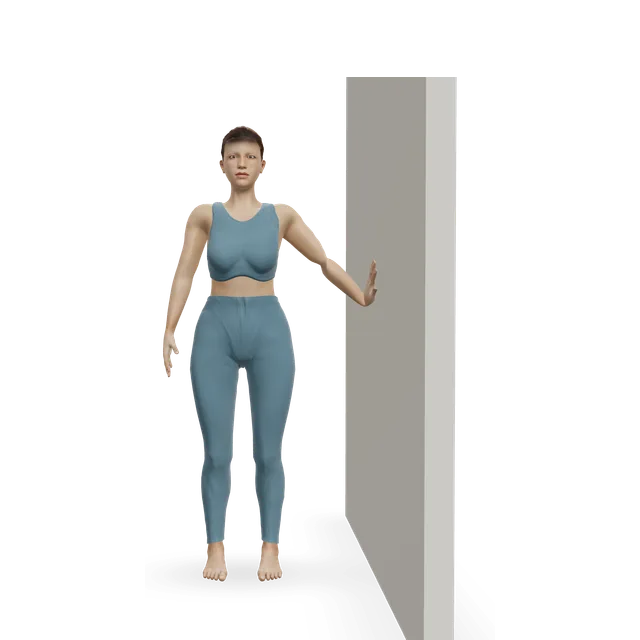
Easier: Isometric ankle eversion against a wall · Harder: Single-leg stand

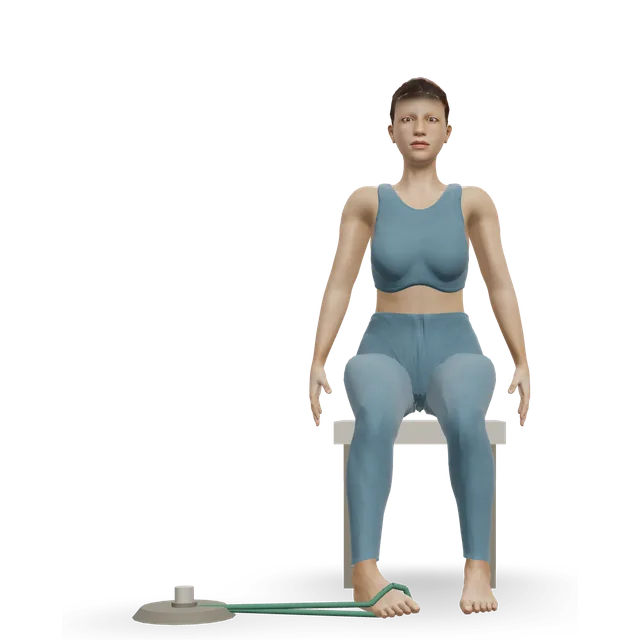
Band ankle inversion demonstrated on the 3D figure3D demoBand ankle inversionBeginner · A band + A bench or box

3 sets of 10 at 3 to 5 s a rep, then 4 sets and 3 sets of 10 in alternate weeks · The Hall 2018 trial's progression, borrowed

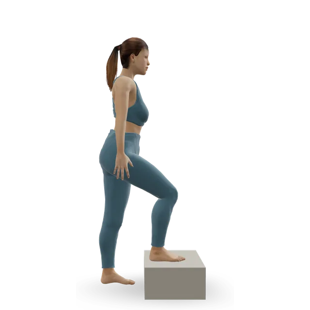
Harder: Single-leg calf raise from a step

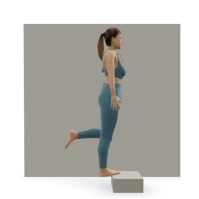
Single-leg calf raise from a step demonstrated on the 3D figure3D demoSingle-leg calf raise from a stepIntermediate · A step + A wall

2 sets of 10, building to 4 sets of 15 by week 6 · The trial's heel-raise progression, borrowed. Start on flat ground, or with slow calf raises on both feet if one leg is too hard

Easier: Slow calf raise · Harder: Single-leg pogo hops

Single-leg stand demonstrated on the 3D figure3D demoSingle-leg standBeginner · A wall

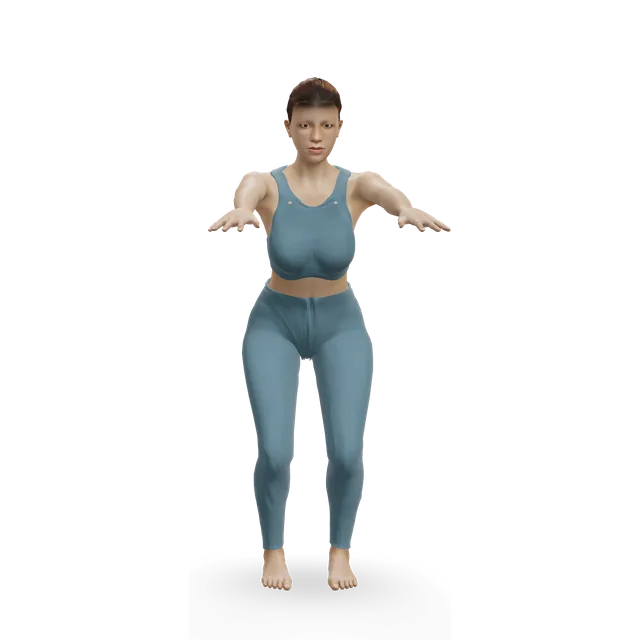
3 holds of 30 s, eyes open, then closed, then on a cushion · Our chronic ankle instability guide's levels

Easier: Tandem stance and heel-to-toe walk · Harder: Single-leg reach matrix

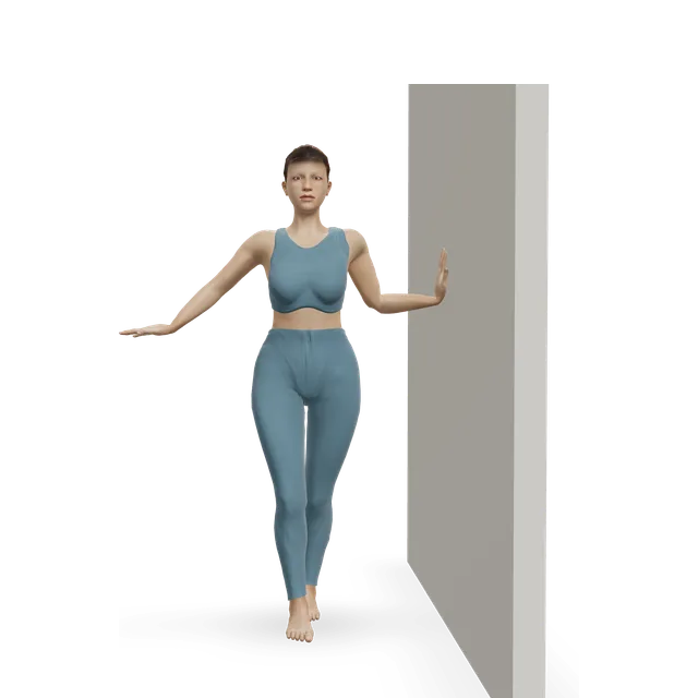
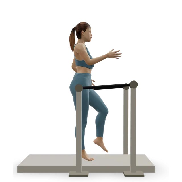
Tandem stance and heel-to-toe walk demonstrated on the 3D figure3D demoTandem stance and heel-to-toe walkBeginner · A wall

Hold 20 s with each foot forward, then 3 walks of 15 steps · Editorial dose, as in our sinus tarsi syndrome guide

Easier: Semi-tandem stance · Harder: Single-leg stand

Knee-to-wall ankle rock demonstrated on the 3D figure3D demoKnee-to-wall ankle rockBeginner · A wall

10 slow rocks on each side, daily, staying pinch-free · The move page's dose

Move on when: Hopping and landing on the sore foot is comfortable (no more than 3 out of 10 for teenagers, settling quickly), and a 30-second single-leg stand with your eyes closed is steady.

Follow along: a timer, a cue and the next move, one step at a time.
Stage 3

Back to sport

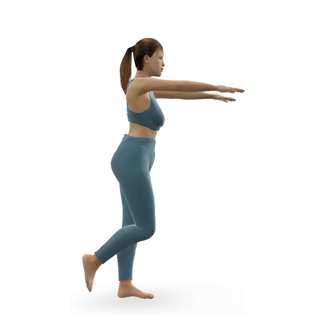
Hop-and-hold drills, then running on the flat, then cutting and uneven ground. Teenagers: pain during sport no more than 3 out of 10, settling quickly afterwards, and restart one activity or training session a week (NHS Lothian's Sever's disease rules, borrowed). Keep balance work going for at least 8 weeks, as in a trial in athletes after a sprain (borrowed), and use a brace or tape for sport if the ankle feels unstable.

Single-leg reach matrix demonstrated on the 3D figure3D demoSingle-leg reach matrixIntermediate · No equipment

3 rounds of 5 reaches in each direction, each leg · The move page's dose

Easier: Standing crossed-leg side reach · Harder: Single-leg Romanian deadlift

Ankle bounces (mini pogo) demonstrated on the 3D figure3D demoAnkle bounces (mini pogo)Beginner · No equipment

2 sets of 20, with quiet landings · Editorial dose, as in our Iselin's disease guide

Harder: Pogo hops

Single-leg hop and stick demonstrated on the 3D figure3D demoSingle-leg hop and stickIntermediate · No equipment

3 sets of 5 in each direction, each leg · Editorial dose; hopping to a steady hold, as in the Hall 2018 trial's balance programme

Easier: Standing broad jump with stuck landing · Harder: Alternate-leg bounding

Lateral cone hops demonstrated on the 3D figure3D demoLateral cone hopsIntermediate · Other / check instructions

3 sets of 10 side to side · Our chronic ankle instability guide's dose

Easier: Lateral bound (skater jump) · Harder: Single-leg cone hops

Treadmill jogging demonstrated on the 3D figure3D demoTreadmill joggingIntermediate · Cardio machine

Walk-jog intervals on the flat, then outdoors on even ground, then trails · Editorial

Easier: Treadmill walking · Harder: Sprint intervals

Move on when: Full training stays within the rule and settles by the next day. Still painful after about 6 months of good non-surgical care, or pain stops you walking comfortably or playing sport: ask your specialist about surgery.

Follow along: a timer, a cue and the next move, one step at a time.

What to change for now

Settling a flare (written steps)

From AAOS, two NHS services and the NHS growing pains page, with our suggestion marked.

  • 1. Take a break from high-impact sport, typically for 3 to 6 weeks. Swimming or cycling are fine if they don't hurt (our suggestion).
  • 2. Wear supportive trainers, with arch supports or insoles if advised. An ankle boot or a rocker-sole shoe can limit painful movement.
  • 3. If it's very painful, your clinician may use a walking boot or a weight-bearing below-knee cast for 4 to 6 weeks.
  • 4. Anti-inflammatory painkillers can help, and a steroid injection may give temporary relief. Follow the dose on the packet, and don't give aspirin to anyone under 16 unless a doctor prescribes it.
  • 5. If the muscles on the outside of the leg go into spasm, stop the activity and see your clinician: it's a protective response to pain.

Sources 1, 3, 2, 22

After coalition surgery, as your team allows (written steps)

From three NHS services and one series. Your own team's plan comes first.

  • 1. After removal of the bar: a non-weight-bearing cast for about 2 weeks, with the foot raised above the knee most of the time, and crutches.
  • 2. At about 2 to 3 weeks: a wound check, then often a walking boot for about 4 weeks, when physiotherapy may start.
  • 3. In one NHS podiatric surgery service, removal of a calcaneonavicular bar is followed by a below-knee cast for 4 to 6 weeks, then physiotherapy.
  • 4. After a fusion: a non-weight-bearing below-knee cast for up to 8 to 10 weeks in that service.
  • 5. Full activities including sport at 3 to 6 months in one NHS children's hospital's leaflet, and 18 weeks on average in one series of 78 people. Swelling and low-level discomfort can last 6 to 12 months; driving usually after about 6 weeks; a desk job after about 3 to 4 weeks.
  • 6. Once your team agrees, pick the plan up at stage 2.

Sources 2, 3, 6, 20

Positions to swap, and why

Habit or positionWhy it mattersInstead
Playing through pain above 3 out of 10 (teenagers)It's above the limit NHS Lothian uses for growing athletes, borrowed here.Cycling or swimming while it settles.Try: Stationary cycling
Forcing the foot to turn in or out against a strong bandThe joint is joined, so that movement isn't there to gain, and forcing it can bring on pain or spasm (our reasoning).Gentle holds first, then band work within the range you have.Try: Isometric ankle eversion against a wall
Hard stretches of the outer-leg muscles while they're in spasmThe spasm is a protective response to pain.Settle the foot (rest, and a boot or cast if advised) and see your clinician.
Thin, floppy shoes or flip-flops for sport and long walksArch supports, ankle boots and rocker soles limit painful movement.Supportive trainers, with insoles if advised.
Cutting sports and uneven ground before your balance is steadyThe stiff foot makes the ankle roll more.Balance work first, then flat ground, and a brace or tape for sport.Try: Single-leg stand
Trusting a normal X-ray when the signs fitX-rays miss many talocalcaneal coalitions.Ask whether a CT or MRI scan is needed.
Expecting surgery to give a normal footThe foot stays stiffer than normal after surgery.Realistic goals: less pain and better function.

Routines to follow

More moves for this area

Questions people ask

What is a tarsal coalition?

A bar of bone, cartilage or fibrous tissue joining two bones at the back or middle of the foot, which formed before birth. The commonest join the heel bone to the navicular or to the talus.

How common is tarsal coalition?

Estimates range from under 1% to about 13%, depending on how carefully it's looked for; scans found one in 6% of people having CT scans of both feet in one hospital. About half of people with one have it in both feet.

What are the symptoms of tarsal coalition?

A stiff flat foot, pain below the ankle or around the back of the foot with activity, ankle sprains that keep happening, difficulty on uneven ground, and sometimes a limp or spasm of the muscles on the outside of the leg. Many people never have symptoms.

Why does it start hurting in the teenage years?

The bar hardens into bone as a child grows, usually between 8 and 16, which makes the foot stiffer. Pain often starts after a sprain.

How is tarsal coalition diagnosed?

An examination shows a flat foot whose arch doesn't come back on tiptoe, and less movement at the back of the foot. X-rays show many coalitions, but a CT or MRI scan is often needed, especially for the talocalcaneal kind.

What helps without surgery?

A break from high-impact sport, insoles, a walking boot or a below-knee cast for 4 to 6 weeks, painkillers and physiotherapy. In one series, about half of non-surgical treatment courses relieved the pain, and casts, boots and supportive measures did equally well.

Do exercises help tarsal coalition?

No exercise separates the bones, and none has been tested for a coalition. Strength and balance work borrowed from ankle instability trials, where it improved balance and cut repeat sprains, can keep the ankle steadier.

How many people with a painful coalition need surgery?

It depends on the study. In one clinic series, surgery was avoided in 79% of calcaneonavicular and 62% of talocalcaneal coalitions, but in a US population study, 74% and 56% had surgery. Non-surgical care is always tried first, usually for at least 6 months.

Does surgery for tarsal coalition work?

Removing a calcaneonavicular bar succeeded in about 83% across 11 studies, and over 85% of people needed no further operation over about 9 years. The foot stays stiffer than normal, and about 1 in 10 bars grow back.

What's the difference between removal and fusion?

Removal keeps the joint moving and is usually tried first in young people without arthritis. Fusion stiffens the joints for good and is kept for large coalitions, arthritis or a failed removal. No trial has compared them.

How long is recovery after tarsal coalition surgery?

After removal, typically about 2 weeks in a non-weight-bearing cast and about 4 weeks in a walking boot with physiotherapy, with sport at 3 to 6 months, as your team allows. Swelling can take 6 to 12 months to settle.

Can adults have tarsal coalition?

Yes. Some people first get pain as adults, often after an injury or from arthritis in the stiff joints, and fusion is more often needed when there's arthritis.

Related exercise guides

Other pain guides

Sources

  1. American Academy of Orthopaedic Surgeons (OrthoInfo) · Tarsal coalition
  2. The Royal Orthopaedic Hospital NHS · Tarsal coalition
  3. Kent Community Health NHS · Tarsal coalitions: a guide for patients (podiatric surgery leaflet, 2025)
  4. Docquier 2019 · Tarsal coalition in children (review, Orthop Traumatol Surg Res)
  5. Park 2022 · Tarsal coalition on 839 CT scans of both feet (imaging study, Sci Rep)
  6. University Hospitals of Leicester NHS · Foot surgery for tarsal coalition: information for parents (2019, now past its review date)
  7. Jackson 2021 · Painful tarsal coalition in children in one US county over 52 years (population study, J Bone Joint Surg Am)
  8. Mascio 2026 · Talocalcaneal coalition: current concepts and management (narrative review, Life)
  9. Di Gennaro 2020 · Surgery or casting for a painful stiff flat foot with talocalcaneal coalition in 81 feet (comparative study, BMC Musculoskelet Disord)
  10. British Society for Children's Orthopaedic Surgery · Consensus statements on the management of flexible flatfeet in children
  11. Li 2026 · Ultrasound or X-ray for suspected talocalcaneal coalition in 86 children (diagnostic study, J Pediatr Orthop)
  12. Shirley 2018 · Non-surgical treatment for 50 painful tarsal coalitions (retrospective series, Cureus)
  13. NHS · Flat feet
  14. Hall 2018 · Balance and strength training for chronic ankle instability in 39 people (randomised trial, J Athl Train)
  15. Hupperets 2009 · A home balance programme after an ankle sprain in 522 athletes aged 12 to 70 (randomised trial, BMJ)
  16. Garg 2023 · Removing the bar in calcaneonavicular coalition: 11 studies, 394 feet (systematic review, Cureus)
  17. Khoshbin 2013 · Long-term results of coalition removal in 24 young people (cohort study, Foot Ankle Int)
  18. Khoshbin 2015 · Further operations after tarsal coalition removal in 304 people (population study, J Foot Ankle Surg)
  19. Williams 2025 · Tarsal coalition removal in children: 165 feet (cohort study, J Foot Ankle Surg)
  20. Saxena 2022 · Tarsal coalition removal: 97 operations in 78 people (retrospective study, J Orthop Surg Res)
  21. NHS Lothian · Sever's disease (Royal Hospital for Children and Young People booklet, 2023)
  22. NHS · Growing pains
  23. NHS · Ankle pain
  24. NHS · Sprains and strains
  25. NHS · DVT (deep vein thrombosis)
  26. NHS · Septic arthritis

Exercise education, not a diagnosis or treatment plan. It is written from the guidelines and studies listed above; review by a clinician is pending. If your pain follows an injury, is severe, is getting worse, or comes with any warning sign listed on this page, get it assessed. Health disclaimer.