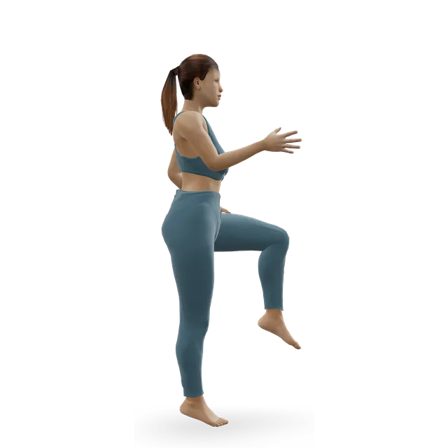
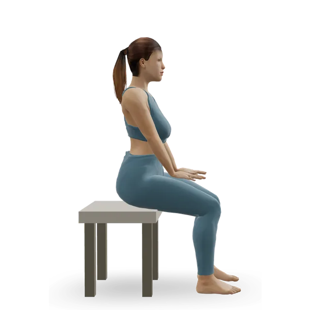
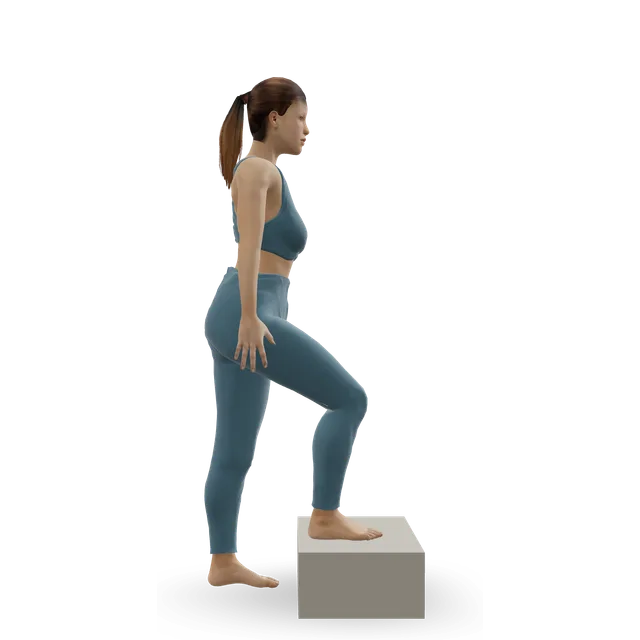
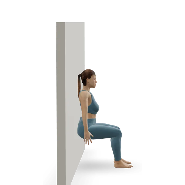
Pain guide · Ankle pain

Osteochondral lesion of the talus (talar dome lesion, ankle OCD): deep ankle pain after a sprain

An osteochondral lesion of the talus (OLT) is a damaged patch of the smooth cartilage and the bone beneath it on the top of the talus, the bone at the bottom of the ankle joint. It's also called a talar dome lesion, or ankle OCD (osteochondritis dissecans, which has nothing to do with obsessive-compulsive disorder). Most follow a sprain or a broken ankle, and the usual sign is deep ankle pain that lingers, sometimes with stiffness, clicking or catching, after the rest of the injury has settled. Teenagers get it too.

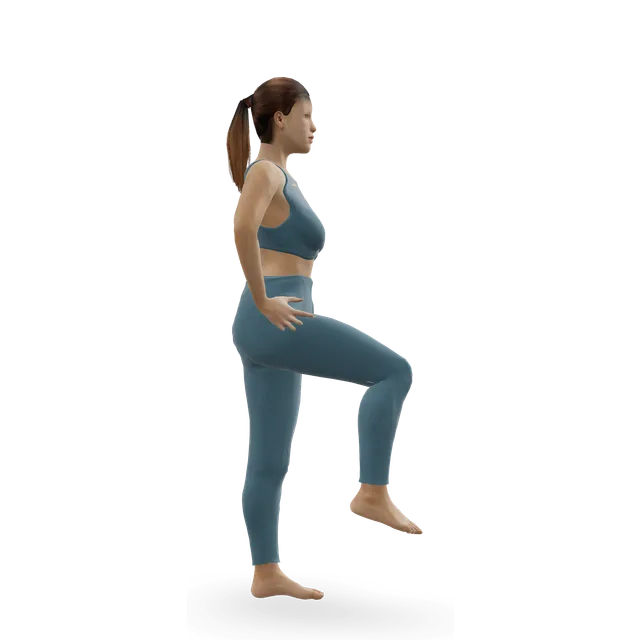
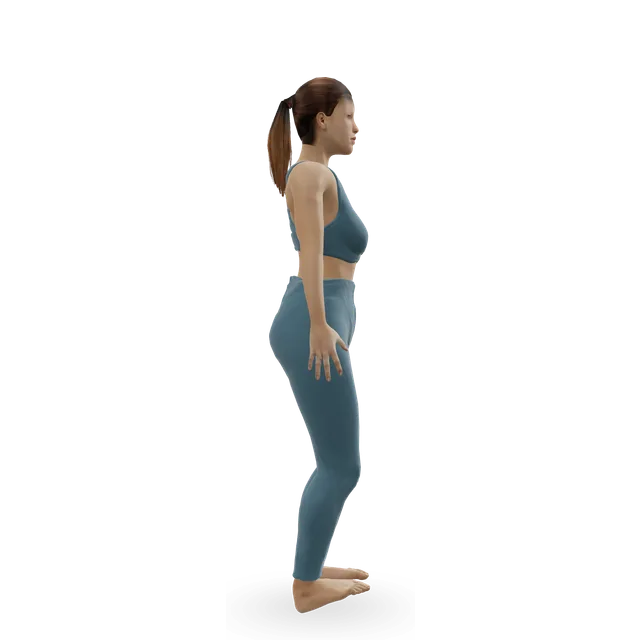
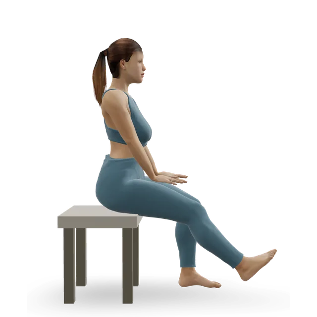
This guide is for teenagers and adults, and parents of teenagers. It explains why X-rays often miss it, what a magnetic resonance imaging (MRI) finding does and doesn't mean, how many settle with rest and rehab (a little under half in the reviews), when surgery such as microfracture is considered and how well it works, and a staged plan with 3D demos back to sport, as your team allows.

When to get help first

Exercise is for pain that has been checked or is clearly mild and settling.

Get urgent medical help if

  • Call 999 or go to A&E: after an injury, you heard a crack, the foot or ankle has changed shape or points at an odd angle, a bone is sticking out, there is a bad cut over it, you're in severe pain, or the toes look blue or white, feel numb or are cold to touch. These can mean a broken bone. Don't drive yourself.
  • Contact NHS 111: after an injury, severe ankle pain, feeling faint, dizzy or sick from the pain, a snap, grinding or popping noise at the time of the injury, the ankle or foot at an odd angle, or not being able to walk. These might be signs of a broken ankle or an infection.
  • Same day: a sudden, very painful, hot, swollen, red joint, often the big toe, especially with a high temperature, feeling shivery or being unable to eat. It may be gout, but it could be a joint infection (septic arthritis), which is urgent.
  • An ankle that suddenly locks and won't move, or swells up quickly and badly after an injury: get help the same day (an urgent GP appointment, NHS 111 or an urgent treatment centre). A loose piece of bone or cartilage can do this.
  • After surgery: redness, swelling, oozing or a smell from the wound, or a fever. Contact your surgical team the same day, or NHS 111 out of hours.
  • Same day: in a cast or boot, pain that is getting worse, toes that are numb or burning, a cast that feels too tight, toes that are swollen, blue or white, or a bad smell from under the cast.
  • Same day (GP or NHS 111): throbbing pain and swelling in one calf, with red or darker skin. It could be a blood clot (DVT). Watch for it after an ankle injury or time in a cast or boot.
  • Call 999: calf or leg pain or swelling with breathlessness or chest pain. It could be a clot that has reached the lungs.

See a doctor or physiotherapist if

  • Ankle pain or swelling still there 6 to 12 weeks after a sprain, or pain that is getting worse rather than better: see your GP. Deep pain inside the joint that lingers after a sprain or a broken ankle can be a damaged patch of cartilage and bone on the talus, and X-rays often miss it.
  • Pain, clicking, locking or swelling that carries on despite a good rehab programme. Cartilage, tendon and other problems are common alongside an unstable ankle and need assessing.
  • See a GP if ankle pain is stopping you doing normal activities, is getting worse or keeps coming back, or hasn't improved after treating it at home for 2 weeks.
  • A child or teenager with ankle pain that comes with sport and keeps coming back, or a deep ache that's hard to pin down: see a GP or physiotherapist. Ankle OCD affects teenagers, and in growing children specialists usually try protection before surgery.
  • After a broken ankle has healed, deep pain or swelling with activity that isn't settling: see your GP or fracture clinic. Cartilage injuries are common with ankle fractures (45% in one review) and are worth asking about.
  • After surgery: pain or swelling that comes back or builds when you increase walking, running or sport, or catching or locking: drop back a stage and contact your team. One NHS trust suggests a referral back to the surgeon at about 3 months if pain is the main barrier.
  • An ankle that keeps giving way months after a sprain.
  • Tingling or loss of feeling in the foot, or a foot that feels weak.

Understand it

What it is

The talus is the bone at the bottom of the ankle joint, and its rounded top (the talar dome) is covered in smooth joint cartilage. An osteochondral lesion is damage to that cartilage and the bone just beneath it. Osteochondral lesion of the talus, shortened to OLT, is the term a 2019 international consensus agreed; older names include osteochondritis dissecans of the talus, osteochondral defect and talar dome lesion.

NHS England's summary says these lesions can cause pain, clicking, grinding or instability of the joint, and that cartilage, which has no blood or nerve supply, has a limited ability to repair itself.

Sources 1, 2

What causes it

Most people with a lesion have had an ankle injury, often a sporting one. Lesions are thought to follow damage to the joint surface or its blood supply after an injury, or to arise without a clear injury, which is called osteochondritis dissecans.

Sources 1, 3

How often it follows an injury: it depends how hard you lookMixed evidence

There's no single figure, because it depends on the injury, the scan and when it's done.

  • NHS England's summary, citing a 2015 review: in 6.5% of new ankle sprains, 20.5% of sprains looked at later, and 57% of ankles with long-term disability.
  • On a detailed (3-tesla) MRI within days of an acute ankle ligament injury in athletes: a lesion on the talus in 24 of 171 injuries (14%), and 85% of all the lesions found were in the cartilage only.
  • In a review of 20 studies of 1,707 broken ankles: 45% had a cartilage or bone-and-cartilage lesion when the joint was examined straight after the injury, and 43% of those lesions were on the talus.
  • An editorial's estimate: up to 70% of sprains and fractures may lead to these lesions. That's higher than the measured figures above.

Sources 1, 4, 5, 3

Who gets itEmerging evidence

Mostly young, active people: NHS England's summary says most lesions occur between the second and fourth decades of life, at an average age of 27, and twice as often in men.

Teenagers get it too. In a US health system with over a million children, ankle OCD was diagnosed in 4.6 per 100,000 children aged 6 to 19 each year: 6.8 per 100,000 at 12 to 19 against 1.1 at 6 to 11, none under 6, and girls 1.6 times as often as boys. 72% were on the inner side of the talus, and none were in both ankles.

Sources 1, 6

The signs

Deep ankle pain that lingers after a sprain or a fracture, or comes on gradually, with stiffness, swelling after activity, less activity than before, and sometimes clicking, catching, grinding or a feeling the ankle might give way.

NHS England's summary calls these lesions one of the most important causes of pain that lingers after a sprain, and says the diagnosis is often delayed. A 2024 editorial says a lesion is easily mistaken for a simple sprain if scans are unclear or not done.

Sources 1, 3, 7

When to suspect it after a sprain

NHS ankle sprain leaflets say most sprains improve within 6 to 12 weeks, though some pain and swelling can last 3 to 6 months, and that pain or swelling still there after 6 to 12 weeks is a reason to contact your GP. The NHS says to see a GP for ankle pain that hasn't improved after 2 weeks of looking after it at home, is getting worse, or keeps coming back.

Sources 8, 9, 10

What scans show, and what a finding does and doesn't meanEmerging evidence

X-rays can show larger lesions but miss many. In 103 people with long-standing ankle pain, computed tomography (CT) and MRI found or ruled out lesions better than the history, examination and X-rays, and the two scans did equally well.

A lesion on a scan isn't automatically the cause of pain. In 108 MRI scans of ankles with no symptoms on the outer side, 16 (15%) showed a lesion on the talus. Bone bruises (swelling inside the bone on MRI) are common after sprains, and a review found they usually fade within 6 to 12 months without any specific treatment.

Your specialist decides with the symptoms, the examination and the scan together, including the lesion's size, which affects how well surgery works.

Sources 11, 12, 13, 14, 15

How it usually goes without surgeryEmerging evidence

In a 2023 review of 30 studies (868 patients) treated without surgery, the 16 studies that could be pooled gave a success rate of 45%. Arthritis progressed on X-ray in 9%, and the lesion looked unchanged on MRI in 84%.

Lesions causing few symptoms didn't get worse in two series: in 48 people followed for over 4 years, 86% of ankles were pain-free or less painful and MRI showed no real progression; and in 142 people who had no immobilisation or activity limits, average pain fell from 3.8 to 0.9 out of 10 over about 6 years, with no arthritis progression, and only 9 reported limits on sport. In children, 44% of 192 lesions did well without surgery. These are mostly small, older series of mixed quality, and people whose pain carried on went on to surgery, which is why the success rates look modest.

Sources 16, 17, 18, 19

What helps, and how strong the evidence is

Non-surgical care first for mostEmerging evidence

Non-surgical care is the first treatment for most lesions: less physical activity and weight-bearing, physiotherapy and insoles, with about 50% success in the studies one review summarised, and 45% in the pooled review above.

In children, the published protocols used about 6 weeks of immobilisation (3 to 8), in a cast in some studies, then about 5 months (4 to 6) of restricted weight-bearing and sport, and 44% of 192 lesions did well. Lesions causing few symptoms didn't worsen over 4 to 6 years whether or not activity was restricted. No trial has compared different non-surgical approaches.

Sources 7, 16, 19, 17, 18

Rehab: no trials, so NHS protocols set the shapeEmerging evidence

No trial has tested exercise for these lesions. NHS protocols after surgery set a shape that non-surgical rehab can borrow: movement first, then strength (heel raises from two legs to one), balance and reach, then impact only once criteria are met.

One NHS trust's criteria before impact are full ankle movement without pain or swelling, an even forwards and backwards reach on each leg on the star excursion balance test, 10 leg presses at body weight, 10 good single-leg squats and 25 pain-free single-leg heel raises, with sport-specific training 4 to 6 weeks after running starts. A review of 227 articles found rehab plans after surgery vary widely and are mostly based on time rather than criteria. The plan below uses the criteria as its stage gates.

Two pieces are borrowed from ankle instability trials and labelled: the heel-raise progression off a step comes from a trial in 39 people with unstable ankles, and the advice to keep balance work going comes from a trial in 522 athletes, where an 8-week home balance programme after a sprain cut repeat sprains from 33% to 22% over a year.

Sources 20, 21, 22, 23, 24

Surgery: marrow stimulation (microfracture)Emerging evidence

For lesions that keep causing symptoms despite non-surgical care, NHS England describes the first operations as clearing the damaged area, with or without drilling or microfracture of the bone underneath, or bone grafting, and more complex resurfacing only if those fail. Marrow stimulation means clearing the damaged area and making small holes in the bone so that healing cells fill the defect with repair tissue.

In adults with first-time lesions, a review of 52 studies found pooled success of 82% in 317 ankles after marrow stimulation, and no operation better than another; an earlier review found 85% success across 18 treatment groups. In 50 people followed for 8 to 20 years (12 on average), 78% had a good or excellent result, 94% had gone back to work and 88% to sport, and arthritis had progressed one grade in a third.

The repair tissue is fibrocartilage, which bears weight less well than the original cartilage and may wear over time, a review in children explains. The trial evidence is thin: a Cochrane review found only one small trial, of 15 people.

Sources 1, 7, 25, 26, 27, 19, 28

Lesion size, and other operationsEmerging evidence

Larger lesions do less well with marrow stimulation. In 120 ankles, lesions of 150 mm² or more failed in 80% (20 of 25), and a review of 25 studies suggests marrow stimulation suits lesions under about 107 mm² or 10 mm across. Larger lesions, or ones that don't heal, may need grafts of bone and cartilage or other resurfacing.

In children, a review found success in 77% after marrow stimulation, 79% after fixing a loose piece back, 95% after drilling through the bone from below (in 22 lesions) and 67% after grafts (in 9), from small series of mixed quality.

Sources 15, 14, 1, 19

Return to sport and work, and complicationsEmerging evidence

After marrow stimulation, 88% went back to sport at any level and 79% to their previous level, usually 13 to 26 weeks after surgery, across 61 studies of 2,347 lesions. Across 18 studies, 93% went back to work, after 2 to 11 weeks. The studies were of low quality.

Complications happened in about 1 in 20 operations, across 6,962 operated lesions; nerve injury was the commonest, and none was life-threatening.

Sources 29, 30, 31

Weight-bearing after microfracture, and microfracture with ligament repairEmerging evidence

In a randomised trial of 81 ankles with small to mid-sized lesions, putting weight through the foot from 2 weeks gave the same results at about 3 years as 6 weeks without weight. NHS protocols differ: one usually allows weight as tolerated from the start, another touching or partial weight at first, building to full weight from about week 6. So the guide says: as your team allows.

In a trial of 71 people with an unstable ankle and a small cartilage lesion, adding microfracture to ligament reconstruction made no difference to function, pain or complications at 2 years, and recovery was slower.

Sources 32, 20, 21, 33

Cell implants and injections: limited evidenceMixed evidence

NHS England doesn't routinely fund autologous chondrocyte implantation (growing your own cartilage cells and implanting them) for these lesions, because the evidence isn't strong enough. A review found only limited comparative evidence for adding biological treatments to marrow stimulation. In a meta-analysis of 4 trials of platelet-rich plasma (PRP), the pooled gain in ankle function was uncertain (its range included no effect) and results varied widely, though the authors were positive. They're add-ons with uncertain benefit, not cures.

Sources 1, 34, 35
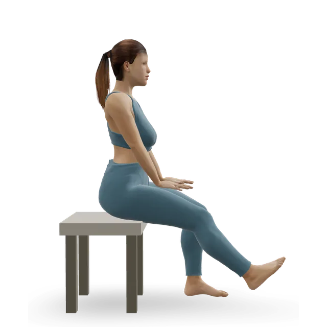
How much pain is OK while you exercise?

No pain in the ankle joint, no swelling, no catching (borrowed from our knee OCD guide, with an NHS trust's criteria)

No study has tested a pain rule for these lesions. This is our knee OCD guide's rule (the same as our elbow OCD guide's), borrowed because the joint surface is healing. If your specialist or physiotherapist has given you a programme, follow theirs.

Weight-bearing, impact and sport: no pain in the ankle joint during, straight after or the next morning. Strength and balance work: no pain in the joint; a working ache in the calf or shin muscles that settles within the hour is fine (editorial).

Swelling, catching, locking or giving way are never part of the rule: stop that activity and drop back a stage, and if they keep happening, contact your team. The NHS protocols set no pain, no swelling and no catching or locking as goals before running and sport.

It's stricter than our general foot and ankle rule, because pain in the joint, not the muscles, is what tells you the healing surface is being overloaded (our reasoning, as in our knee OCD guide).

Sources 20, 21

The exercise plan: 3 stages

Start at the first stage. Each move opens a full guide with a 3D demo.

Stage 1

Protect the joint surface (your specialist sets how long)

No running, jumping or sport, and a boot or cast if your specialist uses one; in children's protocols, about 6 weeks (3 to 8) of protection, then about 5 months of restricted sport. Keep the ankle moving gently in a pain-free range, and the rest of you fit, with cycling or swimming only if allowed. After surgery, the written steps below follow the NHS protocols' phases, as your team allows.

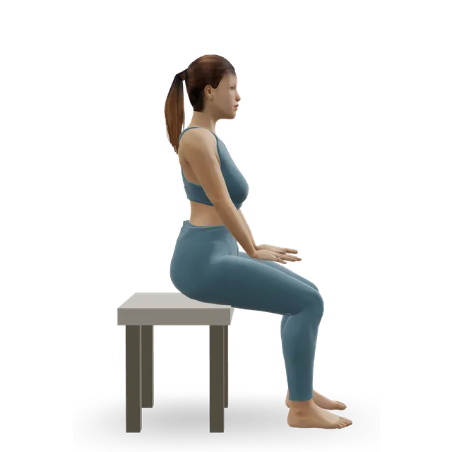
Ankle CARs (controlled articular rotations) demonstrated on the 3D figure3D demoAnkle CARs (controlled articular rotations)Beginner · No equipment

5 slow circles each way, morning and evening, in the pain-free range · The move page's dose

Easier: Ankle circles · Harder: Toes-tucked kneeling (toe squat)

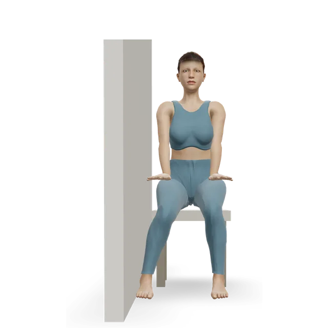
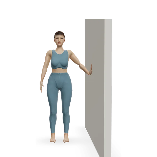
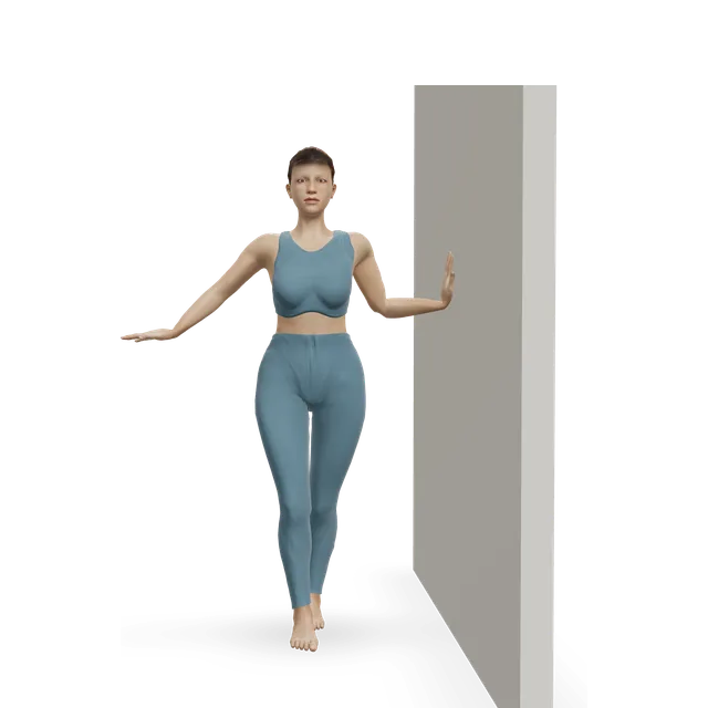
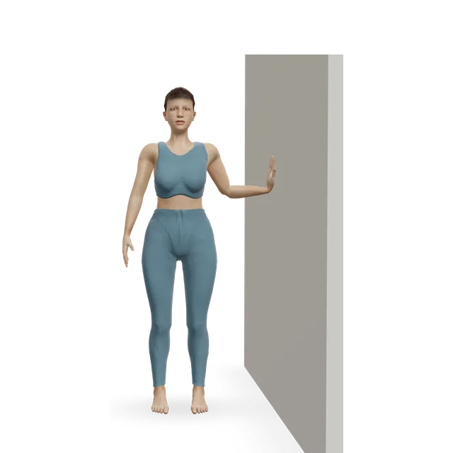
Isometric ankle eversion against a wall demonstrated on the 3D figure3D demoIsometric ankle eversion against a wallBeginner · A wall + A bench or box

8 to 10 holds of about 6 s, 2 to 3 times a day · Press gently. Our peroneal tendinopathy guide's dose

Harder: Band ankle eversion

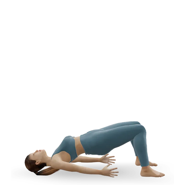
Glute bridge demonstrated on the 3D figure3D demoGlute bridgeBeginner · No equipment

2 sets of 10 · Editorial dose, as in our knee OCD guide

Easier: Pelvic tilt into bridge · Harder: Single-leg glute bridge

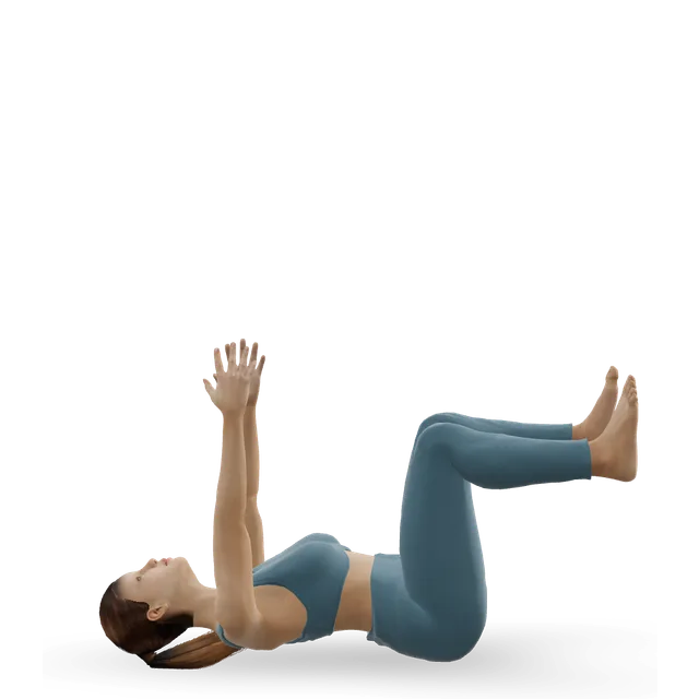
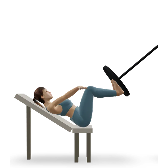
Dead bug demonstrated on the 3D figure3D demoDead bugBeginner · No equipment

2 sets of 6 to 8 on each side, slowly · Editorial dose, as in our knee and elbow OCD guides

Easier: Dead bug heel taps · Harder: Forearm plank

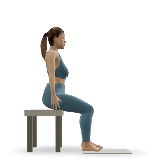
Stationary cycling demonstrated on the 3D figure3D demoStationary cyclingBeginner · Cardio machine

15 to 30 min, if your team allows · Editorial dose

Easier: Recumbent exercise bike · Harder: Air bike intervals

Move on when: Everyday walking is pain-free with no swelling, and your specialist clears strengthening.

Follow along: a timer, a cue and the next move, one step at a time.
Stage 2

Strength and balance without impact

Two or three sessions a week: heel raises from two legs to one, balance and reach, and leg strength, as in the NHS protocols' 6-to-12-week phase after surgery, with low-impact fitness. Still no running or jumping.

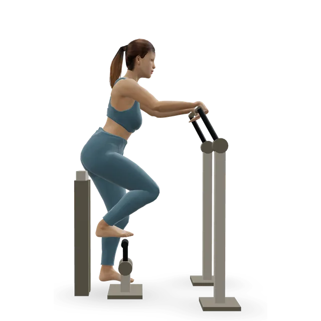
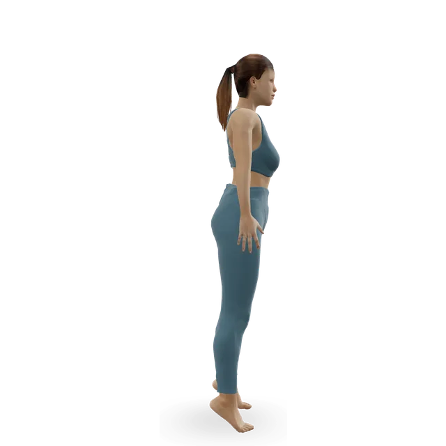
Single-leg calf raise from a step demonstrated on the 3D figure3D demoSingle-leg calf raise from a stepIntermediate · A step + A wall

Slow calf raises on both feet, then one leg on flat ground, building towards 25 pain-free single-leg heel raises; then off a step, 2 sets of 10 building to 4 sets of 15 · The 25 heel raises are an NHS trust's criterion; the step progression is borrowed from an ankle instability trial (Hall 2018)

Easier: Slow calf raise · Harder: Single-leg pogo hops

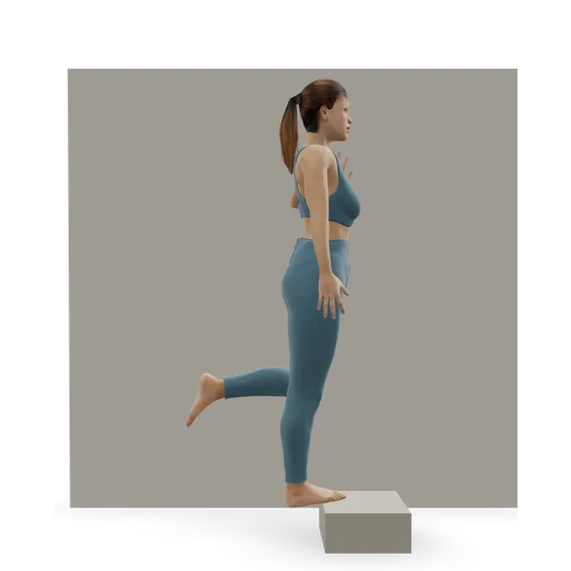
Single-leg stand demonstrated on the 3D figure3D demoSingle-leg standBeginner · A wall

3 holds of 30 s, eyes closed, then on a cushion · Our chronic ankle instability guide's levels

Easier: Tandem stance and heel-to-toe walk · Harder: Single-leg reach matrix

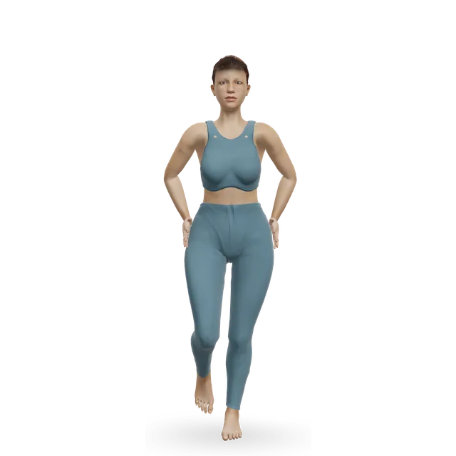
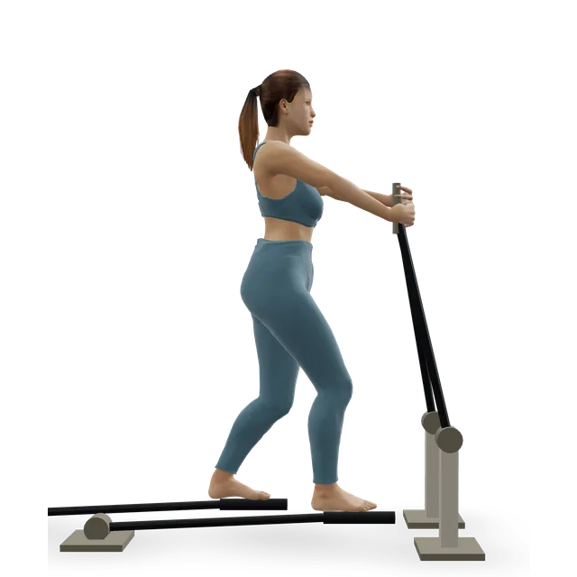
Single-leg reach matrix demonstrated on the 3D figure3D demoSingle-leg reach matrixIntermediate · No equipment

3 rounds of 5 reaches in each direction, each leg · The move page's dose, working towards an even reach on both legs (an NHS trust's criterion)

Easier: Standing crossed-leg side reach · Harder: Single-leg Romanian deadlift

Leg press demonstrated on the 3D figure3D demoLeg pressBeginner · Strength machine

3 sets of 10, building towards 10 reps at your body weight · The target is an NHS trust's criterion; the sets are editorial

Harder: Bodyweight squat

Knee-to-wall ankle rock demonstrated on the 3D figure3D demoKnee-to-wall ankle rockBeginner · A wall

10 slow rocks on each side, daily, with no pinch at the front of the ankle · The move page's dose

Elliptical trainer demonstrated on the 3D figure3D demoElliptical trainerBeginner · Cardio machine

10 to 20 minutes at a moderate effort, building towards 30, if allowed · The move page's dose

Harder: Stair-stepper machine

Move on when: You meet the NHS trust's criteria for impact: full ankle movement without pain or swelling, an even reach on both legs, 10 leg presses at body weight, 10 good single-leg squats and 25 pain-free single-leg heel raises, and your specialist clears impact (often from about 12 weeks after surgery in that protocol).

Follow along: a timer, a cue and the next move, one step at a time.
Stage 3

Impact and return to sport (as your specialist allows)

A walk-to-jog programme, then sport-specific training about 4 to 6 weeks after running starts (one NHS trust's protocol); another protocol starts jogging from month 2, agility and sport drills from month 3, and sport from month 4 when strength, balance and power allow. Hop on alternate days at first (editorial). It can take up to a year to judge the result of surgery.

Treadmill jogging demonstrated on the 3D figure3D demoTreadmill joggingIntermediate · Cardio machine

Walk-jog intervals on the flat, building time before speed · Editorial

Easier: Treadmill walking · Harder: Sprint intervals

Ankle bounces (mini pogo) demonstrated on the 3D figure3D demoAnkle bounces (mini pogo)Beginner · No equipment

2 sets of 20, with quiet landings · Editorial dose

Harder: Pogo hops

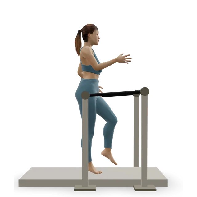
Pogo hops demonstrated on the 3D figure3D demoPogo hopsIntermediate · No equipment

2 to 3 short sets of 10 · Editorial dose, as in our knee OCD guide

Easier: Ankle bounces (mini pogo) · Harder: Single-leg pogo hops

Lateral cone hops demonstrated on the 3D figure3D demoLateral cone hopsIntermediate · Other / check instructions

3 sets of 10 side to side · Our chronic ankle instability guide's dose

Easier: Lateral bound (skater jump) · Harder: Single-leg cone hops

Shuttle runs demonstrated on the 3D figure3D demoShuttle runsAdvanced · No equipment

4 to 6 runs, wide turns first · Editorial dose

Easier: Treadmill jogging

Move on when: Full training causes no pain in the ankle joint, no swelling and no catching or locking for 2 weeks. Keep a balance warm-up going, especially if the ankle has been unstable: in a trial in athletes after a sprain, an 8-week home balance programme cut repeat sprains.

Follow along: a timer, a cue and the next move, one step at a time.

What to change for now

Protection without surgery (written steps)

From a review in children, another review and one series, with our suggestions marked.

  • 1. Your specialist sets how much protection you need. In children, published protocols used a boot or cast for about 6 weeks (3 to 8), then about 5 months of restricted weight-bearing and sport. Adults are usually asked to cut back activity and weight-bearing, with physiotherapy and sometimes insoles.
  • 2. Some people with mild symptoms do well with no boot and no activity limits: follow the advice you're given (our suggestion).
  • 3. Keep fit with upper-body and trunk work, and with cycling or swimming if allowed (editorial, as in our knee OCD guide).
  • 4. Follow-up scans are the specialist's call. A lesion often looks the same on MRI even when the symptoms settle, so judge progress by your symptoms and the stage gates.

Sources 19, 7, 18, 16

After ankle arthroscopy and microfracture, as your team allows (written steps)

From the Derby and Burton and the Robert Jones and Agnes Hunt NHS protocols, and a trial. Your own team's plan comes first.

  • 1. Weeks 0 to 2: weight-bearing as your surgeon advises. One NHS trust usually allows weight as tolerated, with crutches if they help; another starts with touching or partial weight. Keep the foot raised, with about 10 minutes on your feet in each hour at first. Gentle ankle movement without weight, and thigh, hip and circulation exercises. A physiotherapy appointment within 2 weeks.
  • 2. Weeks 2 to 6: wean off the crutches and walk normally; ankle movement and strength, first without weight and then with more weight as pain allows; a light exercise bike. From about week 3 in one protocol, mini-squats and low step-ups with part of your weight. This is stage 1 moving into stage 2.
  • 3. Weeks 6 to 12: a clinic review at about 6 to 8 weeks; full movement, balance work and heel raises from two legs to one; full weight-bearing from about week 6 in one protocol. This is stage 2.
  • 4. From about 12 weeks in one protocol: gentle impact and a walk-to-jog programme once the stage gates are met. The other starts straight-line jogging from month 2, agility and sport drills from month 3, and sport from month 4 onwards when strength, flexibility, balance and power allow. Your team decides which applies to you. This is stage 3.
  • 5. In a trial of 81 ankles with small to mid-sized lesions, putting weight through the foot from 2 weeks did as well as waiting 6 weeks, but follow your own team's plan.

Sources 20, 21, 32

Positions to swap, and why

Habit or positionWhy it mattersInstead
Bouncing, running, jumping or sport while the lesion is being protectedImpact loads the healing joint surface, and protocols restrict sport for months.Cycling or swimming, if allowed.Try: Stationary cycling
Deep, loaded ankle bending into a pinch or joint painPain in the joint is the stop sign in this guide's rule (editorial).Gentle knee-to-wall rocks in the pain-free range.Try: Knee-to-wall ankle rock
Going back to sport by the calendarReturn depends on strength, balance and no pain or swelling, not the date.The NHS trust's criteria before impact.Try: Single-leg reach matrix
Putting more or less weight through the foot than your team allows after surgeryProtocols differ, and your team plans for your lesion.Follow your team's plan.
Assuming a spot on the MRI means surgeryLesions show on 15% of scans of ankles without outer-ankle symptoms, and many settle.Decisions based on your symptoms, the examination and the scan together.
Paying for injections to regrow cartilageThe evidence is limited and mixed.Discuss the options with your surgeon.
Dropping balance work after an unstable ankleLesions often go with instability, and balance training cut repeat sprains in a trial.Keep a balance warm-up going.Try: Single-leg stand

Routines to follow

More moves for this area

Questions people ask

What is an osteochondral lesion of the talus?

A damaged patch of the cartilage and the bone beneath it on the top of the talus, the bone at the bottom of the ankle joint. It's also called a talar dome lesion or ankle OCD.

What causes it?

Most follow an injury such as an ankle sprain or a broken ankle, and some come on without a clear injury. How often depends on how carefully it's looked for: NHS England's summary gives 6.5% of new sprains, a detailed MRI study found one on the talus in 14% of athletes' acute ligament injuries, and a review found cartilage injuries in 45% of broken ankles.

What are the symptoms?

Deep ankle pain that lingers after a sprain or a fracture, stiffness, swelling after activity, and sometimes clicking, catching, grinding or a feeling the ankle might give way. Pain or swelling still there 6 to 12 weeks after a sprain needs checking.

Why didn't my X-ray show it?

X-rays miss many of these lesions. MRI and CT scans find them better, and equally well, which is why lingering pain after a sprain is sometimes scanned.

What does it mean if my MRI shows one?

It depends on your symptoms: lesions turn up on some scans of ankles that don't hurt there, and bone bruises after sprains usually fade within 6 to 12 months. Your specialist weighs the scan with your symptoms and examination.

Will it heal without surgery?

Often: about 45% of people in one review and 44% of children in another did well with non-surgical care, and lesions causing few symptoms didn't get worse over several years. Those whose pain carries on are usually offered surgery.

How long do I need off sport?

Usually months: children's protocols used about 6 weeks in a boot or cast, then about 5 months of restricted sport. Impact comes back when strength and balance tests are met, not by the calendar.

When is surgery needed?

When the ankle keeps hurting despite good non-surgical care. The usual operation clears the damaged area and makes small holes in the bone so new repair tissue grows (marrow stimulation, or microfracture).

How well does surgery work?

Marrow stimulation was successful in 82% of 317 ankles in one review, and results held at an average of 12 years in one series. Larger lesions do less well, and other operations are used for them.

When can I return to sport after surgery?

Usually 13 to 26 weeks after marrow stimulation in the studies, though your team sets your timeline: 88% went back to sport at any level and 79% to their previous level. Most people return to work within 2 to 11 weeks.

Will I get arthritis?

Some people do: arthritis had progressed one grade in a third of people about 12 years after surgery in one series, and in 9% of those treated without surgery in a review. Many still had good function.

Can children and teenagers get it?

Yes, mostly teenagers, and girls more often than boys. Specialists usually try protection first (a review reports an international consensus that children's ankle cartilage lesions may all be managed without surgery at first), and about 4 in 10 children did well that way, much as in adults.

Related exercise guides

Other pain guides

Sources

  1. NHS England · Autologous chondrocyte implantation for osteochondral lesions of the talus (clinical commissioning policy, 2016)
  2. Murawski 2022 · Terminology for osteochondral lesions of the ankle (international consensus, J ISAKOS)
  3. Mangwani 2024 · Osteochondral lesions of the talus (editorial, Bone Joint Res)
  4. Baltes 2025 · Cartilage lesions on MRI in 171 athletes' acute ankle ligament injuries (cohort study, Am J Sports Med)
  5. Martijn 2021 · Cartilage lesions in 1,707 ankle fractures: 20 studies (systematic review, Knee Surg Sports Traumatol Arthrosc)
  6. Kessler 2014 · Ankle osteochondritis dissecans in children and teenagers in a US health system (population study, Am J Sports Med)
  7. Anwander 2022 · Evidence for operations on talus lesions (systematic review, EFORT Open Rev)
  8. King's College Hospital NHS · Ankle sprain (patient leaflet, 2021, now past its review date)
  9. St George's University Hospitals NHS · Ankle sprains (physiotherapy leaflet, 2025)
  10. NHS · Ankle pain
  11. Verhagen 2005 · MRI, CT and examination for talus lesions in 103 people with ankle pain (prospective diagnostic study, J Bone Joint Surg Br)
  12. Galli 2014 · Talus lesions on MRI in 108 ankles without outer-ankle symptoms (imaging review, J Foot Ankle Surg)
  13. Longo 2013 · Bone bruises after acute ankle ligament injury (systematic review, Knee Surg Sports Traumatol Arthrosc)
  14. Ramponi 2017 · Lesion size and results of marrow stimulation: 25 studies, 1,868 ankles (systematic review, Am J Sports Med)
  15. Choi 2009 · Is there a critical size for poor results in talus lesions? 120 ankles (case series, Am J Sports Med)
  16. Buck 2023 · Non-surgical care for talus lesions: 30 studies, 868 patients (systematic review, Knee Surg Sports Traumatol Arthrosc)
  17. Klammer 2015 · Talus lesions treated without surgery in 48 people over 4 years or more (case series, Foot Ankle Int)
  18. Seo 2018 · Few people with talus lesions need surgery: 142 treated without it (case series, Acta Orthop)
  19. Dahmen 2022 · Treating talus lesions in growing children: 20 studies, 381 lesions (systematic review, J Pediatr Orthop)
  20. University Hospitals of Derby and Burton NHS · Ankle protocols: ankle debridement and microfracture (physiotherapy page)
  21. The Robert Jones and Agnes Hunt Orthopaedic Hospital NHS · Rehabilitation guide following microfracture to the ankle
  22. Buck 2024 · Rehabilitation protocols after surgery for talus lesions vary widely: 227 articles (systematic review, Knee Surg Sports Traumatol Arthrosc)
  23. Hall 2018 · Balance and strength training for chronic ankle instability in 39 people (randomised trial, J Athl Train)
  24. Hupperets 2009 · A home balance programme after an ankle sprain in 522 athletes aged 12 to 70 (randomised trial, BMJ)
  25. Dahmen 2018 · No superior treatment for first-time talus lesions in adults: 52 studies (systematic review, Knee Surg Sports Traumatol Arthrosc)
  26. Zengerink 2010 · Treatment of talus lesions: 52 studies (systematic review, Knee Surg Sports Traumatol Arthrosc)
  27. van Bergen 2013 · Keyhole marrow stimulation for talus lesions in 50 people, 8 to 20 years on (case series, J Bone Joint Surg Am)
  28. Loveday 2010 · Interventions for talus lesions in adults (Cochrane review)
  29. Steman 2019 · Return to sport after surgery for talus lesions: 61 studies, 2,347 lesions (systematic review, Orthop J Sports Med)
  30. Emanuel 2026 · Return to work after surgery for talus lesions: 18 studies (systematic review and meta-analysis, Cartilage)
  31. Hollander 2023 · Complications of surgery for talus lesions: 6,962 lesions (systematic review and meta-analysis, Cartilage)
  32. Lee 2012 · Early or delayed weight-bearing after microfracture in 81 ankles (randomised trial, Am J Sports Med)
  33. Figa Barrios 2025 · Adding microfracture to ligament repair for small talus lesions in 71 people (randomised trial, J Orthop Traumatol)
  34. Seow 2022 · Limited evidence for biologic add-ons to marrow stimulation for talus lesions (systematic review, Knee Surg Sports Traumatol Arthrosc)
  35. Peng 2023 · Platelet-rich plasma for talus cartilage repair: 10 studies, 4 trials (systematic review and meta-analysis, BMC Musculoskelet Disord)
  36. NHS · Sprains and strains
  37. NHS · DVT (deep vein thrombosis)
  38. NHS · Septic arthritis

Exercise education, not a diagnosis or treatment plan. It is written from the guidelines and studies listed above; review by a clinician is pending. If your pain follows an injury, is severe, is getting worse, or comes with any warning sign listed on this page, get it assessed. Health disclaimer.