Pain guide · Foot pain

Broken heel (calcaneus fracture): weight limits, surgery or not, and walking again

A broken heel bone (calcaneus fracture) usually comes from a fall from a height, landing hard on the feet, or from a car crash. The bone heals in weeks, but swelling lasts months, and after a bad break recovery can take a year or two. The same fall can break a bone in the back or the other heel, so those need checking too.

Your fracture clinic decides whether you need surgery and how much weight the foot can take, and plans differ. This guide covers the warning signs, what the UK Heel Fracture Trial found about surgery, why you may be off the foot for weeks, managing swelling and stairs, and a staged plan back to walking, work and sport, with a 3D demo of each move.

When to get help first

Exercise is for pain that has been checked or is clearly mild and settling.

Get urgent medical help if

  • Call 999 or go to A&E: after an injury, you heard a crack, the foot or ankle has changed shape or points at an odd angle, a bone is sticking out, there is a bad cut over it, you're in severe pain, or the toes look blue or white, feel numb or are cold to touch. These can mean a broken bone. Don't drive yourself.
  • If you think you might have broken your heel bone, don't try to treat it yourself. Contact NHS 111 if you have severe heel pain after an injury; you feel faint, dizzy or sick from the pain; your ankle or foot has changed shape or is at an odd angle; you heard a snap, grinding or popping noise at the time; you can't walk, walk on tiptoe or walk up the stairs; or you have swelling and bruising in your calf and ankle. These might be signs of a broken heel bone or a broken ankle.
  • Call 999 or go to A&E: back pain that started after a serious accident, such as a car crash or a fall from a height, especially if you landed on your feet. The same fall that breaks a heel can break a bone in the back. Don't drive yourself.
  • Call 999 or go to A&E: loss of feeling or numbness around the genitals or bottom, new trouble peeing or controlling your bladder or bowels, changes in genital sensation during sex or problems with erections or orgasm, or pain, tingling, weakness or numbness in both legs. These are signs of cauda equina syndrome, which needs treating in hospital urgently. Don't drive yourself.
  • Call 999: after a fall, someone may have hurt their head, neck, back or hip, or they cannot get up. Keep them comfortable and warm, and help them up only if they can get up themselves: don't try to lift them yourself.
  • Go to A&E: after a crush injury or several broken bones, a very swollen, tense foot with pain far worse than the injury seems to explain, pain when the toes are moved, and tingling. These can be signs of compartment syndrome, a medical emergency.
  • Same day: swelling or a bony lump pressing on the skin and causing blisters, or skin at the back of the heel that looks tight, pale or broken. A piece of bone pulled off by the Achilles tendon can threaten the skin and may need an early operation. Contact the fracture clinic, or go to A&E out of hours.
  • Same day: in a cast or boot, pain that is getting worse, toes that are numb or burning, a cast that feels too tight, toes that are swollen, blue or white, or a bad smell from under the cast.
  • After an operation, oozing or pus from the wound: ask for an urgent GP appointment or get help from NHS 111. If you feel unwell or feverish and the wound looks infected, go to A&E.
  • Call 999: calf or leg pain or swelling with breathlessness or chest pain. It could be a clot that has reached the lungs. Do not drive yourself to A&E.
  • Same day (GP or NHS 111): throbbing pain and swelling in one calf, with red or darker skin. It could be a blood clot (a deep vein thrombosis, or DVT). Watch for it after a foot or ankle injury or time in a cast or boot.
  • Same day: you have diabetes and a new foot problem, such as a wound, signs of infection, or a hot, swollen foot with a change in colour, even if it doesn't hurt. Contact your diabetes foot team or GP.

See a doctor or physiotherapist if

  • Still significant pain and stiffness about 12 weeks after the injury, or you're struggling to wean out of the boot: contact your fracture clinic.
  • After surgery, numbness, tingling or burning around the scar, along the outer edge of the foot or in the sole: tell your surgeon or physiotherapist. Nerve irritation is a known complication.
  • Pain deep in the heel or under the outer ankle bone that builds on uneven ground months later, plates or screws that rub, or shoes that no longer fit: ask your fracture clinic or surgeon. Heel pads, heel cups, deeper shoes or, for some people, another operation can help.
  • You broke your heel in a fall from standing height or less (not from a height) and you're 50 or over: ask about a bone health check.
  • Pain that is getting worse instead of better, skin that is shiny, red or very sensitive to touch, colour or temperature changes in the foot, or a foot that doesn't feel like yours: contact your fracture clinic or physiotherapist promptly. These can be early signs of complex regional pain syndrome (CRPS), and early treatment may help.

Understand it

What a broken heel is

The heel bone (calcaneus) is the largest bone at the back of the foot. With the talus above it, it forms the subtalar joint, which tilts the foot from side to side, especially on uneven ground. In a fall landing on the feet, the talus is driven down into the heel bone like a wedge; in a car crash, the heel can be crushed against the floor of the car. Either way the heel widens, shortens and can change shape, and the break usually goes into the subtalar joint, damaging its cartilage.

The trials studied displaced breaks into that joint. Breaks outside the joint, and small chip (avulsion) fractures pulled off by a ligament when the ankle twists, are usually simpler: one NHS service treats a chip like a sprain. A piece pulled off the back of the heel by the Achilles tendon is uncommon and may need an early operation to protect the skin. Stress fractures from overuse, for example in long-distance runners, are covered in our stress fracture guide.

Sources 1, 2, 3, 4, 5

Who breaks a heelEmerging evidence

Heel fractures are up to 2% of all fractures; US orthopaedic surgeons (the American Academy of Orthopaedic Surgeons) say breaks of the tarsal bones, the seven bones at the back and middle of the foot, are only about 2% of adult fractures, and half of them are in the heel. In 3,629 Swedish adults, the average age was 48 and 60% were men; 46% fell from a height and 28% had a high-energy injury such as a crash; small chip fractures and breaks into the joint were equally common; and 8% of the fractures were in people who broke both heels.

In US data from 2016 to 2024, heel fractures rose by 41%, with peaks in young men and in women after the menopause. In the UK Heel Fracture Trial, the average age was 46.5, 16% were women and about half smoked.

Sources 3, 1, 5, 6, 2

How it's recognised, and the scans

Pain, bruising, swelling, a heel that looks a different shape, and being unable to put weight on the foot or walk; with some minor breaks you can still walk, with a limp. The NHS heel pain page links sudden sharp heel pain, swelling, a popping or snapping sound at the time and difficulty walking with a broken heel or a ruptured Achilles tendon.

The team checks the skin for wounds, the pulses, the toes' movement and the feeling in the sole, and examines the other leg, the pelvis and the spine. X-rays show the break, and a CT (computed tomography) scan, a detailed 3D X-ray, usually follows to show how bad it is and plan the treatment.

Sources 1, 7

Check the back, and the other heelStrong evidence

NICE (the National Institute for Health and Care Excellence) asks doctors to check for a spine injury after a dangerous mechanism, which includes a fall from more than 3 metres or a heavy landing on the feet or buttocks. Of 208 people with heel fractures, 15 (7.2%) also had a broken vertebra, 12 of them in the lower back. UK orthopaedic standards say a spinal injury should be assumed until it has been ruled out. And 8% of the Swedish heel fractures were in people who had broken both heels.

Sources 8, 9, 10, 5

What the X-ray and CT scan do and don't tell you

The scans show how flattened and widened the heel is, and surgery aims to restore its shape. But in the UK trial, surgery made no difference to how people felt or walked at 2 years, and even heel width, often given as a reason for surgery so that shoes fit, ended up the same. In a Swedish trial, arthritis on the X-ray at 8 to 12 years was less common after surgery, with only a trend towards better function.

Sources 2, 11

How it usually goesEmerging evidence

A simple break heals in about 6 to 12 weeks, but pain and swelling last 3 to 6 months, and mild discomfort and swelling, worse at the end of the day, 6 to 12 months.

After a displaced break into the joint, recovery typically takes about 2 years. In the UK trial, improvement levelled off at about 18 months whatever the treatment, and most people were still affected at 2 years. Average pain and function scores were about 66 to 70 out of 100 at 2 years and 76 to 79 at 5 years, where 60 to 80 means moderate pain and some restriction in walking. US orthopaedic surgeons say minor breaks are back to normal activities in 3 to 4 months, severe ones take 1 to 2 years, and after a severe break people rarely regain normal foot movement.

Later problems can include shoes rubbing, walking differently, trouble on uneven ground, and pain and stiffness in the subtalar joint. An operation to fuse that joint for arthritis was needed by 3% after surgery and 17% after non-surgical care in the largest older trial, and by none of 73 and 3 of 78 within 2 years in the UK trial. Compartment syndrome of the foot, a dangerous build-up of pressure, followed 3.8% of 313 heel fractures, more often the most shattered ones.

Sources 12, 13, 2, 14, 1, 3, 15

What helps, and how strong the evidence is

Surgery or not: the UK Heel Fracture TrialStrong evidence

The UK Heel Fracture Trial randomised 151 people with displaced breaks into the subtalar joint, at 22 UK hospitals, to a plate fitted through a long incision on the outside of the heel or to non-surgical care, with the same weight limits and physiotherapy. At 2 years, pain and function scores were the same (69.8 against 65.7 out of 100), as were general health, walking speed, foot movement and heel width. 23% had a complication or another operation after surgery, against 4% without; wound infections affected 19% against none, and 5 of the 14 infections needed another operation. Three people treated without surgery later had the joint fused for pain.

At 5 years, with 78% followed up, the scores were still the same (79.2 against 76.4), and more people had needed extra operations after surgery (10 against 4). The authors concluded that this operation isn't recommended for these fractures. The trial left out heels so deformed that the bone pressed on the outer ankle bone, and it used the long incision, so it may not apply to smaller-incision techniques.

Sources 2, 14

Older trials and the Cochrane reviewMixed evidence

In a Canadian trial of 424 people, there was no overall difference, though some groups did better after surgery: people not claiming workers' compensation, younger people, those in lighter work, and those whose joint surface was rebuilt to within 2 mm. In a Swedish trial of 82, there was no difference at a year, but a trend towards better scores and less arthritis on X-ray at 8 to 12 years after surgery. A Cochrane review of 4 trials (602 people, before the UK trial) found no clear difference in function or long-term pain, more major complications after surgery (28% against 19%) and fewer later fusions (3% against 17%), from low-quality evidence.

US orthopaedic surgeons say restoring the heel's shape tends to give better outcomes and that, in most cases, only surgery can do it, while surgery risks wound problems, infection and nerve damage, and treatment without surgery can leave pain, arthritis and a limp. Your surgeon will explain which applies to your break.

Sources 16, 11, 3, 1

Smaller incisionsEmerging evidence

Surgery through a smaller incision over the outer side of the foot (the sinus tarsi approach) had fewer wound complications than the long incision used in the UK trial: in 21 studies (2,086 people, mostly not randomised), the long incision carried 2.82 times the risk. The X-ray results were similar. This may shift the balance for some breaks, but no trial has tested it against non-surgical care.

Sources 17, 2

Weight limits: why so long, and why plans differStrong evidence

UK orthopaedic standards ask for your weight-bearing status (none, limited or unrestricted) to be written down for each leg, with the reason and how long it lasts, and reviewed at every visit. Plans differ by break and hospital. In the UK trial, both groups spent 6 weeks with no weight on the foot, then 6 weeks with partial weight. The traditional aftercare after surgery is no weight for up to 8 to 12 weeks, then a quarter more each week. US orthopaedic surgeons say some people start partial weight at 6 to 10 weeks, but most wait 3 months before putting full weight on the heel.

Small, stable breaks seen in NHS virtual fracture clinics may touch the foot down for balance, or take weight as comfort allows, in a walking boot for at least 6 weeks. UK standards also say people over 60 should be considered for unrestricted weight-bearing by default.

Sources 18, 2, 19, 1, 12, 20, 13, 21

Earlier weight after surgery: being testedEmerging evidence

In a 2025 review of 20 studies (1,051 people, mostly looking back at records), putting weight through the foot early after surgery gave good scores, with complications in 13.9%, but there was no comparison group, and no trial has yet compared early and restricted weight-bearing. A Dutch trial of 115 people (the PIONEER trial) is now testing it.

Sources 19, 22

Exercise: early ankle movement, then balance and strengthEmerging evidence

We found no trial of an exercise programme after a heel fracture. In the UK trial, both groups moved the ankle and the joint below it early and had a standard physiotherapy programme. NHS leaflets start ankle and foot movements straight away: the foot up and down, turned in and out, and in circles, 10 of each, 3 to 4 times a day, without pushing into pain. Once weight is allowed and the boot is coming off, they add a calf stretch with a towel and a front-of-ankle stretch, each held for 30 seconds 3 times, balance work from feet together to one leg for 30 seconds, and heel raises, 10 times, all 3 to 4 times a day.

US orthopaedic surgeons say the exercises are often painful at first and progress may be slow, but they're needed to get back to normal activities.

Sources 2, 12, 13, 1

Swelling, wounds and smokingEmerging evidence

Raising the foot above the hip, especially for the first 24 to 72 hours, a cold pack wrapped in a damp towel for up to 15 minutes every few hours, and early ankle movement are the NHS measures for swelling. Smoking slows bone and wound healing, US orthopaedic surgeons say, and in 16 studies (41,944 people), smokers had 1.6 times the odds of a wound infection after ankle or heel surgery, and 2.1 times the odds of a deep one. These are observational studies. NHS leaflets ask you to stop while it heals, and to tell the clinic if you have diabetes, because you may need a specialist boot.

Sources 12, 13, 1, 23

Back to work: most people, often to lighter jobsEmerging evidence

In the UK trial, 84.4% of the people treated with surgery and 88.9% of those treated without it were back at work at 2 years. These figures come from small groups (45 and 36 people analysed for return to work), and most had changed to less physically demanding work. In a Cochrane review, two small trials suggested more people got back to their old job after surgery (27 of 34 against 15 of 27). US orthopaedic surgeons say people whose jobs need a lot of walking or climbing may need to change their work.

Sources 2, 3, 1

Blood clotsStrong evidence

NICE says consider blood-thinning injections for people whose lower leg is immobilised, if their clot risk outweighs their bleeding risk, and consider stopping them if the immobilisation lasts beyond 42 days. UK orthopaedic standards ask for a written clot risk assessment for everyone with a leg or foot injury. Know the signs of a deep vein thrombosis (DVT): throbbing pain and swelling in one calf, with red or darker skin.

Sources 24, 21, 25
How much pain is OK while you exercise?

Your weight-bearing plan comes first; then mild to moderate pain that settles within an hourEmerging evidence

Your written weight-bearing plan overrides any pain rule. Feeling fine isn't a reason to put more weight through the heel early: US orthopaedic surgeons say the pieces can shift, and after surgery the screws can loosen or break.

For the early exercises, NHS leaflets say you don't need to push into pain; move within a comfortable range, and stand on the injured leg only if it doesn't increase your pain.

Once you're walking, use this site's foot and ankle rule: mild to moderate pain, no more than about 5 out of 10, that settles within an hour, with no more swelling or stiffness the next morning. If it's worse the next day, do a little less, not nothing. The 5 out of 10 figure is an editorial starting point, not a tested rule. Swelling worse at the end of the day is normal for months.

For running, start only when 30 minutes of fast walking is pain-free; move up a level after a pain-free week, and down a level if it hurts.

Sources 18, 1, 12, 13, 26

The exercise plan: 4 stages

Start at the first stage. Each move opens a full guide with a 3D demo.

Stage 1

Protect the heel: move the ankle and keep the leg strong (about weeks 0 to 6, longer after surgery, as your fracture clinic allows)

As your fracture clinic allows. Keep to your written weight-bearing plan: no weight, touching down for balance, or as comfort allows in a boot. Raise the foot above hip level as much as you can, especially for the first 24 to 72 hours, and wiggle your toes and bend your knee in the boot or cast. Move the ankle from the start if your plan allows; after surgery, start when your team says, often once the wound has healed. For stairs, see the written steps below.

Ankle circles demonstrated on the 3D figure3D demoAnkle circlesBeginner · No equipment

Point the foot up and down, turn it in and out, then circle it each way: 10 of each, 3 to 4 times a day · Whittington and St Helens and Knowsley NHS leaflets' dose. Don't push into pain

Harder: Ankle CARs (controlled articular rotations)

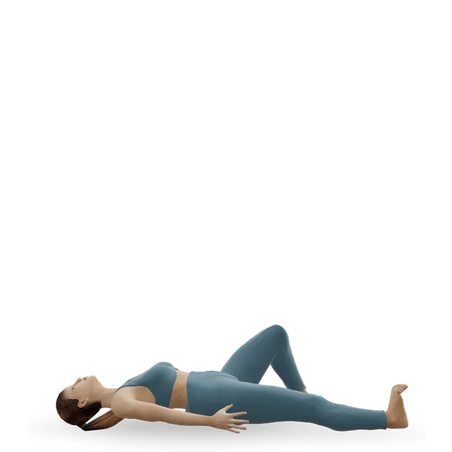
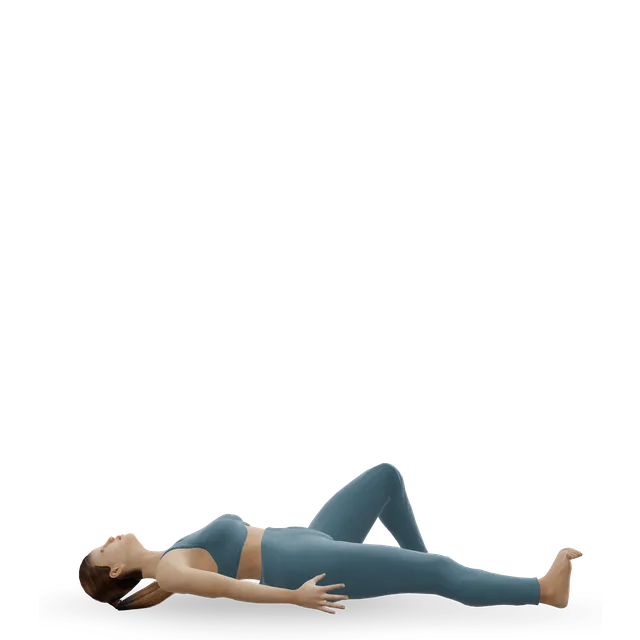
Quad set (static quads) demonstrated on the 3D figure3D demoQuad set (static quads)Beginner · No equipment

10 holds of 5 s, twice a day · Editorial dose, as in our broken foot guide

Harder: Straight-leg raise

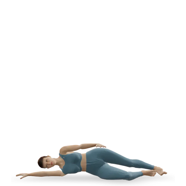
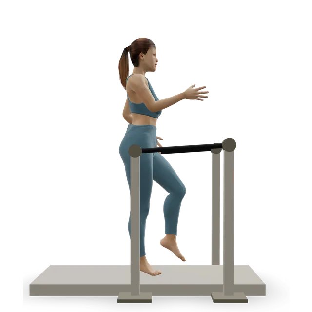
Side-lying leg raise demonstrated on the 3D figure3D demoSide-lying leg raiseBeginner · No equipment

2 sets of 10 each side · Editorial dose. Keeps the hip strong for crutches

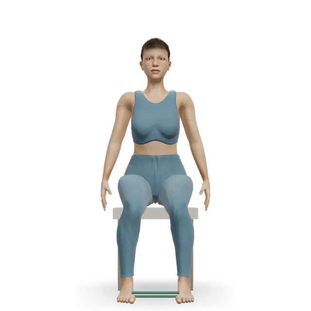
Easier: Lateral band walk · Harder: Lateral band walk, band at the feet

Move on when: The fracture clinic allows weight through the foot. For a small, stable break in a boot this can be from the start; for a displaced break it's often 6 to 12 weeks, sometimes longer.

Follow along: a timer, a cue and the next move, one step at a time.
Stage 2

Taking weight again: off the crutches and out of the boot (often from about 6 to 12 weeks, as your fracture clinic allows)

As your fracture clinic allows. Walk normally again as ankle movement, calf strength and balance come back. Wean out of the boot around the house first, keeping it for longer walks outside at first, and wear a supportive, firm-soled shoe on the other foot while you're in the boot. Add the front-of-ankle stretch below.

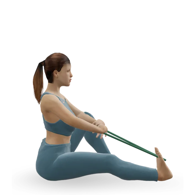
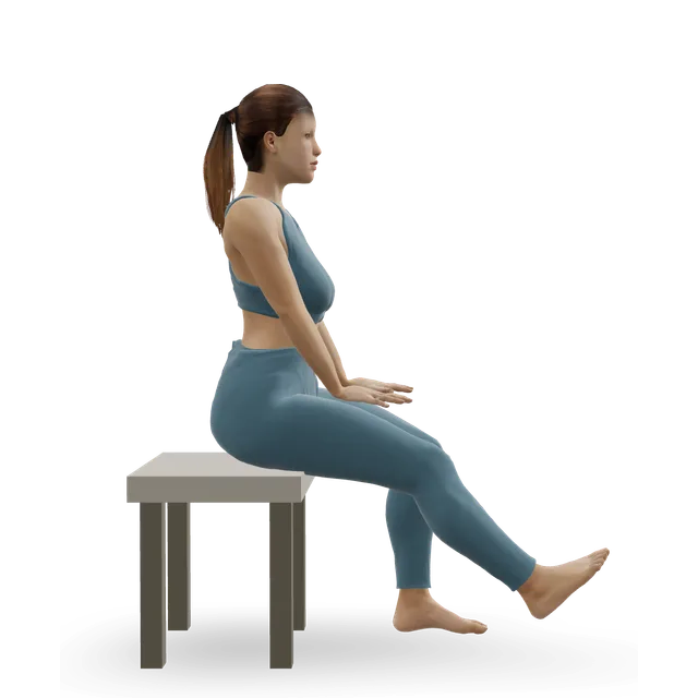
Seated calf stretch with a towel demonstrated on the 3D figure3D demoSeated calf stretch with a towelBeginner · Other / check instructions

Towel round the foot, knee straight: hold 30 s, 3 times, 3 to 4 times a day · A St Helens and Knowsley NHS leaflet's dose

Harder: Wall calf stretch (gastrocnemius)

Knee-to-wall ankle rock demonstrated on the 3D figure3D demoKnee-to-wall ankle rockBeginner · A wall

10 slow rocks per side, daily · The move page's dose. Once full weight is allowed

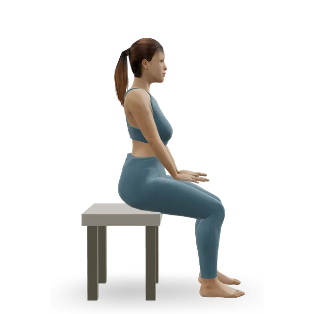
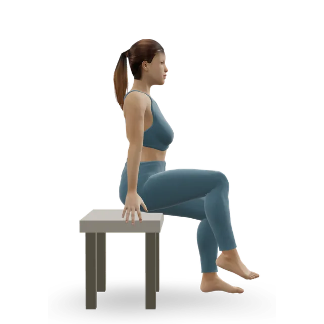
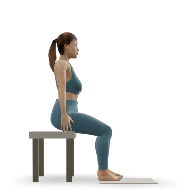
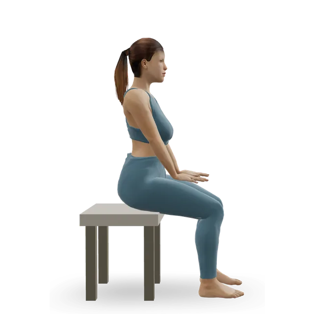
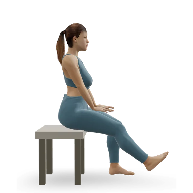
Seated bent-knee calf raise (soleus raise) demonstrated on the 3D figure3D demoSeated bent-knee calf raise (soleus raise)Beginner · A bench or box

2 sets of 10 to 15 · Editorial dose, as in our broken foot guide. Once some weight is allowed

Harder: Slow calf raise

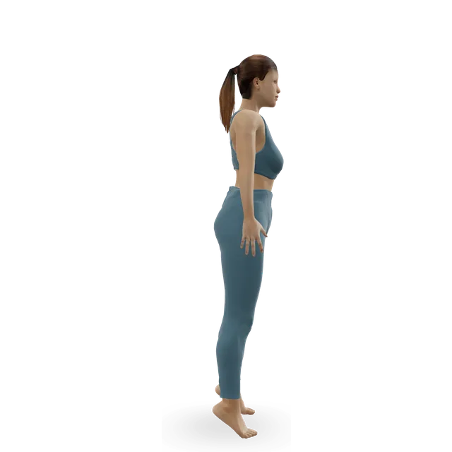
Slow calf raise demonstrated on the 3D figure3D demoSlow calf raiseBeginner · A wall

Both feet, holding on: 10 times, 3 to 4 times a day · A St Helens and Knowsley NHS leaflet's dose. Once full weight is allowed

Easier: Seated bent-knee calf raise (soleus raise) · Harder: Standing dumbbell calf raise

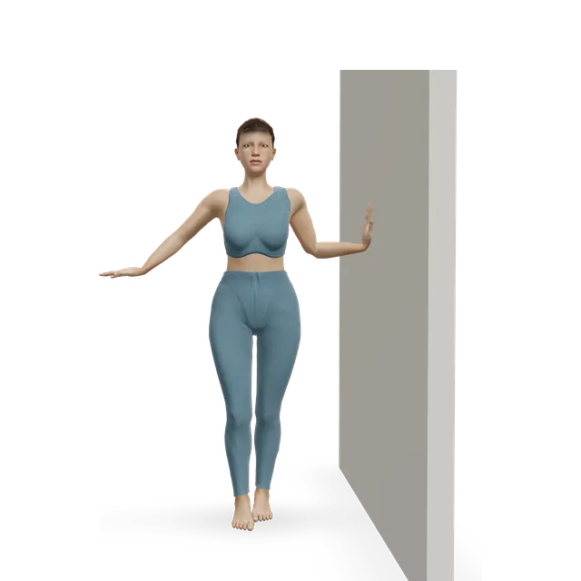
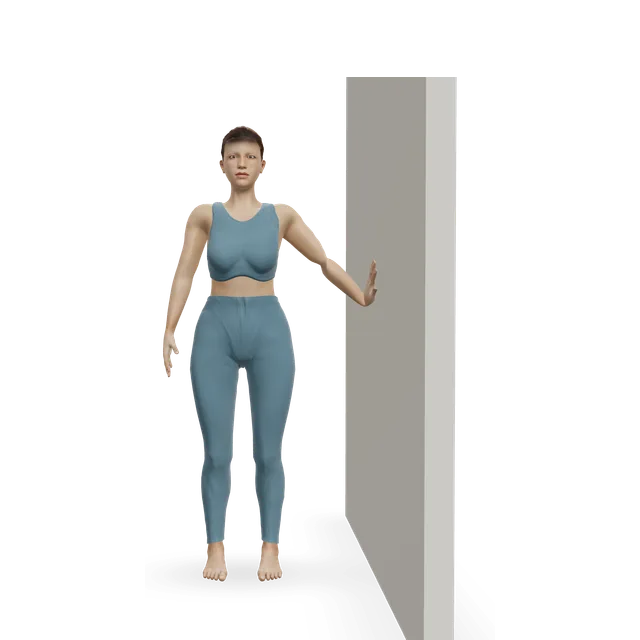
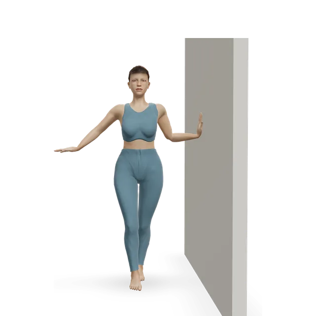
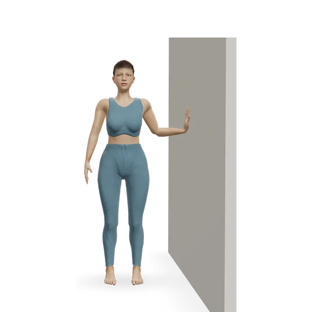
Semi-tandem stance demonstrated on the 3D figure3D demoSemi-tandem stanceBeginner · A wall

Holding on, feet close together, then one foot in front of the other: 30 s each, then without holding on · A St Helens and Knowsley NHS leaflet's dose

Harder: Tandem stance and heel-to-toe walk

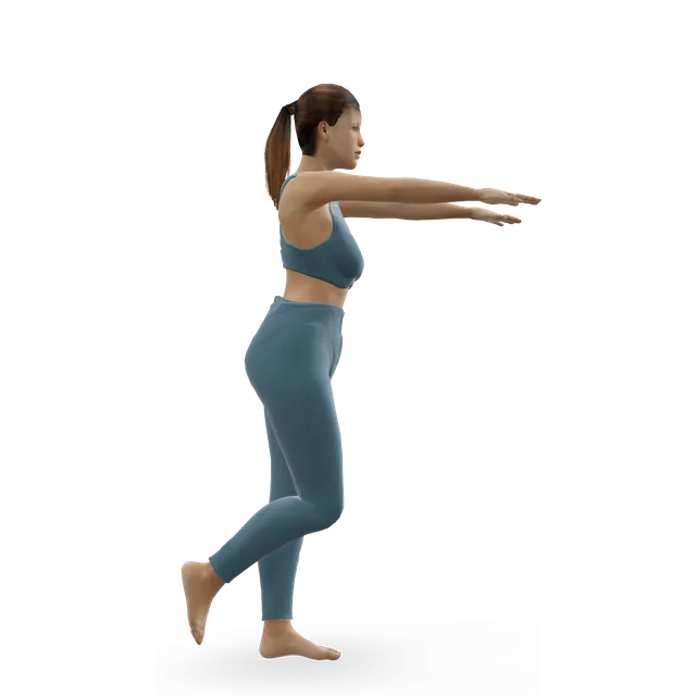
Single-leg stand demonstrated on the 3D figure3D demoSingle-leg standBeginner · A wall

On the injured leg, holding a firm surface: 30 s without more pain; then without holding on; then eyes closed for 10 s · A St Helens and Knowsley NHS leaflet's dose

Easier: Tandem stance and heel-to-toe walk · Harder: Single-leg reach matrix

Move on when: You walk indoors and out without crutches or the boot, and the swelling settles overnight. Simple breaks are largely healed by 8 to 12 weeks.

Follow along: a timer, a cue and the next move, one step at a time.
Stage 3

Strength, balance and longer walks (often from about 3 months, as your fracture clinic allows)

As your fracture clinic allows. Single-leg strength and balance for stairs, slopes and uneven ground. Build longer walks gradually. No running, jumping or dancing for about 3 months after the injury, or longer if your clinic says.

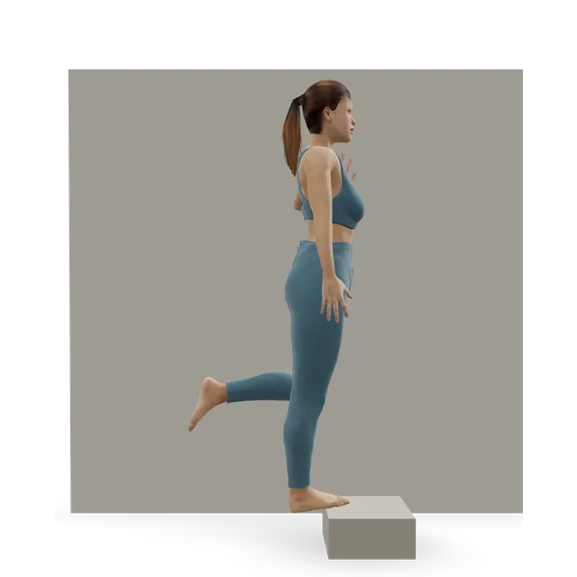
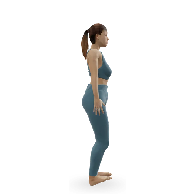
Single-leg calf raise from a step demonstrated on the 3D figure3D demoSingle-leg calf raise from a stepIntermediate · A step + A wall

Build towards 15 to 20 slow raises each leg, on the floor first · Editorial dose, as in our broken foot guide

Easier: Slow calf raise · Harder: Single-leg pogo hops

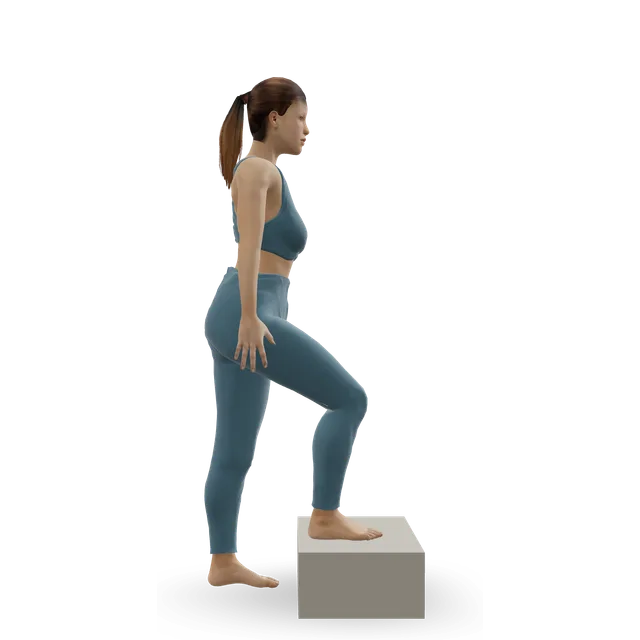
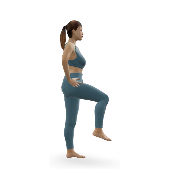
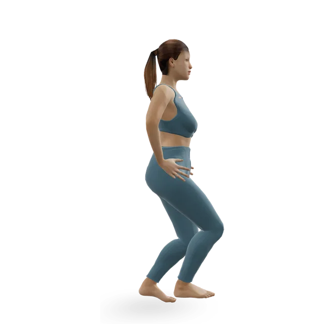
Step-up demonstrated on the 3D figure3D demoStep-upBeginner · A step

3 sets of 8 to 10 each leg · Editorial dose

Easier: Low step-up · Harder: Dumbbell step-up

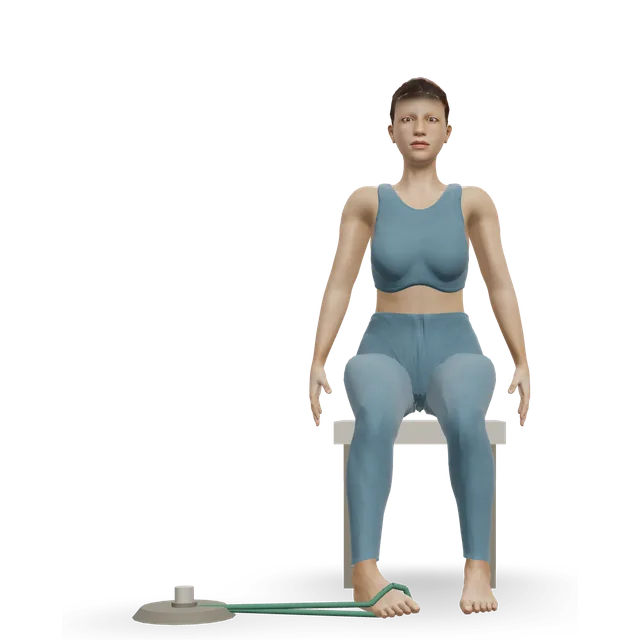
Band ankle inversion demonstrated on the 3D figure3D demoBand ankle inversionBeginner · A band + A bench or box

Light band: 2 to 3 sets of 10 · Editorial dose; the move page's doses are for sprains and tendon pain

Harder: Single-leg calf raise from a step

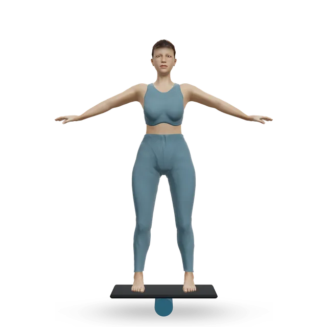
Balance board stand demonstrated on the 3D figure3D demoBalance board standBeginner · Other / check instructions

Several holds of 20 to 30 s on most days, with a wall within reach · The move page's dose. Or stand on a cushion for 30 s, then eyes closed for 10 s (a St Helens and Knowsley NHS leaflet)

Easier: Single-leg stand

Recumbent exercise bike demonstrated on the 3D figure3D demoRecumbent exercise bikeBeginner · Cardio machine

10 to 20 minutes at a moderate effort, building towards 30 · The move page's dose

Harder: Stationary cycling

Move on when: 30 minutes of fast walking is pain-free, single-leg calf raises are close to the other side, and your clinic is happy for impact.

Follow along: a timer, a cue and the next move, one step at a time.
Stage 4

Back to running, jumping and heavier work (from about 3 to 4 months at the earliest, as your fracture clinic allows)

As your fracture clinic allows. Impact and sport for those who want them, through the walk-jog programme below. After a displaced break, many people never get back to high-impact sport, and some need lighter work or different shoes; that's a common outcome, not a failure.

Elliptical trainer demonstrated on the 3D figure3D demoElliptical trainerBeginner · Cardio machine

10 to 20 minutes at a moderate effort, building towards 30 · The move page's dose. A low-impact step before running

Harder: Stair-stepper machine

Ankle bounces (mini pogo) demonstrated on the 3D figure3D demoAnkle bounces (mini pogo)Beginner · No equipment

2 to 3 sets of 20, every other day · Editorial dose

Harder: Pogo hops

Pogo hops demonstrated on the 3D figure3D demoPogo hopsIntermediate · No equipment

3 sets of 20, once ankle bounces are easy · Editorial dose

Easier: Ankle bounces (mini pogo) · Harder: Single-leg pogo hops

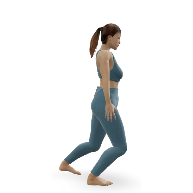
Treadmill jogging demonstrated on the 3D figure3D demoTreadmill joggingIntermediate · Cardio machine

The walk-jog programme, every other day · An Oxford NHS return-to-running leaflet, written for any injury lay-off and used here as a guide; the weeks are in the steps below

Easier: Treadmill walking · Harder: Sprint intervals

Single-leg hop and stick demonstrated on the 3D figure3D demoSingle-leg hop and stickIntermediate · No equipment

3 sets of 5 each leg, before cutting and jumping sports · Editorial dose

Easier: Standing broad jump with stuck landing · Harder: Alternate-leg bounding

Move on when: Running and hopping don't hurt the heel during, after or the next morning; then sport, building up gradually.

Follow along: a timer, a cue and the next move, one step at a time.

What to change for now

Swelling, ice and the boot

Rest and raise the foot above hip level, especially for the first 24 to 72 hours. Use an ice pack or frozen peas wrapped in a damp towel for up to 15 minutes every few hours (one leaflet says 20 minutes 3 to 4 times a day), never straight on the skin. Wear the boot whenever you stand and walk; it can come off at night, at rest and to wash, with a long sock inside. In the boot or cast, keep your toes moving and bend your knee to ease stiff muscles.

Sources 12, 13, 27, 28

Stairs without putting weight on the foot (written steps)

From a University Hospitals Sussex NHS physiotherapy leaflet. Once some weight is allowed, your physiotherapist will show you how to change the steps.

  • 1. A physiotherapist will usually practise this with you before you go home. Hold the rail or banister with one hand and a crutch in the other.
  • 2. Going up: lean on the rail and the crutch, bend the injured knee so the foot clears the step, and hop up with your good leg. Then bring the crutch up.
  • 3. Going down: stand close to the edge of the step. Put the crutch down on the next step, hold the injured leg out in front of you, off the floor, then hop or lower your good leg down to the same step.
  • 4. Take one step at a time. Without a rail, use both crutches the way you were shown.

Source 29

Front-of-ankle stretch (written steps)

From a St Helens and Knowsley NHS leaflet's second stage. It has no 3D demo.

  • 1. Start once your clinic allows weight through the foot. Sit on a chair and cross the injured ankle over the other knee.
  • 2. With your hand over the top of the foot, gently point the toes down to stretch the front of the foot and ankle.
  • 3. Or stand holding on to something firm, with the top of the foot resting on a chair behind you, and point the foot down.
  • 4. Hold for 30 seconds, 3 times, 3 to 4 times a day.

Source 13

The walk-jog programme (written steps)

From an Oxford NHS leaflet written for coming back from any injury lay-off. We use it here as a guide.

  • 1. Start once 30 minutes of fast walking is pain-free, and your team is happy for you to run.
  • 2. Week 1: jog 1 minute, walk 1 minute, for 30 minutes. Do each week's session every other day.
  • 3. Week 2: jog 2 minutes, walk 1 minute, for 30 minutes.
  • 4. Week 3: jog 4 minutes, walk 1 minute, for 30 minutes.
  • 5. Week 4: jog 9 minutes, walk 1 minute, for 30 minutes.
  • 6. Week 5: jog 15 minutes, walk 1 minute.
  • 7. Week 6: jog 30 minutes without stopping.
  • 8. Move up a level after a week without pain, and down a level if it hurts. From week 7, add no more than 10% a week.

Source 26

Driving, shoes and work

Don't drive in the walking boot: a Cambridge NHS leaflet warns it voids your insurance. Other leaflets say drive once you're out of the boot, walking comfortably and able to do an emergency stop without pain, and test it somewhere safe first. A widened heel can make shoes rub: for long-term problems, US orthopaedic surgeons suggest a heel pad, lift or cup, or shoes with extra depth. People whose work needs a lot of walking or climbing may need a phased return or a change of role.

Sources 27, 12, 13, 1

A small chip off the heel bone

A calcaneal avulsion fracture is a small piece of bone pulled off by a ligament when the ankle twists. A Leeds NHS service treats it like a sprain: walk as pain allows, with a walking boot for comfort for at least 4 weeks; it heals in about 6 weeks, and the pain and swelling take about 3 months to settle. Drive once you're permanently out of the boot and can do an emergency stop, and go back to work as soon as you're comfortable.

Source 4

Positions to swap, and why

Habit or positionWhy it mattersInstead
Putting more weight on the foot than your plan allowsThe pieces can move, and after surgery the screws can loosen or break.Follow your written plan and use crutches; the ankle and leg exercises in stage 1 keep you moving.
Sitting with the foot down for long spellsSwelling builds, especially in the first days.Raise it above hip level, especially for the first 24 to 72 hours.
Driving in the walking bootOne NHS leaflet warns it voids your insurance.Drive once you're out of the boot, walking comfortably and able to do an emergency stop without pain.
SmokingIt slows bone and wound healing and raises the risk of a wound infection after surgery.NHS stop-smoking support.
Stairs without a planFalls on crutches are easy.The stairs steps below, practised with a physiotherapist first.
Ankle bounces, hopping, running or jumping before about 3 monthsNHS leaflets say avoid impact, such as running, jumping and dancing, for 3 months.The exercise bike, then the cross-trainer.Try: Recumbent exercise bike
Long walks or standing all day too earlySwelling and pain build, especially at the end of the day.Build up gradually, and rest with the foot up.
Hard, thin-soled or tight shoes once out of the bootA widened heel rubs, and uneven ground is harder.Supportive trainers; ask about heel pads, heel cups or deeper shoes.
Ignoring back pain after the fallThe same fall can break a bone in the back.Get it checked: see the warning signs.

Routines to follow

More moves for this area

Questions people ask

Can you walk on a broken heel?

Sometimes, with a limp, after a minor break, but most heel breaks make it hard or impossible to take weight. The NHS says don't try to treat a suspected broken heel yourself: contact NHS 111. Once it's diagnosed, your fracture clinic decides how much weight it can take.

How long does a broken heel take to heal?

The bone takes about 6 to 12 weeks, but pain and swelling last 3 to 6 months, and mild symptoms 6 to 12 months, even after a simple break. After a displaced break, recovery typically takes about 2 years, levelling off at about 18 months.

Do I need surgery?

It depends on the break. In the UK Heel Fracture Trial, surgery for typical displaced breaks gave no better pain or function at 2 or 5 years and more complications, including wound infections in 19%. Surgery is still used for some breaks, such as a badly deformed heel, an open wound, or a piece pulled off by the Achilles tendon that threatens the skin.

How long will I be off my foot?

Plans differ: in the UK trial, 6 weeks without weight and then 6 weeks with partial weight; traditionally 8 to 12 weeks without weight after surgery; and small, stable breaks may touch down or take weight as comfort allows in a boot. Your fracture clinic should write your weight-bearing plan down.

Why do they want to check my back?

NICE treats falls from more than 3 metres, and heavy landings on the feet, as dangerous for the spine. In one study, 7% of people with heel fractures also had a broken vertebra, mostly in the lower back. Back pain after a serious accident or fall is a 999 call.

How long will the swelling last?

Usually 3 to 6 months, worse at the end of the day, and mild swelling can last 6 to 12 months. Raising the foot above the hip, cold packs and moving the ankle all help.

When can I drive?

Not in the walking boot: one NHS leaflet warns it voids your insurance. Others say you can drive once you're out of the boot, walking comfortably and able to do an emergency stop without pain.

Will I get back to work and sport?

In the UK trial, 84.4% of the people treated with surgery and 88.9% of those treated without it were back at work at 2 years, though these were small groups and most had moved to less physically demanding jobs. After a severe break, people rarely regain normal foot movement or their previous level of sport. Running starts no earlier than about 3 months, with a walk-jog programme, as your fracture clinic allows.

Will I need special shoes or insoles?

Shoes can rub if the heel has widened. For long-term problems, US orthopaedic surgeons suggest a heel pad, lift or cup, or shoes with extra depth.

Do I need blood-thinning injections?

NICE says consider them while the lower leg is immobilised if your clot risk outweighs your bleeding risk, and UK fracture services should assess your clot risk. Know the signs of a clot: throbbing pain and swelling in one calf, with red or darker skin.

Does smoking matter?

Yes. Smoking slows bone and wound healing, and in a review of 41,944 people, smokers had about 1.6 times the odds of a wound infection after ankle or heel surgery.

What is a calcaneal avulsion fracture?

A small chip of bone pulled off by a ligament when the ankle twists. One NHS service treats it like a sprain: walk as pain allows, with a boot for comfort for at least 4 weeks, and it heals in about 6 weeks.

Related exercise guides

Other pain guides

Sources

  1. AAOS OrthoInfo · Calcaneus (heel bone) fractures
  2. Griffin 2014 · Surgery or not for displaced heel fractures: the UK Heel Fracture Trial (RCT of 151, BMJ)
  3. Bruce 2013 · Surgical or conservative treatment for displaced heel fractures (Cochrane review)
  4. Leeds Teaching Hospitals NHS · Calcaneal avulsion: consultant advice (virtual fracture clinic, 2024)
  5. Rubenson 2025 · 3,949 heel fractures in the Swedish Fracture Register (registry study, Foot Ankle Surg)
  6. Shah 2026 · Heel fractures in the United States: 89,434 patients (national database study, J Foot Ankle Surg)
  7. NHS · Heel pain
  8. NICE NG41 · Spinal injury: assessment and initial management (2016)
  9. Walters 2014 · Heel and spine fractures together (cohort of 208, J Foot Ankle Surg)
  10. British Orthopaedic Association · BOAST: assessment of the spine in the trauma patient (standard, 2025)
  11. Ågren 2013 · Surgery or not for displaced heel fractures (RCT of 82, J Bone Joint Surg Am)
  12. Whittington Health NHS · Calcaneal fracture: touch weight-bearing (virtual fracture clinic leaflet, 2022, now past its review date)
  13. St Helens and Knowsley Teaching Hospitals NHS · Calcaneus (heel) fracture (leaflet, now past its review date)
  14. Dickenson 2021 · The UK Heel Fracture Trial at 5 years (long-term follow-up, Bone Joint J)
  15. Park 2018 · Compartment syndrome of the foot after heel fractures (cohort of 313, Bone Joint J)
  16. Buckley 2002 · Surgery or not for displaced heel fractures (RCT of 424, J Bone Joint Surg Am)
  17. Attenasio 2024 · Wound complications with a long or a smaller incision for heel fracture surgery (meta-analysis of 21 studies, Injury)
  18. British Orthopaedic Association · BOAST: mobilisation and weight-bearing after orthopaedic surgery or musculoskeletal injury (standard, 2024)
  19. Verstappen 2025 · Early weight-bearing after heel fracture surgery (systematic review of 20 studies, J Orthop Traumatol)
  20. Whittington Health NHS · Calcaneal fracture: weight-bearing as comfort allows (virtual fracture clinic leaflet, 2022, now past its review date)
  21. British Orthopaedic Association · Outpatient and on-call services for people with fractures or musculoskeletal injury (standard, 2026)
  22. Verstappen 2024 · Permissive or restricted weight-bearing after heel fracture surgery: the PIONEER trial (trial protocol, Trials)
  23. Tran 2024 · Smoking and wound infection after ankle and heel fracture surgery (meta-analysis of 16 studies, EFORT Open Rev)
  24. NICE NG89 · Venous thromboembolism in over 16s: reducing the risk of hospital-acquired deep vein thrombosis or pulmonary embolism (2018, updated 2019)
  25. NHS · DVT (deep vein thrombosis)
  26. Oxford University Hospitals NHS · Return to Running Programme (OxSport patient leaflet, 2024)
  27. Cambridge University Hospitals NHS · Calcaneal fracture (patient information, 2023)
  28. NHS · Broken ankle
  29. University Hospitals Sussex NHS · Managing the stairs (non-weight bearing) (physiotherapy leaflet, 2025)
  30. NHS · Back pain
  31. NHS · Falls
  32. NHS · Compartment syndrome
  33. British Orthopaedic Association · BOAST: compartment syndrome of the extremities (standard, revised 2025)
  34. NICE NG259 · Osteoporosis: risk assessment (2026)

Exercise education, not a diagnosis or treatment plan. It is written from the guidelines and studies listed above; review by a clinician is pending. If your pain follows an injury, is severe, is getting worse, or comes with any warning sign listed on this page, get it assessed. Health disclaimer.