A less common place, with many possible causesEmerging evidence
Pain behind the knee is less common than pain at the front or sides. A review lists tendon and other soft tissue causes as commoner than nerve and blood vessel ones, and a family medicine review says pain only at the back may come from a Baker's cyst; after an injury, from the PCL or the back of the meniscus; and, when it lasts, from a hamstring tendon. Pain can also be felt there from the hip, the lower back and the kneecap joint.
Sources 1, 2
A Baker's cyst usually points to something else in the kneeStrong evidence
A Baker's cyst is a fluid-filled lump at the back of the knee that connects with the joint. It may show more when you stand, and it can cause pain, tightness, stiffness, difficulty bending and clicking, often worse after walking, the NHS says. In adults it usually comes from another knee problem, such as osteoarthritis, a meniscus tear, rheumatoid arthritis or gout.
On 1,001 knee magnetic resonance imaging (MRI) scans, cysts were seen in 4.7%, and 94% came with another problem in the knee, a meniscus tear in 83%, often at the back of the inner meniscus. An NHS trust leaflet says cysts are commonest in children aged 4 to 7 and adults aged 35 to 70.
Sources 3, 4, 5, 6
A burst cyst and a clot look alikeEmerging evidence
About 1 to 2 in 20 cysts are thought to burst, an NHS trust leaflet says, and a burst cyst is hard to tell from a deep vein thrombosis (DVT), a clot in a deep vein of the leg. Of 3,072 people scanned for a suspected clot, 95 had cysts, and 7 of those also had a clot. Cysts don't seem to cause clots, though: across 357,703 leg vein scans, cysts were not a risk factor.
The NHS lists the signs of a DVT separately, and any of them can be a clot: throbbing pain in one leg (rarely both), usually the calf or thigh; swelling in one leg; red, blue or darkened skin around the painful area; or swollen veins. A suspected clot is referred for an ultrasound scan within 24 hours, and the National Institute for Health and Care Excellence (NICE) asks for the result within 4 hours if possible when a clot is likely.
Sources 6, 7, 8, 9, 10
Tendons, the calf and the popliteusEmerging evidence
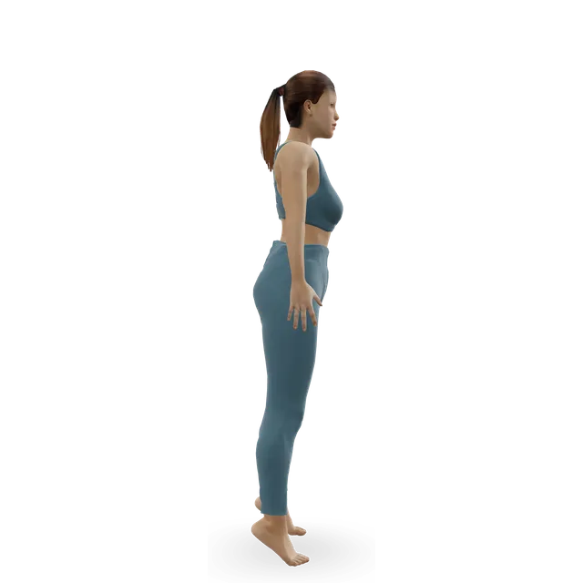
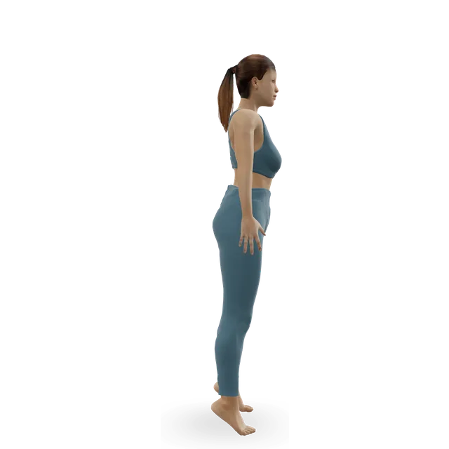
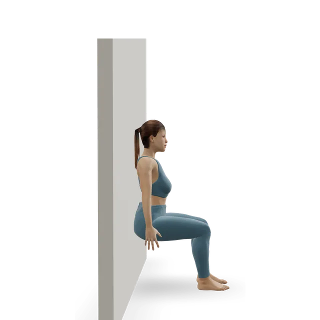
The semimembranosus, a hamstring tendon at the back inner corner, aches with tenderness just below the joint; it's uncommon, often seen in runners, and little studied. The biceps femoris, the outer hamstring, runs to the bony bump below the outer knee. The calf muscle (gastrocnemius) hurts when you bend the knee against resistance or rise onto your toes, and the popliteus, deep at the back outer corner, hurts bearing weight with the knee bent 15° to 30°.
Sources 11, 1
The meniscus and the PCLEmerging evidence
Meniscus tears rarely show up as pain at the back, mostly tears of the back of the inner meniscus. A single painful pop in mid-life can point to a tear at the root of the inner meniscus, as one study of people already having keyhole surgery found; in 234 people whose root tears were repaired, about 80% were women and the average age was 65.
In 494 people with a long-standing PCL injury, traffic accidents (45%) and sport (40%) were the causes, a dashboard injury the commonest way (35%), and 53% had other ligaments injured too. The American Academy of Orthopaedic Surgeons (AAOS) describes pain and swelling that come quickly, a stiff knee, a limp and a knee that may feel it will give out.
Sources 1, 12, 13, 14, 15
Rare but important: the arteriesEmerging evidence
A popliteal artery aneurysm, a swelling of the artery behind the knee, is the commonest aneurysm in the limbs; 95% to 99% are in men, about half are in both legs, and 30% to 50% come with an aneurysm of the aorta. A clot in one can threaten the leg, and a review lists it among the lumps that can be mistaken for a cyst.
Popliteal artery entrapment, where a muscle squeezes the artery, causes calf and foot pain after exercise that eases with rest in young, active people without the usual risk factors for narrowed arteries, sometimes in both legs. It's uncommon, and diagnosing it early helps prevent clots.
Sources 16, 17, 1
When a test helpsStrong evidence
For a lump, an ultrasound scan confirms a cyst and shows what's inside it, and an MRI shows the problems inside the knee that cause it; a GP may arrange either. A possible clot needs an ultrasound scan, within 24 hours, the NHS says. After an injury, the Ottawa knee rule decides on an X-ray, and an MRI looks at the PCL and the meniscus.
Artery problems are checked by comparing the blood pressure at the ankle and the arm, or with a Doppler ultrasound scan while the foot is pushed or pulled, then a scan of the arteries; an aneurysm is followed with ultrasound scans.
Sources 1, 3, 9, 2, 15, 17, 16
How it usually goesEmerging evidence
A Baker's cyst may go away on its own, especially in children, or when its cause is treated, and the NHS says it can take a few months to a few years; a burst cyst makes symptoms worse for a few weeks until the swelling goes down, and cysts often come back after draining. A clot is treated with blood-thinning medicine, usually for at least 3 months. Mild hamstring injuries get better in a few days and severe ones take weeks or months; a cramp lasts from a few seconds to 10 minutes. No one can promise a date.
Sources 3, 6, 9, 18, 19