Pain guide · Foot pain

Pain on the outer side of the foot: the tendons, a fifth metatarsal break, cuboid and old sprains

Pain on the outer side of the foot often follows a rolled ankle: a sprain, a break at the bump halfway along the outer edge of the foot, or an injury to the tendons that run behind the outer ankle bone. Without an injury, the main candidates are those tendons, a stress fracture, cuboid syndrome or a tailor's bunion at the base of the little toe.

This guide starts with the signs that need help today, from a 999 call or accident and emergency (A&E) to the National Health Service's 111 line (NHS 111) or a general practitioner (GP): the bony points that mean an X-ray after a twist, a tendon that slips behind the ankle bone and any new foot problem if you have diabetes. It then maps the causes by the exact spot, explains why an old sprain can still hurt and why six to eight weeks of rest is wrong for most of these, and gives a three-stage plan that ends with walking, running, side-to-side sport and shoes.

When to get help first

Exercise is for pain that has been checked or is clearly mild and settling.

Get urgent medical help if

  • Call 999 or go to A&E: after an injury, you heard a crack, the foot or ankle has changed shape or points at an odd angle, a bone is sticking out, there is a bad cut over it, you're in severe pain, or the toes look blue or white, feel numb or are cold to touch. These can mean a broken bone. Don't drive yourself.
  • Contact NHS 111: after an injury, severe ankle pain, feeling faint, dizzy or sick from the pain, a snap, grinding or popping noise at the time of the injury, the ankle or foot at an odd angle, or not being able to walk. These might be signs of a broken ankle or an infection.
  • Same day (urgent GP appointment or NHS 111): after an injury, you can't put weight on it or walk more than a few steps, the pain is very bad or getting worse, there is a lot of swelling or bruising or it is getting worse, or it feels very stiff. If you think it's broken, don't treat it yourself.
  • Same day: after a twist or a fall, if you can't walk four steps, if pressing on the bony bump on the inner side of the midfoot (the navicular) or the bump halfway along the outer edge of the foot (the base of the fifth metatarsal) hurts, if the foot looks out of shape, or if you're unsure, get it checked. Clinicians use these points, the Ottawa rules, to decide who needs an X-ray.
  • Same day: after an injury, a pop or snap behind the outer ankle bone with swelling, or a tendon you can see or feel slipping over the bone. The tendons may have come out of their groove (dislocated), which is often mistaken for a sprain; experts advise a specialist review.
  • A child who has hurt their foot or may have broken a toe: don't treat it yourself; take them to an urgent treatment centre or A&E.
  • Go to A&E: you think you've broken your big toe, a toe is pointing at an odd angle, the bone is sticking out, or you feel tingling or numbness in the toe or foot. A broken smaller toe without these signs can usually be treated at home.
  • Same day: a sudden, very painful, hot, swollen, red joint, often the big toe but sometimes the middle of the foot (the midfoot) or the ankle, especially with a high temperature, feeling shivery or being unable to eat. It may be gout, but it could be a joint infection (septic arthritis), which is urgent.
  • Same day: skin on the foot that is painful, hot and swollen. It could be a skin infection (cellulitis).
  • Call 999 or go to A&E: a red, hot, swollen foot with a very high temperature, feeling hot, cold or shivery, a fast heartbeat or fast breathing, purple patches on the skin, dizziness or confusion.
  • Call 999 or go to A&E: a foot or leg that is suddenly cold, pale, numb, weak or very painful without an injury. A sudden loss of blood supply is a vascular emergency.
  • Call 999 or go to A&E: you have diabetes and a foot ulcer with a fever or signs of sepsis, an ulcer with poor circulation, or gangrene. NICE asks for these to go straight to hospital.
  • Same day: you have diabetes and a new foot problem, such as an ulcer or wound, signs of infection, or a hot, swollen foot with a change in colour, even if it doesn't hurt. Contact your diabetes foot team or GP: NICE asks for these to reach a specialist foot service within 1 working day, and a suspected Charcot foot is kept off weight until it has been checked.
  • If you have diabetes, see your local foot team urgently, within 24 hours, for a change in the colour or shape of your foot (especially a red, hot, swollen toe, ankle or foot), part of your foot feeling much colder or hotter than usual, new blisters, cuts or wounds (especially ones you can see but don't feel), a cut or wound that smells bad or is leaking fluid or pus, or new or unexplained pain in your foot. Meanwhile sit or lie down, take the weight off it and raise it.
  • Same day: in a cast or boot, pain that is getting worse, toes that are numb or burning, a cast that feels too tight, toes that are swollen, blue or white, or a bad smell from under the cast.
  • Same day (urgent GP appointment or NHS 111): throbbing pain in one leg (rarely both), usually the calf or thigh; swelling in one leg (rarely both); red, blue or darkened skin around the painful area, which may be harder to see on brown or black skin; or swollen veins. It could be a blood clot (DVT). Watch for it after an ankle injury or time in a cast or boot.

See a doctor or physiotherapist if

  • Pain that isn't better after 2 weeks of looking after it yourself, is getting worse, keeps coming back, or stops you doing normal things.
  • One spot on a foot bone that hurts with every step and later at night, or an ache on the outer edge of the foot that built up before any injury: a possible stress fracture. Our stress fracture guide covers it.
  • Pain in the middle of the foot that is hard to pinpoint, on the outer edge of the midfoot, or on the front of the shin bone in a running or jumping athlete. These sites heal less well and need prompt assessment, often with a scan.
  • With a Jones fracture, pain at the outer edge of the foot coming back once you're active again: stop, and get it checked, because these fractures can break again.
  • Clicking or snapping behind the outer ankle bone that keeps happening or hurts.
  • An ankle that keeps giving way months after a sprain.
  • Pain, clicking, locking or swelling that carries on despite a good rehab programme. Cartilage, tendon and other problems are common alongside an unstable ankle and need assessing.
  • Pain on the outer side of the foot or ankle that hasn't settled 6 weeks after a sprain, or a sprain that keeps hurting in one spot: ask for a review. A small fracture or a tendon injury can be missed at first (our suggestion).
  • No better a week or two after treatment for cuboid syndrome, or it keeps coming back: get it re-examined. Tendon problems, a painful small bone in the peroneal tendon (the os peroneum), a stress fracture or a coalition can look like cuboid syndrome.
  • Tingling or loss of feeling in the foot, or a foot that feels weak.
  • Any foot pain if you have diabetes: foot problems can be more serious.
  • A high arch that is new, getting higher or only in one foot, or high arches with weakness, tripping, numbness or ankles that keep giving way: see a GP. It can be the first sign of a nerve condition.
  • Hard skin or a corn over a bump at the base of the little toe: don't treat it yourself if you have diabetes, problems with your circulation or a weakened immune system, or are an older person; see a GP or foot specialist. See a GP too if it bleeds, has pus or discharge, or isn't better after 3 weeks of treating it at home.
  • Pain at the bump on the outer edge of the foot that keeps coming back with sport in a child: see a GP or physiotherapist once, to confirm what it is. If an X-ray is done, ask whether the growth area has been looked at, because it can be mistaken for a fracture.

Where does it hurt?

Location narrows the likely causes; it does not diagnose.

Behind and below the outer ankle bone, along the tendons towards the outer heel, worse walking, running, on uneven ground and turning, often after sprains

The peroneal tendons: tendinopathy or a tear.

Read: Peroneal tendinopathy →
A pop or snap behind the outer ankle bone with swelling, a tendon you can see or feel slipping, or clicking that keeps happening

The tendons slipping out of their groove, which is often taken for a sprain: get it checked.

Read: Peroneal tendinopathy →
The bump halfway along the outer edge of the foot, after rolling the ankle, with bruising and pain walking

A possible avulsion fracture at the base of the fifth metatarsal: the Ottawa rules decide on an X-ray.

Read: Broken foot (metatarsal fracture) →
Just beyond that bump, after a sudden injury in sport, or after an ache that built up first

A possible Jones fracture, which heals more slowly.

Read: Broken foot (metatarsal fracture) →
The outer edge of the midfoot, an ache building over runs or training with no injury, later at rest

A possible stress fracture at the base of the fifth metatarsal, a site that heals less well: stop and get it checked.

Read: Stress fracture →
The outer midfoot over the cuboid, sharp at push-off, hopping or moving side to side, after a sprain or in dancers and runners

Possibly cuboid syndrome, once a fracture is ruled out. The evidence for it is thin.

Read: Cuboid syndrome →
The underside of the outer foot near the cuboid, worse pushing off or on tiptoe

Possibly a painful os peroneum, a small bone inside a peroneal tendon.

Read: Peroneal tendinopathy →
The soft hollow just in front of and below the outer ankle bone, worse on uneven ground, the ankle feeling unstable

Sinus tarsi syndrome.

Read: Sinus tarsi syndrome →
The outside of the ankle in the days and weeks after it rolled inwards

A lateral ankle sprain.

Read: Sprained ankle →
Months or years after a sprain, the ankle giving way, with outer pain or swelling

Chronic ankle instability, often with a second injury alongside it.

Read: Chronic ankle instability →
In front of and below the outer ankle after a twist, not settling as a sprain should

Rarely, a small fracture at the front of the heel bone, which X-rays can miss.

Read: Broken heel →
A bony bump at the base of the little toe that shoes rub, sometimes red, with a corn, the little toe angled inwards

A tailor's bunion (bunionette).

Read: Tailor's bunion →
The little toe swollen and bruised after a stub or a kick

A possible broken toe.

Read: Broken toe →
An active child aged about 8 to 14 with pain at the bump during and after running, jumping and cutting

Iselin's disease, a strained growth area where a tendon attaches.

Read: Iselin's disease →
A teenager's stiff foot with ankles that keep spraining, aching on the outer side

Possibly a tarsal coalition, where two foot bones are joined.

Read: Tarsal coalition →
A high arch with pain on the outer edge, hard skin under the outer foot, and ankles that roll

High arches, which load the outer edge and the peroneal tendons.

Read: High arches →
Burning, tingling or numbness along the outer edge of the foot

Rarely, a squeezed sural nerve, or a nerve in the back: see a GP.

The top of the foot's outer side after rolling the ankle

Our pain on top of the foot guide maps it.

Read: Pain on top of the foot →
A sudden hot, red, swollen joint, a skin infection, or diabetes with a hot, swollen foot

Gout, an infection or an active diabetic foot problem: see the warning signs.

Outer foot pain mostly when you run

Our foot pain when running guide.

Read: Foot pain when running →
Outer foot pain mostly when you walk

Our foot pain when walking guide.

Read: Foot pain when walking →
The outer edge of the foot on the bike

Our numb feet when cycling guide.

Read: Numb feet when cycling →
Pain in the heel rather than the outer side of the foot

Our heel pain guide maps it.

Read: Heel pain →

Understand it

Where it hurts is a clue, not a diagnosisStrong evidence

No study opened for this guide counts how often each cause explains pain on the outer side of the foot. After a twist, the usual stories are a sprain, a break at the base of the fifth metatarsal (the long bone that ends at the bump halfway along the outer edge) or a tendon injury. Without an injury, an imaging review lists the peroneal tendons, a hidden fracture of the cuboid or the heel bone, the joint between the heel bone and the cuboid, a joined pair of bones (a tarsal coalition), a pinch at the front of the ankle, the hollow in front of the outer ankle bone, small extra bones and a nerve.

So this guide says a spot points to a cause rather than proving it, and an examination decides. The NHS says a sprain causes pain, swelling and bruising, and most sprains feel better after 2 weeks; NHS inform expects most new foot problems to begin to settle within 6 weeks.

Sources 1, 2, 3, 4

The tendons behind the outer ankle boneEmerging evidence

Two peroneal tendons run behind the outer ankle bone and along the outer edge of the foot: one to the base of the fifth metatarsal, one under the foot. A specialist review calls their problems an under-appreciated cause of pain on the outer side of the foot and ankle, easily confused with a ligament injury, and says they go with ankles that keep giving way and with high-arched feet whose heels tip inwards. Family-medicine reviews describe tenderness along the outer heel towards the base of the fifth metatarsal.

A tendon that slips out of its groove after an injury, with a pop and swelling behind the ankle bone, is often taken for a sprain, and an international consensus advises a specialist review. Scans often show changes in the tendons of people with no pain, so a scan has to match the examination.

Sources 5, 6, 7, 8

The bump halfway along the outer edge: fifth metatarsal fracturesStrong evidence

The fifth metatarsal most often breaks at its base when the foot and ankle twist downward and inward, and the ligament pulls a fragment of bone away (an avulsion fracture), the American Academy of Orthopaedic Surgeons (AAOS) says. A Jones fracture is a break across the base, just beyond the bump, in an area with a poor blood supply, so it can heal slowly or not at all and is more often treated with surgery, especially in athletes. Breaks at the base of the fifth metatarsal are about 30% of metatarsal fractures, a review says.

A stress fracture there often begins as pain on the outside of the midfoot with no injury, and is especially common in high-level athletes. In 64 elite European football teams over 11 seasons, a team of 25 players might expect one fifth metatarsal fracture every fifth season; only 1 of 38 first fractures was an avulsion, 54% of first X-rays showed stress-type fractures, 45% of players had warning symptoms first, and a third of all the fractures were re-fractures.

Sources 9, 10, 11, 12, 13

After a twist, the Ottawa rules decide who needs an X-rayStrong evidence

The National Institute for Health and Care Excellence (NICE) advises using the Ottawa ankle and foot rules to decide whether someone over 5 needs an X-ray. For the foot, an X-ray is needed if the base of the fifth metatarsal or the navicular (the bump on the inner midfoot) is tender, or you couldn't take 4 steps.

In the study that refined the rules, they picked up all 19 fractures in the midfoot zone. Across 27 studies of 15,581 people, they missed almost no midfoot fractures, and they could cut X-rays by 30% to 40%. They're a guide for clinicians, not a self-test: being able to walk doesn't rule out a break if that bump is tender.

Sources 14, 15, 16

An old sprain that still hurtsEmerging evidence

Across 31 studies of ankle sprains, pain fell quickly in the first 2 weeks, but 5% to 33% of people still had pain a year later, and up to a third felt the ankle was unstable. Ankle sprains are the commonest muscle and joint injury in active people and often recur, and many people go on to have an ankle that keeps giving way (chronic ankle instability). After a first sprain, not being able to jump and land well at 2 weeks went with lasting instability a year later.

A second injury is often there: of 61 people having surgery for an unstable ankle, none had a ligament injury alone; 77% had inflamed peroneal tendon sheaths, 25% a tear in one of the tendons and 23% a damaged patch of cartilage. In 2,577 foot and ankle MRI scans, 14 showed a small fracture at the front of the heel bone, and only 3 of 8 of those had been visible on X-rays.

Sources 17, 18, 19, 20, 21, 22

The outer midfoot: cuboid syndrome, on thin evidenceMixed evidence

Cuboid syndrome is thought to be a subtle disturbance of the joint between the cuboid and the heel bone, with symptoms like a sprain, a review says. There is no definitive test and no evidence-based guideline; X-rays, computed tomography (CT) and MRI scans don't show it, and their job is to rule out a fracture or a coalition. The evidence for treating it is case reports and one series of 7 athletes.

A painful os peroneum is different: a small bone inside the tendon that runs under the foot can crack, or the tendon can tear or catch there, causing pain under the outer side of the foot.

Sources 23, 24, 25, 3

The base of the little toe: a tailor's bunionEmerging evidence

A bunionette, or tailor's bunion, is a bony bump on the outside of the foot at the base of the little toe, much like a bunion, sometimes with an irritated fluid sac or hard skin over it, the AAOS says. In a survey of 2,662 adults in Turkey, 13.8% reported one, most of them with a family history, and tight shoes went with it. Painful bunionettes go with wider angles between the fourth and fifth metatarsals and at the little toe joint.

Sources 26, 27, 28

Children: Iselin's diseaseStrong evidence

In an active child, pain and swelling at the bump halfway along the outer edge, where a tendon attaches to the growth plate of the fifth metatarsal, is usually Iselin's disease. A children's hospital lists growth spurts (especially at 9 to 13), a rolled ankle or a blow to the outer foot, and tight or narrow shoes such as football boots or spikes, among the risks. After a twist, tenderness there still needs an X-ray check for a fracture.

Source 29

A hot foot, a broken toe, or diabetesStrong evidence

A sudden, hot, swollen, painful joint can be gout or a joint infection (septic arthritis), which needs an urgent GP appointment or NHS 111, and red, hot, swollen skin can be a skin infection (cellulitis), the NHS says. A broken smaller toe can usually be treated at home and heals in 4 to 6 weeks, sometimes months, but go to A&E for a suspected broken big toe, a toe at an odd angle, bone through the skin, tingling or numbness, or a child's toe injury.

With diabetes, a hot, swollen foot with a change in colour, painful or not, is an active foot problem that NICE asks to reach a specialist foot service within 1 working day; Diabetes UK says see your foot team within 24 hours.

Sources 30, 31, 32, 33, 34

When a scan or test helpsStrong evidence

After a twist or a fall, the Ottawa rules decide on an X-ray; a foot that looks out of shape or is cold and numb goes straight to A&E. An X-ray shows which kind of fifth metatarsal fracture it is, which decides the treatment. A stress fracture may not show at first, and an MRI or bone scan finds it earlier.

For the peroneal tendons, ultrasound and MRI are both suitable, but a scan has to match the examination. When pain lingers after a sprain, an MRI can find a hidden fracture, a tendon tear or a cartilage injury. Cuboid syndrome doesn't show on scans. X-rays of a tailor's bunion matter when surgery is being considered.

Sources 14, 9, 13, 6, 5, 23, 22, 18, 26

How it usually goesEmerging evidence

Sprains: pain eases fast in the first 2 weeks, but a minority still have pain a year later, and up to a third sprain the ankle again within 3 years. An avulsion fracture of the fifth metatarsal usually heals well, walking as comfort allows, and metatarsal fractures usually heal enough to walk on in 6 to 8 weeks, with swelling for months, the AAOS says. Jones fractures are slower: in elite footballers, those treated with surgery healed faster than those treated without (75% against 33%), with similar time out of football (80 and 74 days).

A stress fracture at the base of the fifth metatarsal usually means at least 6 weeks without weight on it in a cast or boot, and sport not usually before 12 weeks. No one can promise a date.

Sources 17, 11, 10, 9, 12, 13, 35
What helps, and how strong the evidence is

The tendons: load them, don't immobilise themEmerging evidence

An international consensus of 16 specialists advises considering non-surgical care first for all peroneal tendon problems: treating any ligament injury first, then weight-bearing, movement and tendon-loading exercise once it is painless, and avoiding immobilisation. Shockwave therapy can be considered if symptoms last more than 3 months; there isn't enough evidence for platelet-rich plasma (PRP) injections, and the specialists couldn't agree on steroid injections, which carry a rupture risk. This is expert opinion, not trial evidence.

A specialist review uses anti-inflammatory painkillers, rest from what flares it, a change of activity and insoles that tilt the outer forefoot for mild cases, and 6 weeks in a cast or boot only for stubborn ones; its unit doesn't use steroid injections. No trial has tested exercise for peroneal tendon pain, so our peroneal guide's plan borrows from an ankle instability trial and NHS programmes.

Sources 6, 5

Fifth metatarsal fractures: a bandage for an avulsion, the fracture clinic for the restEmerging evidence

In 60 people with an avulsion fracture, 4 weeks in an elastic bandage did as well as a below-knee walking cast at 4 weeks and 6 months, though many people were lost to follow-up. A review of the few trials found avulsions and breaks near them did well with early functional treatment (in one trial, people were back at work after 11 days against 28 with a cast), while breaks just beyond the bump did better with surgery in one trial. Most fifth metatarsal fractures are treated with walking as comfort allows in a cast or boot, the AAOS says, and some Jones fractures in athletes are operated on.

Sources 11, 10, 9, 12

After a sprain: early movement and balance trainingStrong evidence

For the first 2 to 3 days after a sprain, the NHS advises protection, rest, ice for up to 20 minutes every 2 to 3 hours, compression and raising it, then keeping it moving once pain doesn't stop you, and avoiding strenuous exercise such as running for up to 8 weeks. In 522 athletes after a sprain, an unsupervised 8-week home balance programme cut repeat sprains in the next year from 33% to 22%: about 9 people needed to do it to prevent one sprain. Not being able to jump and land well at 2 weeks went with lasting instability, so hops and landings are part of the last stage.

Sources 2, 36, 20, 19

Cuboid syndrome: manipulation, on case-report evidenceMixed evidence

Case reports suggest cuboid syndrome often responds to manipulation by a trained clinician, or to taping or a pad. In the only series, 7 athletes seen 1 to 8 weeks after an ankle sprain all returned to sport after one or two manipulations, with no recurrence over about 6 months. That is too little to know how often it works; a fracture must be ruled out before any manipulation.

Sources 23, 24

A tailor's bunion: shoes and padding firstEmerging evidence

Changing footwear comes first: wide, deep shoes and a pad over the bump, the AAOS says, and surgery is considered only if pain and trouble walking continue after that. Tight shoes went with bunionettes in a large survey, though a survey can't show they cause them.

Sources 26, 27

Iselin's disease: rest from what hurts, and coldEmerging evidence

A children's hospital advises cold for 10 to 15 minutes every 2 to 3 hours and after any activity that makes it worse, properly fitting shoes, and sometimes a brace or walking boot to ease the pain. Our Iselin's guide has the plan back to sport.

Source 29

How much pain is OK while you exercise?

Up to about 5 out of 10, settled by the next morning; an old break or a stress fracture stays pain-free for impact; a child's growth area keeps to 3 out of 10Emerging evidence

This guide uses the foot hub's rule. Some discomfort during exercise is fine. Aim for pain you would call mild to moderate, no more than about 5 out of 10, that settles within an hour, with no more swelling or stiffness the next morning. If it's worse the next day, do a little less (a smaller range, fewer reps, a lighter band), not nothing. NHS inform's foot exercises use the same 0 to 5 limit.

For the tendons behind the outer ankle bone, the 5 out of 10 figure borrows the pain-monitoring approach from an Achilles trial, in which carrying on with running and jumping under the rule did no harm; it hasn't been tested in these tendons. If it's worse the next morning, or flares up, lighten the load for a few sessions rather than stopping, as our peroneal guide says.

After a fifth metatarsal fracture or a stress fracture, the old break should be pain-free during hopping, running and sport, afterwards and the next morning; with a Jones fracture, pain at the outer edge coming back is a reason to stop and get it checked. Health New Zealand says that as long as you can feel the pain, the bone is still fragile. For several days after a cuboid manipulation, avoid vigorous weight-bearing such as running, as reviews advise. A child with Iselin's disease keeps pain during sport to 3 out of 10 or less, as our Iselin's guide explains.

Pain from a shoe pressing on a tailor's bunion or on the bump isn't a training signal: change the shoe or the padding, not the exercise (our suggestion). The Ottawa signs after a twist, a pop with a slipping tendon, a hot, swollen foot and any new foot problem with diabetes aren't pain-rule questions: see the warning signs. In trials of long-lasting muscle and joint pain, exercise that allowed some pain did no worse than pain-free exercise.

Sources 37, 38, 35, 39, 23, 40

The exercise plan: 3 stages

Start at the first stage. Each move opens a full guide with a 3D demo.

Stage 1

Settle and start loading

About the first 2 weeks: after a twist, check the Ottawa points first. For a sprain, the NHS advises rest, ice for up to 20 minutes every 2 to 3 hours, compression and raising it for the first 2 to 3 days, then keeping it moving once pain doesn't stop you. Keep walking in supportive trainers and swap running and jumping for cycling or swimming, as our peroneal guide says, and wear shoes that don't press on a bump. Don't put a tendon problem in a boot unless a clinician advises it. With a fracture, a tendon tear or dislocation, a sprain, cuboid syndrome, a tailor's bunion or Iselin's disease, that guide's plan replaces this one, and a fracture follows the fracture clinic's plan.

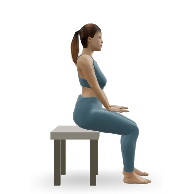
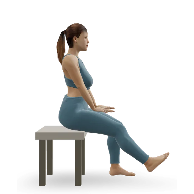
Ankle circles demonstrated on the 3D figure3D demoAnkle circlesBeginner · No equipment

10 slow circles each way with each foot, a few times a day · The move page's dose, an editorial starting point; the page notes an NHS leaflet's advice to move a stiff ankle regularly through the day

Harder: Ankle CARs (controlled articular rotations)

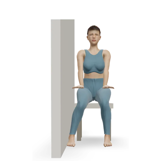
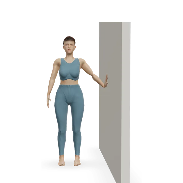
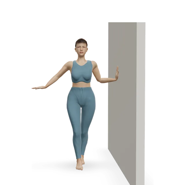
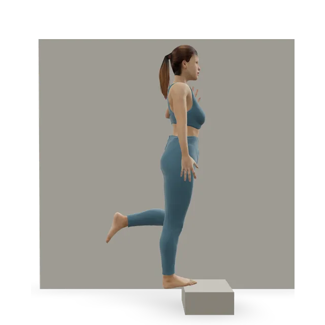
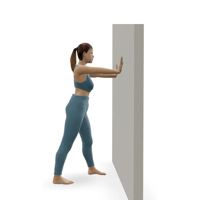
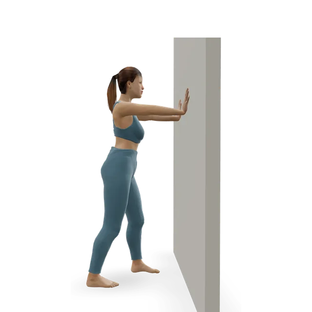
Isometric ankle eversion against a wall demonstrated on the 3D figure3D demoIsometric ankle eversion against a wallBeginner · A wall + A bench or box

8 to 10 holds of about 6 s, 2 to 3 times a day · As in our peroneal guide (an NHS leaflet's belt push). Not until a fifth metatarsal base fracture has healed (our broken foot guide's caution), and gently, with no sharp pain at the bump, in a child (our Iselin's guide)

Harder: Band ankle eversion

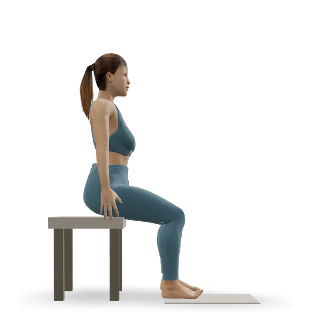
Seated bent-knee calf raise (soleus raise) demonstrated on the 3D figure3D demoSeated bent-knee calf raise (soleus raise)Beginner · A bench or box

2 to 3 at a time, little and often, building to sets of 8, up to 2 sets of 15 · NHS inform's seated heel raise, with its pacing

Harder: Slow calf raise

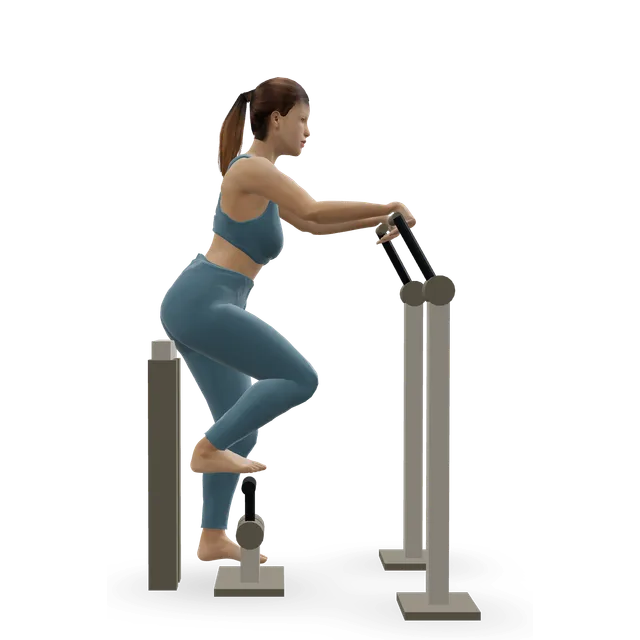
Towel toe curl demonstrated on the 3D figure3D demoTowel toe curlBeginner · Other / check instructions + A bench or box

2 to 3 at a time, building to sets of 8, up to 2 sets of 15 · NHS inform's toe curl, with its pacing

Easier: Big toe press (seated toe-flexor hold) · Harder: Short foot + toe splay

Stationary cycling demonstrated on the 3D figure3D demoStationary cyclingBeginner · Cardio machine

15 to 30 min · As in our peroneal guide, for low-impact fitness; the time is editorial

Easier: Recumbent exercise bike · Harder: Air bike intervals

Move on when: Walking is comfortable, two-leg calf raises stay within the pain rule the next morning (the peroneal guide's check), and pressing the bump halfway along the outer edge no longer hurts (our suggestion).

Follow along: a timer, a cue and the next move, one step at a time.
Stage 2

Build: the outer-ankle muscles, the calf and balance

About weeks 2 to 6: load the tendons with bands and single-leg raises, and train balance. An unsupervised 8-week home balance programme cut repeat sprains from 33% to 22% in a trial.

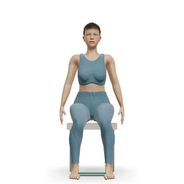
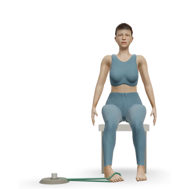
Band ankle eversion demonstrated on the 3D figure3D demoBand ankle eversionBeginner · A band + A bench or box

3 sets of 10 at 3 to 5 s a rep, rising to 4 sets of 10, with a heavier band every 2 weeks, 3 times a week · As in our peroneal and chronic ankle instability guides (an ankle instability trial's progression). Only once any fifth metatarsal base fracture has healed, and pain-free at the bump in a child

Easier: Isometric ankle eversion against a wall · Harder: Single-leg stand

Band ankle inversion demonstrated on the 3D figure3D demoBand ankle inversionBeginner · A band + A bench or box

3 sets of 10 at 3 to 5 s a rep, rising to 4 sets of 10 with a heavier band, 3 times a week · The move page's dose for an ankle that keeps giving way, from the trial it credits

Harder: Single-leg calf raise from a step

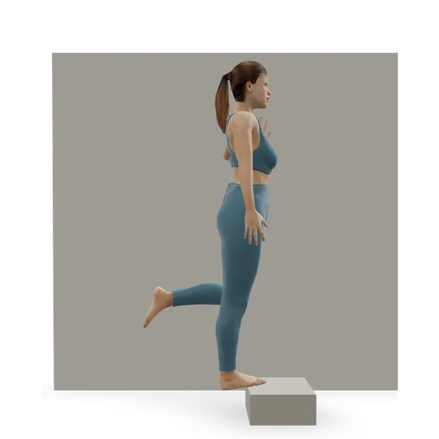
Single-leg calf raise from a step demonstrated on the 3D figure3D demoSingle-leg calf raise from a stepIntermediate · A step + A wall

Up to 10, building to 2 sets of 10, then 4 sets of 15 by about week 6; on flat ground first · As in our peroneal guide

Easier: Slow calf raise · Harder: Single-leg pogo hops

Single-leg stand demonstrated on the 3D figure3D demoSingle-leg standBeginner · A wall

3 holds of 30 s each leg: eyes open near a support first, then eyes closed or on a cushion · As in our peroneal guide (editorial there)

Easier: Tandem stance and heel-to-toe walk · Harder: Single-leg reach matrix

Single-leg reach matrix demonstrated on the 3D figure3D demoSingle-leg reach matrixIntermediate · No equipment

3 rounds of 5 reaches in each direction, each leg · The move page's dose, an editorial starting point. Shallow reaches in the first weeks after a sprain, as the move page says

Easier: Standing crossed-leg side reach · Harder: Single-leg Romanian deadlift

Move on when: 3 sets of 15 single-leg raises and your heaviest band stay within the pain rule (the peroneal guide's check), and you can stand on the sore leg for 30 seconds with your eyes closed without touching down (the chronic ankle instability guide's check).

Follow along: a timer, a cue and the next move, one step at a time.
Stage 3

Hops, then walking, running and side-to-side sport come back

From about week 6: impact and side-to-side movement in small steps, building time before speed, while everyday activities come back by the written steps below. After a fracture, only once your team says it has healed and the old break is pain-free.

Ankle bounces (mini pogo) demonstrated on the 3D figure3D demoAnkle bounces (mini pogo)Beginner · No equipment

3 sets of 20 · As in our peroneal guide. Only once any bone pain has gone, as the move page says

Harder: Pogo hops

Pogo hops demonstrated on the 3D figure3D demoPogo hopsIntermediate · No equipment

3 sets of 20 · As in our peroneal guide, once ankle bounces are easy

Easier: Ankle bounces (mini pogo) · Harder: Single-leg pogo hops

Single-leg hop and stick demonstrated on the 3D figure3D demoSingle-leg hop and stickIntermediate · No equipment

3 sets of 5 each direction · As in our peroneal and chronic ankle instability guides

Easier: Standing broad jump with stuck landing · Harder: Alternate-leg bounding

Lateral cone hops demonstrated on the 3D figure3D demoLateral cone hopsIntermediate · Other / check instructions

3 sets of 10 side to side · As in our peroneal guide; side to side last

Easier: Single-cone quick-feet drill · Harder: Single-leg cone hops

Treadmill jogging demonstrated on the 3D figure3D demoTreadmill joggingIntermediate · Cardio machine

Every other day for 30 min: jog 1 and walk 1 minute in week 1, 2 and 1 in week 2, 4 and 1 in week 3, 9 and 1 in week 4, 15 and 1 in week 5, then 30 minutes' jogging in week 6 · The NHS sports medicine service's programme: up a level after a week without pain, down a level with pain. Flat ground outdoors counts

Easier: Treadmill walking · Harder: Sprint intervals

Single-leg calf raise from a step demonstrated on the 3D figure3D demoSingle-leg calf raise from a stepIntermediate · A step + A wall

3 sets of 15, twice a week · As in our peroneal guide, to keep

Easier: Slow calf raise · Harder: Single-leg pogo hops

Move on when: Our suggestion: walking on uneven ground, 30 minutes' continuous jogging and hopping side to side stay within the pain rule the next morning; then your sport. Keep the balance work going 3 times a week; if a busy week flares it, drop back a stage for a few days rather than stopping.

Follow along: a timer, a cue and the next move, one step at a time.

What to change for now

What to change for now (written steps)

Our 3D figure can't show these changes. They come from the NHS, the peroneal consensus, the AAOS, a children's hospital and Diabetes UK; where a step is our suggestion, it says so.

  • 1. After a roll: check the Ottawa points first. For a sprain, rest, ice for up to 20 minutes every 2 to 3 hours, compress and raise it for the first 2 to 3 days, then keep it moving once pain doesn't stop you (the NHS).
  • 2. Shoes for the tendons: supportive, running-style trainers or walking boots with a slight heel rather than flat, hard or slip-on shoes or sandals (our peroneal guide's NHS leaflet advice).
  • 3. Shoes for a bump: wide, deep shoes and a pad over a tailor's bunion (the AAOS); properly fitting shoes, not tight or narrow boots or spikes, for a child's bump (a children's hospital).
  • 4. Surfaces: flat ground before cambered roads and rough trails while the tendons are sore (our suggestion, as our peroneal guide advises).
  • 5. A tendon problem: keep moving and loading it once it's painless; the international consensus advises against keeping the ankle still, so a boot only on a clinician's advice.
  • 6. A fracture: follow your fracture clinic. Most fifth metatarsal fractures are treated with walking as comfort allows in a cast or boot (the AAOS), and an avulsion often in a bandage or supportive shoe.
  • 7. Diabetes: check your feet every day, and see your foot team within 24 hours for any new problem (Diabetes UK).

Sources 2, 1, 6, 26, 29, 9, 34

Walking, running, side-to-side sport and shoes come back (written steps)

No trial has tested how to bring these back after pain on the outer side of the foot. These steps are our suggestion unless a source is named.

  • 1. Shoes: keep the supportive trainers, and replace running shoes every 300 to 500 miles (the OxSport leaflet).
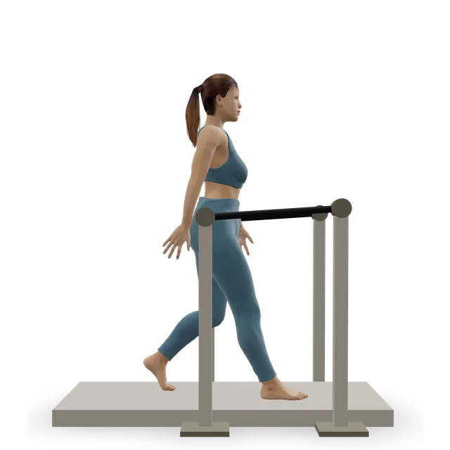
  • 2. Walking: add distance before slopes, cambers and rough ground, one change at a time.
  • 3. Running: start once 30 minutes of brisk walking is pain-free, then follow the walk-jog weeks in stage 3; after week 6, no more than about 10% more a week (the OxSport leaflet).
  • 4. Side-to-side sport: straight lines before cutting, then hops and landings in each direction, then sport. Not being able to jump and land well 2 weeks after a first sprain went with lasting instability, so the hops come before the matches.
  • 5. Keep the balance work going: a home balance programme cut repeat sprains in a trial, and our chronic ankle instability guide keeps a balance warm-up 3 times a week, with a brace for court and field sports if the ankle keeps going over.
  • 6. After a fifth metatarsal fracture: the old break should be pain-free during, after and the next morning before any hopping. With a Jones fracture in an athlete, the specialist's plan decides; in elite footballers, time out was similar with or without surgery.
  • 7. If it isn't improving: see a GP if ankle or foot pain isn't better after 2 weeks of looking after it at home, is getting worse or keeps coming back (the NHS), and ask for a review if one spot still hurts 6 weeks after a sprain, because a small fracture or a tendon injury can be missed at first.

Sources 40, 20, 36, 12, 1

Positions to swap, and why

Habit or positionWhy it mattersInstead
Walking off a twist when the bump halfway along the outer edge is tenderTenderness there, or not managing 4 steps, is one of the Ottawa signs for an X-ray, and avulsion and Jones fractures happen at that bump.A same-day check.
Resting a sprain or a tendon problem for six to eight weeksThe NHS keeps a sprain moving once pain doesn't stop you, and the peroneal consensus advises against keeping the ankle still.The staged plan from the first week.Try: Ankle circles
A boot or cast for outer ankle tendon pain without a clinician's adviceThe consensus advises against immobilisation; a specialist review keeps a boot for stubborn cases.Keep walking in supportive trainers, and load the tendon gently.Try: Isometric ankle eversion against a wall
Flat, hard or slip-on shoes or sandals with outer ankle tendon painOur peroneal guide's NHS leaflet advises against them for this problem.Running-style trainers or walking boots with a slight heel.
Narrow, tight or pointed shoes with a tailor's bunionChanging footwear comes before surgery, the AAOS says, and tight shoes went with bunionettes in a large survey.A wide, deep toe box and a pad over the bump.
Turning the foot out against a band before a fifth metatarsal base fracture has healedThe muscle that turns the foot out attaches near the break (our broken foot guide's caution).Band work turning the foot in.Try: Band ankle inversion
Hopping or side-to-side drills with one-spot bone pain on the outer edgeThe base of the fifth metatarsal is a stress fracture site that heals less well, and as long as you can feel the pain, the bone is still fragile.Get it checked; cycle only if it's pain-free.Try: Stationary cycling
Cambered roads or rough trails early onThey put extra load on the outer ankle (our peroneal guide's caution).Flat surfaces or a treadmill first, trails last.Try: Treadmill jogging
Dropping the balance exercises once a sprain stops hurtingA home balance programme cut repeat sprains from 33% to 22% in a trial.Keep a balance warm-up going 3 times a week.Try: Single-leg stand
Asking for a steroid injection into the outer ankle tendonsInjections there carry a rupture risk; one specialist unit doesn't use them, and the consensus couldn't agree.Loading exercise, and shockwave after 3 months if a clinician advises it.

Routines to follow

More moves for this area

Questions people ask

What causes pain on the outside of the foot?

After a twist, the usual causes are a sprain, a break at the base of the fifth metatarsal (the bump halfway along the outer edge) or a tendon injury. Without an injury, the main candidates are the peroneal tendons behind and below the outer ankle bone, a stress fracture of the fifth metatarsal, cuboid syndrome and a tailor's bunion at the base of the little toe. The exact spot points to the cause, which is what the map on this page is for.

Why does the outside of my foot hurt when I walk?

Pain behind and below the outer ankle bone that's worse walking, on uneven ground and turning points to the peroneal tendons; pain at the base of the little toe in tight shoes is often a tailor's bunion. An ache on the outer edge that builds with walking or running, then at rest, can be a stress fracture and needs checking.

Why does the outside of my foot hurt after running?

The common running causes are the peroneal tendons, a stress fracture at the base of the fifth metatarsal and cuboid syndrome. An ache in one spot that builds over runs, or hurts at night, needs checking before you run again: 45% of professional footballers with a fifth metatarsal fracture had warning symptoms first. Come back with the NHS walk-jog programme once 30 minutes of fast walking is pain-free.

Is it a stress fracture or tendonitis?

A stress fracture usually hurts in one spot on a bone that you can point to with a fingertip, builds with activity and can ache even at night. Peroneal tendon pain runs along the tendons behind and below the outer ankle bone towards the outer edge of the foot, often after sprains, and can come with clicking. If you're not sure, stop the activity that hurts and get it checked, because early X-rays often miss a stress fracture.

Can you walk on a broken fifth metatarsal?

Often, yes, which is why being able to walk doesn't rule out a break: tenderness on the bump halfway along the outer edge is enough for the Ottawa rules to ask for an X-ray. Most fifth metatarsal fractures are treated with walking as comfort allows in a boot or cast, and an avulsion fracture of the bump healed as well in an elastic bandage as in a cast in a trial. A Jones fracture, just beyond the bump, heals more slowly and is sometimes operated on.

Why does the outside of my foot still hurt months after a sprain?

It's common: 5% to 33% of people still had pain a year after a sprain. A second injury is often the reason: in 61 people having surgery for an unstable ankle, none had a ligament injury alone, and 77% had inflamed peroneal tendon sheaths, while small fractures of the heel bone can be missed on X-rays. Ask for a review if one spot still hurts 6 weeks after a sprain.

What is the bony bump on the outside of my foot?

At the base of the little toe, it's usually a tailor's bunion (bunionette), much like a bunion and often rubbed by narrow shoes. Halfway along the outer edge, it's the base of the fifth metatarsal: tenderness there after a twist needs an X-ray check, and in an active child aged about 8 to 14, pain there can be Iselin's disease.

What is cuboid syndrome?

It's thought to be a subtle disturbance of the joint between the cuboid and the heel bone, causing pain on the outer side of the midfoot that resembles a sprain. There's no definitive test, and the evidence for treating it with manipulation is case reports and one series of 7 athletes, all of whom returned to sport after one or two treatments. A fracture has to be ruled out first.

What exercises help pain on the outside of the foot?

Once the warning signs are ruled out, gentle movement and NHS inform's foot exercises first, then strength for the muscles that turn the foot out and in, calf raises and balance work: a home balance programme cut repeat sprains from 33% to 22% in a trial. The international consensus on peroneal tendon problems recommends tendon-loading exercise once it's painless and advises against keeping the ankle still.

Should I rest my foot for 6 to 8 weeks?

Usually not. Some popular pages advise six to eight weeks of rest for a sprain, peroneal tendonitis and cuboid syndrome, but the NHS keeps a sprain moving after the first 2 to 3 days, and the peroneal consensus advises against keeping the ankle still. Fractures and stress fractures are the exception and follow the fracture clinic's plan.

What shoes help pain on the outside of the foot?

Supportive, running-style trainers with a slight heel rather than flat, hard or slip-on shoes while the tendons are sore; a wide, deep toe box and a pad for a tailor's bunion; and shoes that don't press on the bump for a child with Iselin's disease. Insoles that tilt the outer forefoot can help mild peroneal problems, a specialist review says.

When should I worry about pain on the outside of my foot?

Call 999 or go to A&E after an injury if you heard a crack, the foot or ankle has changed shape or points at an odd angle, it's numb or tingling, or the skin has turned blue or grey or is cold. Contact NHS 111 for severe pain, feeling faint, dizzy or sick from the pain, a snap, grinding or popping noise at the time, or not being able to walk, and after a twist, get pain on the bump halfway along the outer edge checked for an X-ray. See a GP if it hasn't improved after 2 weeks, keeps coming back, or you have diabetes.

Related exercise guides

Other pain guides

Sources

  1. NHS · Ankle pain
  2. NHS · Sprains and strains
  3. Crim 2022 · The painful outer column of the foot (imaging review, Skeletal Radiol)
  4. NHS inform · Foot and toe problems
  5. Davda 2017 · Peroneal tendon disorders (review, EFORT Open Rev)
  6. van Dijk 2018 · The ESSKA-AFAS international consensus on peroneal tendon problems (consensus statement, Knee Surg Sports Traumatol Arthrosc)
  7. Tu 2018 · Heel pain: diagnosis and management (clinical review, American Family Physician)
  8. Adams Morancie 2025 · Heel pain: diagnosis and management, the December 2025 update (clinical review, American Family Physician; first part read)
  9. American Academy of Orthopaedic Surgeons (OrthoInfo) · Toe and forefoot fractures
  10. Herterich 2021 · Treating fractures of the base of the fifth metatarsal (systematic review, Dtsch Arztebl Int)
  11. Akimau 2016 · An elastic bandage against a cast for avulsion fractures of the fifth metatarsal in 60 people (RCT, Bone Joint J)
  12. Ekstrand 2013 · Fifth metatarsal fractures in male professional footballers (cohort study, Br J Sports Med)
  13. American Academy of Orthopaedic Surgeons (OrthoInfo) · Stress fractures of the foot and ankle
  14. NICE NG38 · Fractures (non-complex): assessment and management (2016)
  15. Stiell 1993 · Decision rules for X-rays in acute ankle injuries: refinement and prospective validation (JAMA)
  16. Bachmann 2003 · Accuracy of the Ottawa ankle rules to exclude fractures of the ankle and midfoot (systematic review, BMJ)
  17. van Rijn 2008 · How acute ankle sprains go: 31 studies (systematic review, Am J Med)
  18. DiGiovanni 2000 · Injuries found alongside chronic ankle instability in 61 people having surgery (case series, Foot Ankle Int)
  19. Gribble 2016 · How common lateral ankle sprains are, and their long-term effects (consensus evidence review, Br J Sports Med)
  20. Doherty 2016 · Recovery from a first ankle sprain, and what predicted lasting instability, in 82 people (cohort study, Am J Sports Med)
  21. Martin 2021 · Lateral ankle ligament sprains (clinical practice guideline, J Orthop Sports Phys Ther; abstract read)
  22. Ouellette 2006 · Fractures of the front of the heel bone on MRI in 2,577 people with foot and ankle symptoms (imaging series, Skeletal Radiol)
  23. Durall 2011 · Examination and treatment of cuboid syndrome (literature review, Sports Health)
  24. Jennings 2005 · Treating cuboid syndrome after ankle sprains in 7 athletes (case series, J Orthop Sports Phys Ther)
  25. Sobel 1994 · Painful os peroneum syndrome (case review, Foot Ankle Int)
  26. American Academy of Orthopaedic Surgeons (OrthoInfo) · Bunions (and bunionettes)
  27. Şaylı 2018 · Forefoot deformities in 2,662 adults in Turkey (survey, Acta Orthop Traumatol Turc)
  28. Nestor 1990 · The X-ray anatomy of the painful bunionette (comparative study, Foot Ankle)
  29. Nationwide Children's Hospital · Apophysitis of the 5th metatarsal (Iselin's disease)
  30. NHS · Septic arthritis
  31. NHS · Cellulitis
  32. NHS · Broken toe
  33. NICE NG19 · Diabetic foot problems: prevention and management (2015, updated 2019)
  34. Diabetes UK · Signs of serious foot problems and foot infections
  35. Health New Zealand · Stress fractures of the foot and shin bone
  36. Hupperets 2009 · An unsupervised home balance programme after an ankle sprain in 522 athletes (RCT, BMJ)
  37. NHS inform · Exercises for foot problems
  38. Silbernagel 2007 · Carrying on with sport under a pain-monitoring model for Achilles tendon pain in 38 people (RCT, Am J Sports Med)
  39. Smith 2017 · Should exercises be painful in chronic musculoskeletal pain? (systematic review and meta-analysis, Br J Sports Med)
  40. Oxford University Hospitals NHS Foundation Trust (OxSport) · Return to running programme (patient leaflet, September 2024)

Exercise education, not a diagnosis or treatment plan. It is written from the guidelines and studies listed above; review by a clinician is pending. If your pain follows an injury, is severe, is getting worse, or comes with any warning sign listed on this page, get it assessed. Health disclaimer.